Brazilian Journal of Oral Sciences
ISSN 1677-3225
Braz. J. Oral Sci. vol.10 no.4 Piracicaba oct./dic. 2011
ORIGINAL ARTICLE
Bone marrow mesenchymal cell adhesion to polished and nitrided titanium surfaces
Luciana Bastos AlvesI; Fernanda GinaniII; José Sandro Pereira da SilvaIII; Clodomiro Alves JuniorIV; Carlos Augusto Galvão BarbozaV
I PhD candidate, Department of Dentistry, Federal University of Rio Grande do Norte, Brazil
II MSc candidate; Department of Morphology, Federal University of Rio Grande do Norte, Brazil
III PhD, Department of Dentistry, Federal University of Rio Grande do Norte, Brazil
IV PhD, Department of Mechanical Engineering, Federal University of Rio Grande do Norte, Brazil
V PhD, Department of Morphology, Federal University of Rio Grande do Norte, Brazil
ABSTRACT
Aim: To evaluate the adhesion of mouse bone marrow mesenchymal cells (MBMMC) on different titanium surfaces. Methods: Grade II titanium discs (ASTM F86) received two different surface treatments: polished (S1) and cathodic cage plasma nitriding (S2). MBMMC were cultured on titanium discs in 24-well cell culture plates, at a density of 1 x 10⁴ cells per well. After 24 h, the adhesion was evaluated using a hemocytometer. Results: The mean adhesion was greater on S2, though without statistically significant difference from S2 (p>0.05). Conclusions: It was demonstrated that titanium surface treatment with ionic nitriding in a cathodic cage is biocompatible since it preserved the integrity of the cultivated MBMMC for a period of 24 h, allowing their adhesion.
Keywords: mesenchymal stem cells, bone marrow, titanium.
Introduction
Titanium (Ti) is currently considered the biomaterial of choice for the manufacture of intra-osseous implants because this metal has exceptional physical and chemical properties that allow its installation in living tissues with no incompatibility among them. Ti is a very stable metal, although slight oxide formation occurs on its surface. This formation helps deposition and adhesion of the extracellular matrix on the bone-implant interface. These oxides form during the cutting, cleaning, and implant sterilization process and remain adherent to the surface. The foregoing properties allow the scarring and maintenance of tissue structure adhesion to the Ti surface1.
A wide range of approaches have been developed to thoroughly investigate cellular behavior on Ti surfaces. Maeda et al.2 (2007) observed that cell adhesion and proliferation, as well as the osteogenic differentiation of mouse mesenchymal stem cells (MSCs) to Ti discs were significantly similar to those on the plastic surface of the culture, indicating Ti as an excellent material for repairing hard tissue in the field of bone tissue engineering.
However, the interaction between cells and some biomaterials, or the biocompatibility, depends on the material surface properties, such as energy, texture, roughness, and chemical composition. These properties determine the adhesion and behavior of cells in contact with the surface. The term "adhesion" to the biomaterial refers to the most important phase, since the quality of it will influence morphology and the capacity of cell proliferation and differentiation3.
The physicochemical properties of Ti implant surfaces are fundamental to the success of osseointegration. To improve the biological responses for obtaining rapid osseointegration, the surfaces have been modified by a wide range of process involving mechanical, chemical, and physical surface treatment methods, such as: the Ti plasma spray treatment, Ti oxide blasting, laser deposition of Ti carbide, and acid conditioning4.
Ionic nitriding is a surface treatment method which exhibits excellent mechanical properties, chemical stability, and biocompatibility when applied to Ti. This process, also known as plasma nitriding, consists of an ionizing gas or a gaseous mixture containing nitrogen using glow-discharge generated by a difference in potential between the sample (cathode) and the anode in a low pressure atmosphere. The ions produced in the plasma are accelerated towards the sample (cathode), colliding with it, and supplying enough energy to heat it to the nitriding temperature5.
The basic concept in using ionic nitriding to improve Ti surface properties is based on the possibility of forming nitrides or carbides below the alloy surface. Ti nitrides and carbides are brittle materials that improve tribologic surface properties; that is, they increase resistance to corrosion and surface roughness6.
Accordingly, the association of Ti implants with bone tissue culture may contribute to bone tissue regeneration7. Therefore, the present study aimed to evaluate the adhesion capacity of mouse bone marrow MSCs to smooth and plasmanitrided Ti surfaces in the cathodic cage configuration.
Material and methods
The present study was approved by the Research Ethics Committee of the Federal University of Rio Grande do Norte (UFRN; protocol 008/09) and was divided into two stages. First, sample preparation was carried out using the surface treatment of Ti plates. A test was then performed in vitro with isolated mouse bone marrow cells cultivated on Ti discs.
Sample preparation
Grade II titanium discs (ASTM F86), 15 mm in diameter and 1.5 mm thick, were prepared according to the protocol established by Alves Jr et al.8 (2006). All the discs (n=12) were polished and then six discs were submitted to cathodic cage nitriding. A study of these surface characteristics have been previously published by da Silva et al.9 (2011). The discs were subsequently sterilized by gamma radiation (25 kGy dose) released at a mean dose of 8.993 kGy/h (2h 46 min at a distance of 50 mm), in a GAMMACELL 220 Excel irradiator (MDS Nordion, Ottawa, ON, Canada).
Bone marrow cell culture
Bone marrow was extracted from two male Swiss albino mice in accordance with the protocol established by Maniatopoulos et al.10 (1988). After anesthesia, the animals were dissected under aseptic conditions for femur and tibia removal.
The marrow cavity was flushed out with α-MEM medium containing 50 mg/L of gentamicin sulfate and 2 mg/L of amphotericin B (Cultilab, Campinas, Brazil) and supplemented with 10% fetal bovine serum (FBS; Gibco, Carlsbad, CA, USA). The extracted marrow was cultivated in basic medium (α-MEM 10% FBS) for 72 h in a humid atmosphere with 5% CO2 at 37ºC. After this stage, the medium was changed, thus making it possible to remove the non-adhered cells from the culture, and subsequent medium changes were performed every 3-4 days.
In order to confirm the multi-lineage differentiation potential of periodontal ligament cells, aliquots of P1 cells were cultured in osteogenic, chondrogenic, or adipogenic differentiation media (StemPro® Differentiation Kits, Invitrogen Corp., Carlsbad, CA, USA) for up to 21 days. By light microscopy, the cells showed typical osteoblast/ osteocyte, chondroblast, and adipocyte morphology and produced characteristic extracellular matrix components.
Bone marrow cell culture on Ti discs
Bone marrow cells were cultivated in two 24-wells plates (TTP®), with a density of 1x10⁴ cells per well. Twelve Ti discs were used, six from each group (polished and cathodic cage). The same cell density was cultivated in six wells without discs, as a positive control of cell proliferation. The disks are the same size of the well, so the growth area of disks and controls are the same.
Cell viability and adhesion
Data obtained by counting the cells that adhered to the Ti surfaces (the polished group [S1], and the cathodic cage group [S2]), in the 24-h period after plating were used to analyze cell adhesions. The number of cells collected from each well was obtained from a viable cell count using a hemocytometer and the trypan blue dye exclusion method. All the titanium samples were also evaluated by scanning electron microscopy to check the reproducibility of the results, according to the protocol established by Guerra Neto et al.11 (2009).
Statistical Analysis
The data were subjected to non-parametric analysis. Each counting value corresponds to the mean ± standard deviation of the mean (SD) of six samples per group. The differences between the groups were compared by the Mann Whitney statistical test. A statistical difference was considered when p<0.05.
Results
Results of mouse bone marrow cell adhesion to different titanium surfaces shows that the mean adhesion among mouse bone marrow cells was greater in the cathodic cage group [S2] (1.83 ± 0.83) than in the polished group [S1] (1.03 ± 0.26). However, no statistically significant difference was observed (p=0.12) between the two groups ( Table 1).
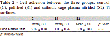
The control surface (plastic) showed the best result (2.32 ± 0.78), since it is the gold standard surface for cell adhesion and proliferation. However, no statistically significant difference was observed either (p=0.16) among the three groups ( Table 2).


Discussion
Mesenchymal cells were first isolated from a cell suspension of bone marrow by Friedenstein and collaborators in the early 1970's, and classified as adherent, fibroblastic and clonogenic cells, and it were initially called colony forming units – fibroblastic (CFU-F)12. This type cell is found in the bone marrow and it is called MSC13-15. From the extraction and culture of bone marrow of mouse is possible to obtain a population of adherent cells with fibroblastic, elongated, spindle-shaped and pointed, called multipotent mesenchymal stromal cells, according to the nomenclature proposed by the International Society for Cellular Therapy in 200516.
In the present experiment, the adhesion capacity of mouse bone marrow mesenchymal cells to different Ti surfaces was analyzed under conditions of cell cultivation. The plastic surface or polystyrene cell culture plate was used as a positive control, since it is the standard surface used in cell cultivation owing to its excellent hydrophilic characteristics2.
Material surface characteristics play an important role in cell adhesion because they provide the necessary adhesion conditions to adsorb and help in the cell adhesion process3. Experimental studies7,17 comparing different surface types concluded that the best results were those obtained with textured surfaces, and that bone marrow cells and osteoblasts cultivated on different Ti surfaces adhere and respond better on rougher surfaces18-23. However, other evaluations of in vitro biocompatibility of Ti using cell culture have indicated that cell attachment was not affected by surface roughness24-26.
In this study, ionic nitriding or the plasma nitriding technique was used. Experiments11,22 with a cell line model (osteo-1 lineage and L929 mouse fibroblasts) demonstrated that cell adhesion to the Ti surface was favored by the low energy ion irradiation surface treatment (plasma).
In agreement with the related literature, it was possible to observe that adhesion and an initial interaction between cell and substrate occurred irrespective of the surface. The best cell adhesion results were obtained by the control surface (plastic) according to the results obtained by Santiago et al.25 (2005) and Resende et al.27 (2010), who showed no statistically significant difference among the different titanium surfaces and a larger number of cells on the polystyrene surface.
Even though more experiments are needed to explain the Ti cell adhesion mechanism, the results suggest that Ti surface characteristics are similar to those of a plastic surface, resulting in good cell adhesion capacity, in accordance with Maeda et al.2 (2007). This result reinforces the argument that ionic nitriding treatment to the surface (S2) may contribute to better adhesion of bone marrow mesenchymal cells, corroborating a number of studies on roughness and wettability3.
Further studies, analyzing the capacity of proliferation and differentiation of these types of cells when in contact with the biomaterial may contribute to an understanding of the osseointegration process. Furthermore, molecular studies that analyze the types of adhesion bonds involved might be important in explaining the mechanism by which each cell type adheres to different surfaces.
References
1. Hansson HA, Albrektsson T, Branemark PI. Structural aspects of the interface between tissue and titanium implants. J Prosthet Dent. 1983; 50: 108-13. [ Links ]
2. Maeda M, Hirose M, Ohgushi H, Kirita T. In vitro mineralization by mesenchymal stem cells cultured on titanium scaffolds. J Biochem. 2007; 141: 729-36.
3. Stiehler M, Lind M, Mygind T, Baatrup A, Dolatshahi-Pirouz A, Li H, et al. Morphology, proliferation, and osteogenic differentiation of mesenchymal stem cells cultured on titanium, tantalum, and chromium surfaces. J Biomed Mater Res A. 2008; 86: 448-58.
4. Silva MAM, Martinelli AE, Alves Jr C, Nascimento RM, Távora MP, Vilar CD. Surface modification of Ti implants by plasma oxidation in hollow cathode discharge. Surf Coat Technol. 2005; 200: 2612-26.
5. O'Brien JM, Goodman D. Plasma (ion) nitriding. In: ASM International Handbook Committee, editors. ASM Handbook: heat treating. Utah: International Library Service; 1991. p.420-4.
6. Yilbas BS, Sahin AZ, Al-Garni AZ, Said SAM, Ahmed Z, Abdulaleem BJ, et al. Plasma nitriding of Ti-6Al-4V alloy to improve some tribological properties. Surf Coat Technol. 1996; 80: 287-92.
7. Franzolin SOB, Francischone CE, Bittencourt REC, Felisbino SL, Deffune E. Diferenciação de célula-tronco hematopoética periférica humana em osteoblasto sobre diferentes superfícies de implantes de titânio. Rev Dent Press Periodont Implantol. 2008; 2: 68-79.
8. Alves Jr C, de Araújo FO, Ribeiro KJB, da Costa JAP, Souza RRM, de Sousa RS. Use of cathodic cage in plasma nitriding. Surf Coat Technol. 2006; 201: 2450-4.
9. da Silva JS, Amico SC, Rodrigues AO, Barboza CA, Alves C Jr, Croci AT. Osteoblastlike cell adhesion on titanium surfaces modified by plasma nitriding. Int J Oral Maxillofac Implants. 2011; 26: 237-44.
10. Maniatopoulos C, Sodek J, Melcher AH. Bone formation in vitro by stromal cells obtained from bone marrow of young adult rats. Cell Tissue Res. 1988; 254: 317-30.
11. Guerra Neto CLB, da Silva MAM, Alves Jr C. In vitro study of cell behaviour on plasma surface modified titanium. Surf Eng. 2009; 25: 146-50.
12. Friedenstein AJ, Piatetzky-Shapiro II, Petrakova KV. Osteogenesis in transplants of bone marrow cells. J Embryol Exp Morphol. 1966; 16: 381-90.
13. Pittenger MF, Mackay AM, Beck SC, Jaiswal RK, Douglas R, Mosca JD et al. Multilineage potential of adult human mesenchymal stem cells. Science. 1999; 284: 143-7.
14. Caplan AI. The mesengenic process. Clin Plast Surg. 1994; 21: 429-35.
15. Donzelli E, Salvadè A, Mimo P, Viganò M, Morrone M, Papagna R et al. Mesenchymal stem cells cultured on a collagen scaffold: In vitro osteogenic differentiation. Arch Oral Biol. 2007; 52: 64-73.
16. Horwitz EM, Le Blanc K, Dominici M, Mueller I, Slaper-Cortenbach I, Marini FC et al. Clarification of the nomenclature for MSC: The International Society for Cellular Therapy position statement. Cytotherapy. 2005; 7: 393-5.
17. Buser D, Schenk RK, Steinemann S, Fiorellini JP, Fox CH, Stich H. Influence of surface characteristics on bone integration of titanium implants. A histomorphometric study in miniature pigs. J Biomed Mater Res. 1991; 25: 889-902.
18. Anselme K, Bigerelle M, Noel B, Iost A, Hardouin PJ. Effect of grooved titanium substratum on human osteoblastic cell growth. J Biomed Mater Res. 2002; 60: 529-40.
19. Deligianni DD, Katsala N, Ladas S, Sotiropoulou D, Amedee J, Missirlis YF. Effect of surface roughness of the titanium alloy Ti-6A1-4V on human bone marrow cell response and on protein adsorption. Biomaterials. 2001; 22: 1241-51.
20. Keller JC. Tissue compatibility to different surfaces of dental implants: in vitro studies. Implant Dent. 1998; 7: 331-7.
21. Perizzolo D, Lacefield WR, Brunette DM. Interactions between topography and coating in the formation of bone nodules in culture for hydroxyapatiteand titanium- coated micromachined surfaces. J Biomed Mater Res. 2001; 56: 494-503.
22. Abidzina V, Deliloglu-Gurhan I, Ozdal-Kurt F, Sen BH, Tereshko I, Elkin I, et al. Cell adhesion study of the titanium alloys exposed to glow discharge. Nucl Instr and Meth Phys Res B. 2007; 261: 624-6.
23. Vasconcellos LM, Leite DO, Oliveira FN, Carvalho YR, Cairo CA. Evaluation of bone ingrowth into porous titanium implant: histomorphometric analysis in rabbits. Braz Oral Res. 2010; 24: 399-405.
24. Rosa AL, Beloti MM. Effect of cpTi surface roughness on human bone marrow cell attachment, proliferation, and differentiation. Braz Dent J. 2003; 14: 16-21.
25. Santiago AS, Santos EA, Sader MS, Santiago MF, Soares Gde A. Response of osteoblastic cells to titanium submitted to three different surface treatments. Braz Oral Res 2005; 19: 203-8.
26. Silva TS, Machado DC, Viezzer C, Silva Júnior AN, Oliveira MG. Effect of titanium surface roughness on human bone marrow cell proliferation and differentiation: an experimental study. Acta Cir Bras. 2009; 24: 200-5.
27. Resende CX, Lima IR, Gemelli E, Granjeiro JM, Soares GA. Cell adhesion on different titanium-coated surfaces. Materia 2010; 15: 386-91.
 Correspondence:
Carlos Augusto Galvão Barboza
Universidade Federal do Rio Grande do Norte
Centro de Biociências – Departamento de Morfologia
Av. Salgado Filho, 3000 – Campus Universitário Natal/RN, Brasil
59072-970
E-mail: cbarboza@cb.ufrn.br
Received for publication: July 07, 2011
Accepted: December 07, 2011


