Serviços Personalizados
Artigo
Links relacionados
Compartilhar
Revista Brasileira de Odontologia
versão On-line ISSN 1984-3747versão impressa ISSN 0034-7272
Rev. Bras. Odontol. vol.74 no.1 Rio de Janeiro Jan./Mar. 2017
Short Communication/Oral Pathology
Unusual association between cheilitis glandularis and actinic cheilitis
Juliana Tristão WerneckI; Taiana Campos LeiteI; Ana Maria de Oliveira MirandaI; Eliane Pedra DiasI; Karin Soares Gonçalves CunhaI; Arley Silva JúniorI
I Postgraduate Program in Pathology, Medical School, Universidade Federal Fluminense, Niterói, RJ, Brasil
ABSTRACT
Objective: our aim is to report the simultaneous occurence of cheilitis glandularis and actinic cheilitis on the lower lip of a middle-aged female patient. Case Report: the patient presented clinical features compatible with these two lesions, confirmed by histopathological exam. Conclusion: the importance of the present case is the rare concomitant occurrence of both conditions, with special concern towards the malignancy potential related to both diseases.
Keywords: Actinic cheilitis; Cheilitis glandularis; Potentially malignant disorder; Squamous cell carcinoma.
Introduction
Cheilitis Glandularis (CG) is a rare chronic inflammatory condition of unknown etiology affecting the minor salivary glands of the lips.1,2 It usually affects the lower lip and presents itself as redness and dilatation of the ostia of minor salivary glands on the vermilion, with variable degrees of macrocheilia, which can be associated with eversion of the lip area.1-3 Under stimulation, the ostia may secrete a thick mucoid material, and, in rare occasions, purulent secretion, as a result of secondary infection of the gland.2,4 Minor salivary glands in the deep and superficial tissues are often palpable.3,5
Another disease that affects the lower lip is Actinic Cheilitis (AC), which is more common than CG, and has chronic sun exposure as its main cause, leading to alterations of the epithelium and of the collagen and elastic fibers.6-8 It is a potentially malignant 9disorder, which can develop into squamous cell carcinoma.7,9 Clinically, it is characterized by the presence of pale areas, erythema, white plaques, and atrophy on the lip vermilion, predominantly on the lower lip.6,7 In later stages, erosion, ulcers, crusting, fissures, and blurring of the vermilion border may develop.8,9
Simultaneous occurrence of these two conditions is rare and their presence in female patients is very uncommon.1,2 Our aim is to report a case of CG, associated with AC with mild epithelial dysplasia in a female patient.
Case Report
A 56-year-old nonsmoker, former-alcoholic, Hispanic female patient sought treatment at our Oral Medicine Clinic, and signed an informed consent before beginning treatment. She complained of itching and burning in the entire lower lip vermilion, and reported that the lesions of the lower lip were present ten years earlier, when she noticed redness on the lower lip, which improved with acyclovir. The lesions appeared and disappeared sporadically, but after eight years, there were no signs of improvement, despite the use of topical triamcinolone acetonide and sun protection factor (SPF) 30 lip sunscreen. She also reported that the lip was often swollen, with crust formation, and that she used to be chronically exposed to sunlight.
Clinical examination revealed no extraoral alterations. The lower lip presented dryness, scaling, edema, blurring of the vermilion border, induration, and eversion. It also presented various lesions, such as erythemas, erosion, ulcers, crusts, fissures, and brown and white plaques, as well as many reddish points of non-purulent secretion in the entire everted mucosa (Figure 1A). Intraoral examination revealed poor oral hygiene.
A clinical diagnosis of AC was established, and the patient was instructed to use lip sunscreen and to avoid excessive sun exposure. During the first appointment, as part of the clinic protocol, scrapes of the lower lip for cytopathological examination, as well as toluidine blue test (TBT), and videoroscopic examination in 50-fold magnification were performed.
The TBT was retentive in the area of glandular ostia and delimitated the everted lip area (Figure 1B). The videoroscopic examination clearly revealed the areas with dryness and crusts, as well as mucous drainage through enlarged ostia (Figure 1C). Cytopathological exam revealed candidiasis associated with mild acute inflammation.
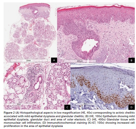
After candidiasis treatment with topical 100,000 units per mL nystatin oral suspension four times a day, for fifteen days, a biopsy was performed. The histopathological exam revealed a squamous metaplastic salivary gland duct, under which there were seromucous minor salivary glands and lymphoplasmatic inflam-

matory infiltrate, which was predominantly perivascular and periductal, characterizing CG.4 There was also solar elastosis in the connective tissue, an important feature of AC (Figure 2A-2C).4 Immunohistochemical staining with anti-ki-67 was performed and showed excessive epithelial cell proliferation in the basal and parabasal layers, which is seen in mild epithelial dysplasia (Figure 2D). Therefore, the final diagnosis was CG associated with AC with mild epithelial dysplasia.

The treatment of choice was topical dexpanthenol ointment four times a day, during 15 days, and daily lip sunscreen during the day, to be reapplied whenever necessary. The CG symptoms disappeared 15 days after the end of the treatment. The patient was seen one year after the end of the treatment and presented no CG lesions.
Discussion
The etiology for CG is unclear, but it has been suggested that it may be an autosomal dominant disease. Excessive sunlight, wind exposure, smoking, poor oral hygiene and compromised immune system may also play a role in its etiopathogenesis.3,4 It remains uncertain, however, whether the origin of CG lies in the gland parenchyma, or if it is in the lip epithelium, in which case the glandular alterations would be secondary to the epithelial damage.1,2 It affects mainly middle-aged and elderly males, and only a few cases have been reported in children and women.3,4
Age, poor oral hygiene, a past history of sun exposure, and tobacco use are factors that could explain the etiology of both conditions in our patient, despite being a female patient, which is an uncommon finding.1,3,5 Nico et al.1 (2010) have found CG associated with AC in the clinical and histopathological exams of three albino patients, however in the literature review we did not find any other case.
Cheilitis glandularis is classified into three types: simplex type, superficial suppurative type, and deep suppurative type.3 However, there are no well-defined clinical criteria, and several clinicians consider them as a progression of the same entity, rather than separate types.2 Because of the difficulty in determining clinical criteria for classification of CG, in the present case, we observed swelling, erosion, dryness, and eversion of the lip, as well as the glandular mucus discharge, and so it was classified as the simplex CG type. This corresponds to a mild behavior, and a better clinical control, despite induration of the lip, which can also be attributed to AC. Patient monitoring becomes essential, considering that CG of deep suppurative type is often considered as a premalignant lesion, with some published cases with development to a squamous cell carcinoma.1,5
Clinically, CG resembles other conditions, and the differential diagnoses include minor salivary gland inflammatory disorders, factitious cheilitis, AC, granulomatous cheilitis, angioedema, and benign and malignant minor salivary gland tumors. Therefore, it is very important that additional exams are performed in order to achieve the correct diagnosis.2,5
The histopathological features of CG are not well defined and usually include unspecific aspects of chronic sialadenits, with ectasic and metaplastic ducts, mucin accumulation, as well as vascular congestion, chronic inflammatory infiltrate and fibrosis.1-3 AC, on the other hand, may present various degrees of epithelial dysplasia, and the connective tissue usually shows basophilic degeneration of collagenous and elastic fibers, called solar elastosis, as well as occasional mild chronic inflammatory cell infiltrate.7,9 The histopathological diagnosis of our case was based on those criteria, although not all findings were present.
The recommended treatment for CG is based on the clinical presentation, and may include the use of topical or intralesional steroids, sunscreen, surgery and antibiotics, if the lesion is infected. 1,4,5 Recurrence after surgery is rare.1,4 There are also several forms of treatment for AC, and patients should always be advised to use lip balm with sunscreen to prevent further damage.6,9 In more severe cases without malignant diagnosis, vermilionectomy, the use of topical tretinoin 5-fluorouracil or topical imiquimod, chemical exfoliation with trichloroacetic acid, or photodynamic therapy may be performed.7,8 The treatment in the present case was conservative, because of the mild histopathological features of both diseases, and also because the patient was cooperative.
Conclusion
In conclusion, the association between AC and CG is extremely rare; moreover, the occurrence in a female patient makes this case even more unique. It is important to perform the appropriate complementary exams in order to achieve the correct diagnosis. These two conditions must not be neglected, considering the possibility of malignant transformation. In the present case, an early diagnosis was performed, with the mildest forms of both lesions. Thus, monitoring, as well as a good care of the lip, avoiding sun exposure and maintaining lubrication, are essential procedures for a good prognosis, preventing the need for surgery.
Acknowledgment
This work has received funding from Conselho Nacional de Desenvolvimento Científico e Tecnológico (Brazil) in the form of grant.
References
1. Nico MMS, de Melo JN, Lourenço SV. Cheilitis glandularis: a clinicopathological study in 22 patients. J Am Acad Dermatol. 2010;62:233-8. [ Links ]
2. Reiter S, Vered M, Yarom N, Goldsmith C, Gorsky M. Cheilitis glandularis: clinico-histopathological diagnostic criteria. Oral Dis. 2011;17:335-9.
3. Stoopler ET, Carrasco L, Stanton DC, Pringle G, Sollecito TP. Cheilitis glandularis: an unusual histopathologic presentation. Oral Surg Oral Med Oral Pathol Oral Radiol Endod. 2003;95:312-7.
4. Musa NJ, Suresh L, Hatton M, Tapia JL, Aguirre A, Radfar L. Multiple suppurative cystic lesions of the lips and buccal mucosa: a case of suppurative stomatitis glandularis. Oral Surg Oral Med Oral Pathol Oral Radiol Endod. 2005;99:175-9.
5. Carrington PR, Horn TD. Cheilitis glandularis: a clinical marker for both malignancy and/or severe inflammatory disease of the oral cavity. J Am Acad Dermatol. 2006;54:336-7.
6. Neville BW, Damm DD, Allen C, Bouquot J. Oral and Maxillofacial Pathology. 3rd ed, St. Louis: Saunders; 2008.
7. Cavalcante ASR, Anbinder AL, Carvalho YR. Actinic cheilitis: clinical and histological features. J Oral Maxillofac Surg. 2008;66:498-503.
8. Kaugars GE, Pillion T, Svirsky JA, Page DG, Burns JC, Abbey LM. Actinic cheilitis: a review of 152 cases. Oral Surg Oral Med Oral Pathol Oral Radiol Endod. 1999;88:181-6.
9. Markopoulos A, Albanidou-Farmaki E, Kayavis I. Actinic cheilitis: clinical and pathologic characteristics in 65 cases. Oral Dis. 2004;10:212-6.
 Endereço para correspondência:
Taiana Campos Leite
e-mail: taiana_leite@yahoo.com.br
Recebido: 10/21/2016
Aceito: 12/01/2016


