Serviços Personalizados
Artigo
Links relacionados
Compartilhar
RGO.Revista Gaúcha de Odontologia (Online)
versão On-line ISSN 1981-8637
RGO, Rev. gaúch. odontol. (Online) vol.62 no.3 Porto Alegre Out./Dez. 2014
ORIGINAL / ORIGINAL
Maxillary complete denture rehabilitation of a patient with marked maxillomandibular discrepancy: a clinical case report
Confecção de prótese total superior em paciente com acentuada discrepância maxilomandibular: relato de caso clínico
Roque Alécio PEGORAROI; Helder Luis DETTENBORNI; Vânia BERGESCHI
I Universidade de Santa Cruz do Sul, Curso de Odontologia. Av. Independência, 2293, Universitário, 96815-900, Santa Cruz do Sul, RS, Brasil
ABSTRACT
Dentistry is constantly concerned with enabling better living conditions for patients, as well as providing comfort so that functions such as chewing, speaking, and swallowing may be executed properly and esthetically satisfactory. In edentulous patients, many of these functions are lost, but can be restored by prosthodontics. Within this context, the present article seeks to report a clinical case in which a maxillary complete denture was made for a patient presenting with maxillomandibular discrepancy and marked maxillary prognathism.
Indexing terms: Complete denture. Dental esthetics. Dental prosthesis retention.
RESUMO
Uma constante preocupação da odontologia é de possibilitar melhores condições de vida aos pacientes, oferecendo-lhe conforto para que funções como mastigação, fonação e deglutição possam ser exercidas de maneira adequada e que apresente uma estética satisfatória. Com a perda dentária, muitas destas funções são perdidas, mas podem ser restabelecidas através do uso de prótese. Neste contexto, o presente artigo visa descrever a execução de um caso clínico onde foi confeccionada uma prótese total superior em paciente com discrepância maxilomandibular apresentando projeção acentuada da maxila em relação à mandíbula.
Termos de indexação: Prótese total. Estética dentária. Retenção em prótese dentária.
INTRODUCTION
Edentulism, i.e., partial or complete tooth loss, is the result of a process of wear1 that often occurs due to a lack of awareness of proper dental care, particularly regarding dental hygiene practices, as well as normal factors such as advancing age2. Edentulism can be corrected with the use of dental prosthetics, which provide functional and esthetic rehabilitation by replacing lost teeth and restoring resorbed alveolar bone3.
As the fabrication of dental prostheses depends on a series of factors, good knowledge of the anatomy and physiology of the patient's oral cavity is essential3. The objective is to obtain good retention and fit of the prosthetic appliance and balanced occlusion, in perfect harmony with all structures of the masticatory system4. The achievement of balanced occlusion will contribute to the preservation of supporting structures and joints, by improving the distribution of masticatory forces5.
According to Georgetti et al.5 and Figueiredo et al.6, in the search for these objectives, the teeth of complete dentures should ideally have simultaneous contact (balanced occlusion) so as not to compromise the stability and retention of one or both dentures. Denture retention can thus be defined as the resistance to vertical movements in opposition to tissues in the basal area, and depends on displacement forces along the path of insertion7.
Prosthetic retention can be achieved through physical phenomena, such as atmospheric pressure, and the adhesion, cohesion, surface tension, and viscosity of saliva3. It is also obtained during the impression procedure, with the use of appropriate materials and methods, selected according to the local environment, taking into account quantity and quality of salivary flow, resilience of the mucosa in the denture-bearing area, ridge height, tone of muscle attachments, and the posterior palatal seal, defined by the vibrating line8-9.
Anatomic variation is a very important issue in prosthodontics, as variants can often hinder treatment. According to Turano & Turano10, the relationship between the upper and lower ridges is favorable when they are harmoniously aligned and unfavorable in the event of a protruding maxilla or mandible; maxillary protrusion is the most unfavorable of these circumstances. Disharmony between the maxillary and mandibular arches may have negative implications for prognosis, by creating mechanical11 and esthetic problems.
In patients with Angle Class II malocclusion or a very large anterior ridge, because the overjet remains excessive, there will be difficulties when incising (cutting) foods, causing functional impairment11 and adverse esthetic outcomes if a conventional denture is fabricated12. This can sometimes be minimized by buccal displacement of the lower teeth11 or by setting the upper teeth lingualized in relation to the natural dentition13.
According to Batista et al.14, artificial teeth for a complete denture should be set so as to meet the esthetic and functional needs of the patient. Therefore, patients should be invited to participate in the choice, arrangement, and assembly of their artificial anterior teeth15.
Another option to improve occlusal and esthetic relationships is orthognathic surgery, which is designed to produce an Angle Class I occlusion, with a good relationship among the facial bones, so as to provide a harmonious facial appearance16. Restoration of maxillomandibular relations by orthognathic surgery will enhance mastication, speaking, breathing, and esthetics.
The present report describes the planning and fabrication of a maxillary complete denture for a patient with marked maxillomandibular discrepancy.
CASE REPORT
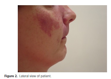
R.A.B., a 36-year-old woman from Santa Cruz do Sul, state of Rio Grande do Sul, presented to the complete denture clinic of the Universidade de Santa Cruz do Sul (UNISC) School of Dentistry with esthetic and functional complaints due to tooth and flange wear in her dentures (Figure 1). History, physical examination, and radiographs revealed that the patient had Angle Class II malocclusion, characterized by marked maxillary prognathism in relation to the mandible (Figure 2). Therefore, we planned a maxillary complete denture without a labial flange.
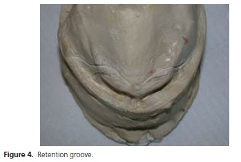
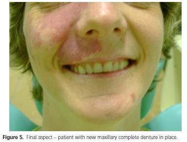
Initial impressions were obtained with irreversible hydrocolloid material (Jeltrate®, Dentsply, Petrópolis, Brazil), using a Vernes-type stock tray9. The denture-bearing area was delimited as clearly as possible on the stone model (Herodent®, Vigodent, Rio de Janeiro, Brazil). The denture was relieved over pressure and retention areas. A custom tray was made from self-curing acrylic resin (Jet®, Artigos Odontológicos Clássico, São Paulo, Brazil), the peripheries were sealed with low-fusing impression wax, and a functional impression was obtained with polyether material (Impregum Soft®, 3M ESPE). Once the working model had been relieved, a trial baseplate was produced from self-curing acrylic resin and waxed up with #7 wax (Wilson, Polidental Indústria e Comércio Ltda., Cotia, Brazil). The models were mounted onto a semi-adjustable articulator with the aid of a facebow (Figure 3). After placement of reference lines, artificial teeth were selected (Vita Triostat®, VITA, Pasadena, CA, USA) and set in accordance with the principles of balanced occlusion for fully edentulous patients10. After the try-on, the labial flange of the denture was cut out to minimize the patient's maxillary discrepancy and obtain a more satisfactory esthetic outcome. To ensure that retention would not be compromised, a small undercut was made on the working model, 4 mm from the cut edge of the anterior flange of the baseplate (Figure 4). The denture was placed into a flask, which, after wax and baseplate removal, was filled with heat-curing resin (Jet®, Artigos Odontológicos Clássico, São Paulo, Brazil). To increase the strength of the denture, a metal framework reinforcement was fabricated and included during this stage. After insertion of the denture, the patient was given instructions on its use, with particular emphasis on awareness of hygiene and aftercare practices (Figure 5). During subsequent visits, adjustments were made as necessary and the patient was given further instructions on denture use and cleaning.


DISCUSSION
According to Pucca Junior,1 edentulism may be defined as partial or complete loss of the teeth. Dental losses have a series of body-wide repercussions, affecting such aspects as esthetics, pronunciation, digestion and, particularly, mastication. Based on this knowledge, and in agreement with Galatti3, who states that the masticatory ability lost after dental extractions can be partly restored by prosthodontics, in this clinical case, we sought to rehabilitate the esthetics and function lost due to edentulism.
In patients with maxillomandibular discrepancies requiring complete prosthetic rehabilitation, the recommended procedures include orthognathic surgery, conventional dentures, or complete dentures without a labial flange. In keeping with these recommendations, and as our patient was unable to undergo any other procedures due to financial constraints, we decided on a complete denture without labial flange3,9-10,12,16.
The objective of orthognathic surgery is to correct dentofacial deformities and produce an Angle Class I occlusion, with a good relationship among the facial bones, so as to provide a harmonious facial appearance16. Therefore, after correction of bony structures, these patients become candidates for conventional complete dentures. However, only some patients are able to undergo the procedure, not only due to financial constraints but also due to other contraindications, such as systemic disorders, that may preclude orthognathic surgery.
It bears stressing that, according to Marzolla16, failure to correct a discrepancy may lead to increased deformity after denture placement, distending the lip. Therefore, we chose to fabricate a maxillary complete denture without a labial flange, to address these unfavorable aspects, restore function and improve esthetics. According to Figueiredo et al.,6 one disadvantage of this type of prosthesis is loss of retention due to absence of peripheral seal in the anterior region. Hence, the smaller the area covered by the denture, the worse its retention.
Denture retention can be achieved through physical factors, but the presence of saliva is essential; the greater the number of molecules in the saliva pool, the greater the viscosity of the saliva and the area of contact between the base of the denture and the alveolar ridge3,8. Accordingly, to ensure that retention would not be jeopardized, a small undercut was made 4 mm from the edge of the anterior flange of the base plate.
When using complete dentures, it is imperative that the opposing arch have teeth, whether natural or artificial, to enable a proper balanced occlusion that can contribute to the retention and stability of the denture as well as preserve supporting structures4-5,6. Therefore, in the present case, the denture teeth were lingualized in relation to the natural dentition, so as to restore occlusal balance. With constant, judicious care, this patient has every chance at achieving complete satisfaction, comfort, and proper function with dentures.
CONCLUSION
As demonstrated in this report, complete dentures without a labial flange can be a very effective tool for functional and esthetic rehabilitation in patients with maxillomandibular discrepancy. Patients should always be reminded of the importance of denture care, as well as of the need for periodic follow-up visits to ensure better preservation of the outcome obtained.
Collaborators
RA PEGORARO, HL DETTENBORN and V BERGESCH took part in all stages of article writing.
REFERENCES
1. Pucca Junior GA. A saúde bucal do idoso: aspectos demográficos e epidemiológicos. Med Center [periódico na internet]. 2002 Abr [citado 2009 Nov 5]. Disponível em: <http://www.odontologia. com.br/artigos.asp?id=81> [ Links ].
2. Gebran MP. As mudanças na qualidade de vida em pacientes edêntulos após a implantação de próteses totais: uma contribuição da ergonomia [dissertação]. Florianópolis: Universidade Federal de Santa Catarina; 2001.
3. Galati A. Prótese total: manual de fases laboratoriais. São Paulo: SENAC; 1996.
4. Jorge JH, Nogueira SS, Varjão FM. Oclusão lingualizada para próteses totais. RGO, Rev Gaúch Odontol. 2003;51(2):105-11.
5. Georgetti MP, Georgetti BA, Corrêa GA, Magalhães Filho O. Aspectos para a estabilidade das próteses totais. Rev Odontol Univ Santo Amaro. 2000;5(2):71-5.
6. Figueiredo AR, Paes Junior TJA, Seraidarian PI. Tratamento por prótese total imediata em caso de acentuada discrepância maxilomandibular. PCL: Rev Bras Prót Clín Labor. ano;1(4):293-302.
7. Nakamae AEM, Cunha EFS, Tamaki R, Guarnieri TC. Avaliação da retenção de próteses totais bimaxilares em função das características da área basal. RPG - Rev Pós Grad. 2006;13(1):69-76.
8. Cunha EFS. Avaliação da retenção de prótese total bimaxilar em função das características da área basal [tese]. São Paulo: Universidade de São Paulo; 2004.
9. Grant AA, Heath JR, Mccord JF. Prótese odontológica completa: problemas, diagnóstico e tratamento. Rio de Janeiro: MEDSI; 1996.
10. Turano JC, Turano LM. Fundamentos de prótese total. 5a ed. São Paulo: Editora Santos; 2000.
11. Genari Filho H. O exame clínico em prótese total. Rev Odontol Araçatuba. 2004;25(2):62-71.
12. Brito MLG, Goulart VL, Cunha VPP. Prótese total sem face labial: relato de um caso clínico. Rev Assoc Paul Cir Dent. 2003;57(1):5-9.
13. Hayakawa I. Principles and practices of complete dentures: creating the mental image of a denture. Tókio: Quintessence Publishing; 2001.
14. Batista MAC, Guerra VVAS, Fonseca DA, Mesquita AE. Estética em prótese total. PCL: Rev Bras Prót Clín Labor. 2000;1(5):81-6.
15. Gomes T, Mori M, Corrêa GA. Atlas de caracterização em prótese total e prótese parcial removível. São Paulo: Editora Santos; 1998.
16. Marzola C. Fundamentos de cirurgia bucomaxilofacial: a cirurgia ortognática. [citado 2009 Maio 26]. Disponível em: <http:// www.clovismarzola.com/textos/CAP_XXXI.pdf>.
 Correspondence to:
V BERGESCH
e-mail: vaniabergesch@hotmail.com
Received on: 23/5/2011
Final version resubmitted on: 13/6/2013
Approved on: 14/2/2014


