Serviços Personalizados
Artigo
Links relacionados
Compartilhar
RSBO (Online)
versão On-line ISSN 1984-5685
RSBO (Online) vol.9 no.2 Joinville Abr./Jun. 2012
Original Research Article
Antimicrobial activity of sodium hypochlorite, chlorhexidine and MTAD® against Enterococcus faecalis biofilm on human dentin matrix in vitro
Cristiana Francescutti Murad I; Luciana Moura Sassone I; Monica Cristina Souza II; Rivail Antonio Sergio Fidel I; Sandra Rivera Fidel I; Rafael Hirata Junior II
I Proclin Department, Rio de Janeiro State University – Rio de Janeiro – Brazil.
II Department of Microbiology, Immunology and Microbiology, Rio de Janeiro State University – Rio de Janeiro – Brazil.
ABSTRACT
Objective: This study evaluated the antimicrobial efficacy of 2.5% and 5.25% NaOCl, 2% gel and liquid CHX and MTAD® against Enterococcus faecalis biofilms on human dentin. Material and methods: E. faecalis biofilms grown on dentin matrix of 216 root sections were submerged in test irrigants for 1, 5, 15 and 30 minutes. The antimicrobial activity of the test irrigants were assessed through CFU counts. Biofilm formation over the dentin surface was ensured by SEM analysis.Results: Results showed no statistic difference among CHX gel, 2.5% and 5.25% NaOCl. However, the CHX liquid and MTAD were less effective than 2.5% and 5.25% NaOCl. Only CHX liquid and MTAD had differences in its efficacy depending on the time.Conclusion: The most effective irrigants in eliminating E. faecalis biofilms were 2.5% and 5.25% NaOCl and 2% CHX gel, at all the tested time intervals, in comparison to CHX liquid and MTAD.
Keywords: root canal irrigants; Enterococcus faecalis; biofilms; antimicrobial action.
Introduction
Bacteria constitute the primary etiological agent for pulpal and perirradicular disease 11. Accordingly, elimination of root canal infection is required for success in endodontic treatment. However, root canal treatment, may fail due to viable bacteria resisting treatment or microorganisms invading the canal via coronal leakage 10. Enterococcus faecalis is one of the most prevalent species isolated from teeth enduring primary 18 and persistent infection 7,10,25. E. faecalis has demonstrated a high resistance 20 and ability to inactivate antimicrobial agents 14, survival capacity in harsh environments, with scarce nutrient supply and extreme alkaline pH 28, and the capacity for growth as a biofilm on root canal walls 21. Therefore, several laboratory studies have been conducted in order to test the susceptibility of E. faecalis to endodontic procedures.
Recent studies have demonstrated biofilm growth over root surfaces on teeth with chronic apical periodontitis and teeth refractory to root canal treatment 12. Biofilms have been found to be one of the most common growth conditions for bacteria in nature. They enhance the ability of bacteria to survive in a hostile environment, increasing their resistance to host systemic defenses and antimicrobial agents 24,29.
The cleaning and shaping process provided by a high-quality instrumentation of the root canal system, associated with the use of antibacterial irrigating solutions, promotes the microbiologic control of the root canal system. Such instrumentation process becomes the most important factor in preventing or healing endodontic disease 26. Despite the variety of cleaning and shaping techniques that had been developed and improved through many decades, none of them have attained an ideal cleaning of root canal.
Several irrigating solutions might be used during chemomechanical preparation of infected root canals, to increase bacterial elimination and facilitates removal of necrotic pulp tissue and dentine chips from root canal. Sodium hypochlorite (NaOCl) is the most popular irrigating solution. This halogenated compound is excellent tissue solvent for vital and necrotic pulp tissues, displaying an effective antimicrobial activity 17. However, it is cytotoxic to periapical tissues especially at high concentrations. Chlorhexidine digluconate (CHX) has also been recommended as a root canal irrigant and medicament 5, for its potent broad spectrum of antimicrobial action 17,30, substantivity and high biocompatibility 5. However, the inability of chlorhexidine to dissolve pulp tissue remains a problem.
A new irrigating solution, MTAD, contains a mixture of a tetracycline isomer, an acid and a detergent. Recent investigations demonstrated that MTAD has ability to eradicate E. faecalis, to reduce the bacterial contamination of the root canal and also to remove the smear layer 23.
This study aimed to evaluate the antibacterial activity, ex vivo, of 2.5% and 5.25% NaOCl, 2% CHX at gel and liquid formulations, and MTAD? irrigating solutions on E. faecalis biofilm during different exposure times.
Material and methods
For this study, 108 single-rooted human mandibular incisors were selected and stored in 0.5% thymol solution. This study was approved by the Ethics Committee, Nucleus of Collective Health Studies (1810-CEP/HUPE), Rio de Janeiro State University (UERJ).
The tooth specimens (n=108) were sectioned 3mm above the apex and below the coronal portion, in order to obtain a standard root length of 5mm. All these roots fragments were instrumented using Gates Glidden drills, with sizes varying from #1 to #3 (Maillefer Instruments SA – CH 1338, Ballaigues, Switzerland). Aiming at exposing the dentinal surface and the dentinal tubules, all roots were sectioned longitudinally with a diamond disc (K.G. Sorensen Ind. Com. Ltda, São Paulo, SP, Brazil) resulting in 212 root sections and had the cementum removed by using 500 grit sandpaper. All the root sections were immersed in 17% EDTA (Biodinâmica Quím. e Farm. Ltda., Paraná, Brazil) for 3 minutes, followed by a final flush of sterile distilled water in order to remove the smear layer. Then, the sections were immersed in the same solution during 24 hours for total removal of 0.5% thymol.
Bacterial Strain
Strains of E. faecalis (ATCC 29212), obtained from the American Type Culture CollectionTM (ATCC, Rockville, MD, EUA), were taken from frozen stock culture and inoculated into Tryptcase Soy Broth (TSB, Difco Lab, Detroit, MI, USA), grown overnight at 37°C, and streaked onto Columbia blood agar plates. Single colonies were used to inoculated TSB broth cultures that were grown for 24h at 37°C and the bacterial suspension was adjusted to a count of 1x10⁸ colony forming units (CFU), which is equivalent to 0.2 absorbance intensity determined at 640nm (B 295II-Spectrophotometer, MicronalTM, São Paulo, SP, Brazil). The cultures were checked for purity by Gram stain and colony morphology.
Biofilm formation and exposure to test agents
Biofilm cultivation method used in this study was adapted from that described previously by Sena et al. 22. Each root section was placed in one vial containing 3mL TSB broth, and autoclaved. Next, aliquots of 100µL of E. faecalis suspension were inoculated in each vial and grown for 24h at 37°C. In order to allow the formation of a biofilm on the dentin surface, sterile TSB broth (TSB, Difco Lab) and new aliquots of 100µL of E. faecalis suspension were replaced every 24 hours. Microorganisms were incubated at 37°C for 72 hours 2.
The root sections were randomly divided in 5 experimental groups containing 40 samples each, and 2 control groups, with 3 samples each. Negative control samples were not inoculated with bacteria, in order to test the sterility of the teeth specimens and solutions. Positive control samples were inoculated with bacteria and placed in Phosphate buffered saline (PBS – Sigma Chemical Company, St Louis, MO, USA), to verify the bacterial growth.
Each experimental group was assigned to one antibacterial solution, as follows: 2.5% NaOCl, 5.25% NaOCl, 2% CHX gel, 2% CHX liquid and MTAD and was divided into 4 subgroups with 10 samples each. The subgroups were tested in 4 different exposure times: 1, 5, 15 and 30 minutes. Neutralizing agents were used to inactivate the antibacterial activity of the tested solutions, to avoid false-negative results during microbiologic culturing. After an agar plate diffusion testing, NaOCl was neutralized using 5% sodium thiosulphate; CHX, using 3% Tween 80 and 0,3% L-a-lecithin (Microbial Content Test Agar – Becton Dickinson Microbiology Systems, Sparks, MD, USA); and MTAD, using calcium chloride (CaCl2 – Merck, Ind. Quim. Rio de Janeiro, RJ, Brazil).
Under the laminar airflow (Bioprotector Plus 09, Veco do Brazil, Campinas, SP, Brazil), all root sections were placed in vials containing 4mL of test solutions. After being submitted to the antimicrobial solutions during the different exposure times, all samples were aseptically washed in 2mL of neutralizing agents to prevent potential carry-over of the irrigants, and then transferred into another vial containing 1mL of neutralizing agents and glass beads (Sigma). The vials were vortexed for 1 min, to disrupt the biofilm structure and to resuspend the microorganisms.
Tenfold serial dilutions were made from the 100µL bacterial suspension and plated out on Petri dishes with Columbia Agar Base – CAB (Difco Lab). The plates were then incubated at 37°C for 24 hours. The number of CFU per milliliter was calculated, in order to evaluate the antibacterial activity of the test solutions.
SEM analysis
Aiming at ensuring the biofilm formation over the dentin surface of the root sections and its removal with the technique used in this study, ten root sections were prepared and submitted to the SEM analysis. All ten samples were cultivated in the same conditions previously described. Five samples were immersed in PBS to verify the bacterial growth and five samples were immersed in a test solution only to verify the removal and resuspension of the biofilm. As NaOCl is well documented in scientific literature, this study used 5.25% NaOCl for 5min in all control samples. All samples were then fixed in 2.5% glutaraldehyde solution, buffered by sodium cacodilate 0.1M for 2 hours. The roots sections were dried with ethanol, in increasing concentrations, and dehydrated to its critical point, using a critical point apparatus (Pelco CPD-2, Critical Point Dryer, Clovis, CA, USA). Each sample was coated with a layer of gold palladium and root surface observations were performed by using a 25kv scanning electron microscopy (Jeol, model JSM 5310, Itaberaba, São Paulo, SP, Brazil) at 500, 2000 e 7500 magnifications.
Statistical analysis
The effect of each test agent on the E. faecalis biofilm was determined by calculating the percentage kill of viable bacteria following treatment with a test agent. The percentage kill was calculated for each test agent, considering the bacterial reduction related to its initial CFU count (positive control). Microbiologic data were standardized using the Reed and Müench formula 3.
Analysis of the bacterial reduction percentage in terms of time and solution was performed by analysis of variance (Anova). Correlations between the percentage of bacterial reduction and exposure time to each irrigating solution were performed separately by Kruskal-Wallis e Mann-Whitney non parametric tests. Significance level was set at 5%.
Results
Biofilm formation
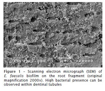
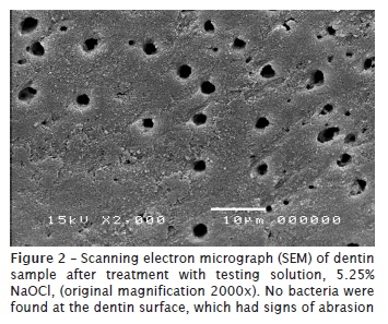
The E. faecalis biofilm was found on dentin surface in the 5 samples examined by SEM analysis. Typical cocci-shaped bacterial cells were aggregated, colonized the dentin surface and invaded the dentinal tubules (figure 1). SEM analysis showed total removal of the biofilm, which was observed on the other 5 samples, treated with 5.25% NaOCl, ensuring that the methodology applied was efficient in disrupting the biofilm structure and resuspending the microorganisms. Despite careful analysis, no bacteria could be found at the dentin surface, which had signs of great abrasion, probably due to the action of the glass beads (Sigma) (figure 2).


Antibacterial activity of tested solutions
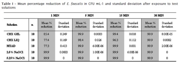
All irrigating solutions tested showed antibacterial activity against E. faecalis. Table I summarizes the average percentage reduction of E. faecalis in CFU mL-1 after exposure to test solutions.

The analysis of variance of the percentage of bacterial reduction due to different solutions and exposure times demonstrated no statistic difference among CHX gel, 2.5% NaOCl and 5.25% NaOCl (p>0.05). However, CHX liquid and MTAD were less effective than 2.5% and 5.25% NaOCl (p<0.05) and similar to CHX gel. The Kruskal-Wallis and Mann-Whitney tests demonstrated that only CHX liquid and MTAD presented statistical differences (p<0.05) in efficacy through different exposure times. MTAD was less effective at exposure time of 1min (p<0.05) and CHX liquid was less effective at 1 and 5 minutes.
Discussion
Endodontic failure is usually caused by microorganisms that resisted to conventional endodontic treatment or invaded root canal system via coronal leakage 10. If bacteria in infected root canals can invade the extraradicular area, via the apical foramen and the dentinal tubules, they may form bacterial biofilms and play an important role in refractory and chronic apical periodontitis 12.
The gap left by studies that evaluate the susceptibility of E. faecalis planktonic cultures to antimicrobial agents and the studies that closely examine the biofilm behavior led us to study the antimicrobial activity of endodontic irrigants, at different concentrations, against E. faecalis biofilms.
A biofilm is a complex aggregation of microorganisms that secrete a protective and adhesive exopolymeric matrix, called extracellular polymeric substance or exopolysaccharide (EPS). The EPS protects the biofilm cells and facilitate communication and nutrient distribution among them. Organizations of microorganisms within biofilms are characterized by surface attachment, structural heterogeneity, complex community interactions and the presence of EPS 9,19. Biofilm formation process, in general, includes 4 steps: initial surface adherence, microcolony formation, depth of the mature biofilm, biofilm architecture growth 19,24.
Bacteria growing in biofilms may survive starvation periods and recover rapidly, and also may exhibit new and more virulent types. Furthermore, bacteria within biofilms have inherently increased resistance to antimicrobial agents, compared with the same bacteria grown under planktonic conditions 29,31. Preliminary studies demonstrated that after 72h the biofilm was uniformly present over the substrate 2,19. These studies showed that E. faecalis cells were fully capable of biofilm formation on human dentin after 72h. Although culture techniques do not entirely simulate clinical conditions, they offer an adequate method to evaluate antimicrobial activity of endodontic irrigants on bacterial biofilm over dentin surface. Also, they allow systematic comparisons among different solutions at different exposure times, are practical and easy to reproduce.
A study about E. faecalis biofilm development showed that after 24 hours of growth there was little or none EPS formation, and after 36h of growth, the presence of EPS uniformly distributed, consolidated the biofilm formation 19. The study suggests that at this stage the biofilm surface was indicative of a young biofilm, corresponding to the maturation of the microcolonies with EPS production. After 72h of growth, it was possible to observe the appearance of an EPS net consisting of filaments and spaces formed from air-dried EPS on bacterial cells after 72 h. This phase may represent early stages of biofilm growth and maturation 19. Our study evaluated E. faecalis biofilm susceptibility to antimicrobial agents after 72 hours of growth, however, considerations must be made regarding the physiological and metabolic phases of biofilm development. Concerning literature suggests that the resistance of biofilm to antimicrobial agents may be influenced by the aging of biofilm and the physiological state of cells. As reported by Shen et al. 24 mature biofilm and biofilm with limited nutrient supply are more resistant to CHX killing than in young biofilms.
The results of their study emphasize the influence of biofilm age on the antimicrobial action of disinfecting agents against biofilm bacteria 24. The results of our study demonstrated that the antimicrobial efficacy of irrigating solutions varies according to the exposure time, type and concentration of the substance used. Statistical analysis showed no difference among CHX gel, 2.5% and 5.25% NaOCl (p>0.05). However, CHX liquid and MTAD were less effective than 2.5% and 5.25% NaOCl (p<0.05) and similar to CHX gel. Only CHX liquid and MTAD had differences in its efficacy depending on time. CHX was less effective at 1 and 5 minutes (p<0.05), and MTAD at 1 minute.
In agreement with other studies 5,13 we found that 2% CHX gel and 5.25% NaOCl significantly reduced E. faecalis counts. Similar results were obtained by Sassone et al. 17, who demonstrated, through contact test, that 0.5 to 5% NaOCl and 0.12 to 1% CHX solutions presented antibacterial action against facultative and strict anaerobes and by Vianna and Gomes 30. Our results also corroborate those reported by Radcliffe et al. 16, who demonstrated that the NaOCl effectiveness varies through time and concentration. Specifically, 2.5% NaOCl was less effective in bacterial kill and needed more exposure time than 5.25% NaOCl.
This study results also concur with those reported by Abdullah et al. 1. In their study, different phenotypes of E. faecalis were submitted to root canal irrigants. Three percent NaOCl was more effective than 0.2% CHX, achieving 100% kill for all presentations of E. faecalis after 2 minutes of contact time. However, CHX was significantly more effective in killing bacteria in planktonic suspension than in biofilm. Previous reports of higher efficiency of 5.25% NaOCl and 2% liquid CHX in eliminating microorganisms within 30 seconds were obtained using a different substrate for biofilm growth, as cellulose nitrate membrane. The nature of the substrate may influence the biofilm formation, once biofilm growth and adherence may differ on distinct structures 9,27. The specific mechanisms for E. faecalis cells to invade and adhere to dentin walls may account for the biofilm formation on dentin. E. faecalis exhibit widespread genetic polymorphisms and it possesses serine protease, gelatinase, and collagen-binding protein (Ace), which help it bind to dentin 28.
Spratt et al. 27 and Dunavant et al. 6, using a similar methodology reported that different concentrations of NaOCl (varying from 1% to 6%) were more effective than 0.2% CHX 27 and 2% CHX and MTAD 6 in the elimination of E. faecalis biofilm. Williamson et al. 31 reported that 6% NaOCl and 6% NaOCl with surface modifiers reduced number of bacteria within the biofilms significantly more than either 2% CHX or 2% CHX with surface modifiers. Although, the results of these studies are consistent with our findings, direct comparisons cannot be made because of the differences in the biofilm assays. The use of CHX at higher concentrations may also be considered.
In the present study, MTAD exhibited excellent antimicrobial action at all time points except 1 minute. This finding is consistent, at some extent, with those reported in a previous study 23, which showed that MTAD is more effective than 5.25% NaOCl as an irrigating solution. A possible explanation for the excellent antimicrobial action of MTAD, even on dentin surface, is its antibiotic activity promoted by the doxycycline and its ability to remove smear layer due to citric acid and doxycycline. Removal of smear layer and opening of dentinal tubules may allow higher penetration of the antimicrobial agent into dentin matrix, promoting a better cleaning of root canal system 8.
When analyzing the antibacterial action of an endodontic irrigating solution, it is necessary to consider its ability to wet the dentin and penetrate dentinal tubules and also its capacity to disrupt the biofilm community. If biofilm is not disrupted and remains on the surface of dentinal tubules, it may prevent an adequate seal of root canal and facilitate endotoxin release. The poor results of CHX, when compared to MTAD and NaOCl, may possibly be explained by different patterns of inhibition of antibacterial activity caused by distinctive compounds of dentin and the presence of bovine serum albumin (BSA) 15.
Clegg et al. 4 reported that 6% NaOCl, 2% CHX and MTAD associated with 1% NaOCl possessed antibacterial activity in culture assessment. However, only 3% and 6% NaOCl were capable of disrupting and removing the biofilm and 2% CHX was not capable of disrupting the biofilm when assessed by SEM analysis. The results obtained by Clegg et al. 4 must be considered, acknowledging that they tested only the antimicrobial activity of the agents, but not their capacity to disrupt the biofilm structure; and the SEM analysis was conducted only to confirm the presence of the biofilm on dentin surface and its removal by the glass beads.
Based on the results obtained in our study, we can conclude that 2.5% and 5.25% NaOCl and 2% CHX gel were the most effective solutions for elimination of E. faecalis biofilm at all exposure times tested and MTAD was more effective after 5 minutes.
The biofilm model used in this study was effective in determining the ex vivo antimicrobial efficacy of root canal irrigants. Since biofilms form on root surfaces in vivo and present high resistance to antimicrobial agents and to host defense, it is more clinically relevant to assess antimicrobial activity against this bacterial phenotype. However, we must be cautious with conclusions withdrawn by an ex vivo study where a single species biofilm was tested, since it is well-known that endodontic infections are often associated with complex microbial interactions.
References
1. Abdullah M, Ng Y-L, Gulabivala K, Moles DR, Spratt DA. Susceptibilities of two Enterococcus faecalis phenotypes to root canal medications. ?J Endod. 2005;31:30-6. [ Links ]
2. Bhuva B, Patel S, Wilson R, Niazi S, Beighton D, Mannocci F. The effectiveness of passive ultrasonic irrigation on intraradicular Enterococcus faecalis biofilms in extracted single-rooted human teeth. Int Endod J. 2010;43:241-50.
3. Bier O. Bacteriologia e imunologia. 17. ed. São Paulo: Melhoramentos; 1976.
4. Clegg MS, Vertucci FJ, Walker C, Belager M, Britto LR. The effect of exposure to irrigant solutions on apical dentin biofilms in vitro. J Endod. 2006;32:434-7.
5. Dametto FR, Ferraz CCR, Gomes BPFA, Zaia AA, Teixeira FB, Souza-Filho FJ. In vitro assessment of the immediate and prolonged antimicrobial action of chlorhexidine gel as an endodontic irrigant against Enterococcus faecalis. Oral Surg Oral Med Oral Pathol. 2005;99:768-72.
6. Dunavant TR, Regan JD, Glickman GN, Solomon ES, Honeyman AL. Comparative evaluation of endodontic irrigants against Enterococcus faecalis biofilms. J Endod. 2006;32:527-31.
7. Gomes BPFA, Pinheiro ET, Jacinto RC, Zaia AA, Ferraz CCR, Souza-Filho FJ. Microbial analysis of canals of root-filled teeth with periapical lesions using polymerase chain reaction. J Endod. 2008;34:537-40.
8. Larsen T. Susceptibility of Porphyromonas gingivalis in biofilms to amoxicillin, doxycycline and metronidazole. Oral Microbiol Immunol. 2002;17:267-71.
9. Liu H, Wei X, Ling J, Wang W, Huang X. Biofilm formation capability of Enterococcus faecalis cells in starvation phase and its susceptibility to sodium hypochlorite. J Endod. 2010;4:630-5.
10. Molander A, Reit C, Dahlén G, Kvist T. Microbiological status of root-filled teeth with apical periodontitis. Int Endod J. 1998;31:1-7.
11. Nair PNR, Sjögren U, Krey G, Kahnberg K-E, Sundqvist G. Intraradicular bacteria and fungi in root-filled, asymptomatic human teeth with therapy-resistant periapical lesions: a long-term light and electron microscopy follow-up study. J Endod. 1990;16:580-8.
12. Noiri Y, Ehara A, Kawahara T, Takemura N, Ebisu S. Participation of bacterial biofilms in refractory and chronic periapical periodontitis. J Endod. 2002;28:679-83.
13. Oliveira DP, Barbizam VB, Trope M, Teixeira FB. In vitro antibacterial efficacy of endodontic irrigants against Enterococcus faecalis. Oral Surg Oral Med Oral Pathol Oral Radiol Endod. 2007;103:702-6.
14. Portenier I, Haapasalo H, Orstavik D, Yamauchi M, Haapasalo M. Inactivation of the antibacterial activity of iodine, potassium iodide and chlorhexidine digluconato against Enterococcus faecalis by dentin, dentin matrix, type-I collagen, and heat-killed microbial whole cells. J Endod. 2002;28:634-7.
15. Portenier I, Waltimo T, Orstavik D, Haapasalo M. Killing of Enterococcus faecalis by MTAD and chlorhexidine digluconate with or without Cetrimide in the presence or absence of dentin powder or BSA. J Endod. 2006;32:138-41.
16. Radcliffe CE, Patouridou L, Qureshi R, Habahbeh N, Qualtrough A, Worthington H et al. Antimicrobial activity of varying concentrations of sodium hypochlorite on the endodontic microorganisms Actinomyces israeli, A. naeslundii, Candida albicans and Enterococcus faecalis. Int Endod J. 2004;37:438-46.
17. Sassone LM, Fidel R, Fidel S, Vieira M, Hirata Jr R. The influence of organic load on the antimicrobial activity of different concentrations of NaOCl and chlorhexidine in vitro. Intl Endod J. 2003;36:848-52.
18. Sassone LM, Fidel R, Figueiredo L, Fidel S, Faveri M, Feres M. Evaluation of the microbiota of primary endodontic infections using checkerboard DNA-DNA hybridization. Oral Microbiol Immunol. 2007;22:390-7.
19. Santos RP, Arruda TTP, Carvalho CBM, Carneiro VA, Braga LQV, Teixeira EH et al. Correlation between Enterococcus faecalis biofilms development stage and quantitative surface roughness using atomic force microscopy. Microsc Microanal. 2008;14:150-8.
20. Sedgley CM, Lennan SL, Clewell DB. Prevalence, phenotype and genotype of oral Enterococci. Oral Microbiol Immunol. 2004;19:95-101.
21. Sedgley C, Nagel A, Dahlén G, Reit C, Molander A. Real-time quantitative polymerase chain reaction and culture analyses of Enterococcus faecalis in root canals. J Endod. 2006;32:173-7.
22. Sena NT, Gomes BPFA, Vianna ME, Berber VB, Zaia AA, Ferraz CCR et al. In vitro antimicrobial activity of sodium hypochlorite and chlorhexidine against selected single-species biofilms. Int Endod J. 2006;39:878-85.
23. Shabahang S, Pouresmail M, Torabinejad M. In vitro antimicrobial efficacy of MTAD and sodium hypochlorite. J Endod. 2003;29:450-2.
24. Shen Y, Stojicic S, Haapasalo M. Antimicrobial efficacy of chlorhexidine against bacteria in biofilms at different stages of development. J Endod. 2011;37:657-61.
25. Siqueira Jr JF, Rôças IN. Clinical implications and microbiology of bacterial persistence after treatment procedures. J Endod. 2008;34:1291-301.
26. Siqueira Jr JF, Rôças IN, Santos SRLD, Lima KC, Magalhães FAC, De Uzeda M. Efficacy of instrumentation techniques and irrigations regimens in reducing the bacterial population within root canals. J Endod. 2002;28:181-4.
27. Spratt DA, Pratten J, Wilson M, Gulabivala K. An in vitro evaluation of the antimicrobial efficacy of irrigants on biofilms of root canal isolates. Int Endod J. 2001;34:300-7.
28. Stuart CH, Schwartz SA, Beeson TJ, Owatz CB. Enterococcus faecalis: its role in root canal treatment failure and current concepts in retreatment. J Endod. 2006;32:93-8.
29. Svensäter G, Bergenholtz G. Biofilms in endodontic infections. Endodontic Topics. 2004;9:27-36.
30. Vianna ME, Gomes BPFA. Efficacy of sodium hypochlorite combined with chlorhexidine against Enterococcus faecalis in vitro. Oral Surg Oral Med Oral Pathol Oral Radiol Endod. 2009;107:585-9.
31. Williamson AE, Cardon JW, Drake DR. Antimicrobial susceptibility of monocultures biofilms of a clinical isolate of Enterococcus faecalis. J Endod. 2009;35:95-7.
 Correspondence:
Sandra Rivera Fidel
Dr. Otávio Kelly, 63 – ap. 301 – Tijuca
CEP 20511-280 – Rio de Janeiro – RJ – Brasil
E-mail:sandrafidel@gmail.com
Received for publication: August 28, 2011.
Accepted for publication: December 5 2011.


