Serviços Personalizados
Artigo
Links relacionados
Compartilhar
RSBO (Online)
versão On-line ISSN 1984-5685
RSBO (Online) vol.10 no.3 Joinville Jul./Set. 2013
ORIGINAL RESEARCH ARTICLE
Ethanol is inefficient to remove endodontic sealer residues of dentinal surface
Keli Regina VictorinoI; Edson Alves de CamposI; Marcus Vinicius Reis SóI; Milton Carlos KugaI; Norberto Batista Faria-JuniorI; Katia Cristina KeineI; Fábio Augusto de Santi AlvarengaI
IAraraquara Dental School – Araraquara – SP – Brazil.
ABSTRACT
Introduction: Endodontic sealer residues on dentinal surface have negative effects on adhesion of adhesives system and/or can cause discoloration of the dental crown.
Objective: To evaluate the efficacy of 95% ethanol in removal of residues of epoxy-based (AH Plus), methacrylate-based (Epiphany SE) or calcium-based (Sealapex) sealers on dentinal surface.
Material and methods: Thirty-two bovine incisor dental crown fragments (0.5 mm x 0.5 mm) were treated with 17% EDTA and 2.5% NaOCl. The specimens were divided into three experimental groups (n = 10): G1 (AH Plus), G2 (Epiphany SE) and G3 (Sealapex). In each group was applied a coating of one endodontic sealer type and were left undisturbed for 5 minutes. After this period, the specimens were cleaned with 95% ethanol. The control group was composed by two specimens that did not receive any sealer or cleaning treatment. The sealer residues persistence after cleaning with 95% ethanol was evaluated by scanning electron microscopy (x500) and a score system was applied. Data obtained were analyzed by Kruskal-Wallis and Dunn tests (α = 5%).
Results: Moderate amount of endodontic sealer residues were observed in all groups, regardless of the endodontic sealer compositions. G1, G2 and G3 presented similar amount of sealer residues on dentinal surface after cleaning with 95% ethanol (p > 0.05).
Conclusion: 95% ethanol was inefficiency to remove completely AH Plus, Epiphany SE and Sealapex residues of sealer-contaminated dentin.
Keywords: endodontics; image analysis; SEM; surface analysis.
Introduction
An essential procedure of the endodontic treatment is to provide a complete obturation of root canal and to develop an adequate fluid-tight seal mainly at the apical third 2. Actually, different sealers have been proposed, mainly containing calcium, methacrylate or epoxy resin in their compositions 6,21,24.
Sealapex (SybronEndo, Romulus, MI, USA) is a calcium-based sealer, composed of two pastes: a catalyzer paste (isobutyl salicylate resin, silicon dioxide, bismuth trioxide, titanium dioxide pigment) and a base paste (N-ethyltoluene sulfonamide resin, silicon dioxide, zinc oxide, and calcium oxide). This sealer has acceptable biological compatibility, but may lead to slight crown discoloration over time 4,8.
AH Plus (Dentsply De Trey, Konstanz, Germany) is an epoxy-based cement, also composed of two pastes: paste A (bisphenol-A epoxy resin, bisphenol-F epoxy resin, calcium tugstate, zirconium oxide, sílica and iron oxide pigments) and paste B (dibenzyldiamine, aminoadamantane, tricyclodecane-diamine, calciun tungstate, zirconium oxide, silica, and silicone oil). This material is routinely used as gold standard material for testing endodontic sealer 1. Notwithstanding, the persistence of this sealer on pulp chamber dentine reduces the microtensile bond of self-etching adhesives 22.
Following recent advances in adhesives systems, methacrylate-based resin sealers were developed to be used in radicular obturation. Epiphany (Pentron Clinical Technologies, Wallingford, CT) was the first methacrylate-based sealer used in endodontics. This sealer is basically composed by resins (Bis-GMA, UDMA, PEGDMA, EBADMA), fillers (barium sulphate, bismuth oxychloride, calcium hydroxide, silica, and silane-treated barium-aluminosilicate glass), colouring pigment, dual-cured initiators (cumene hydroperoxide, thiosinamine, champhorquinone), and stabilizer (butylated hydroxytoluene [2,6-di-tert-butyl-4-methylephenol]) 11. In first generation, this system had a core material (Resilon), a dual-curing resin-based sealer (Epiphany) and a self-etching primer 21. In second generation, Epiphany self-etch (SE) system has only two components: Epiphany self-adhesive sealer (Epiphany SE; Parkell, Farmigton, NY, USA) and Resilon. Acid resin monomers that are originally found in SE primers have been incorporated into the self-adhesive resin-based sealers, reducing the application time and the possibility of errors during adhesive procedures 12. The system has possible capability to create a "monoblock" between the radicular dentin wall and root canal obturation 12.
Presence of residues of endodontic materials interferes in the prognosis of the endodontic treatment 13,19. Presence of endodontic sealer residues on the pulp chamber dentin may cause crown discoloration and/or negatively affect the bond strength of dentin adhesives 4,20,22. To prevent these adverse effects, an appropriate cleaning of the pulp chamber dentin should be carried out. Several solutions containing ethanol, ethyl acetate and acetone have been recommended for removal of debris and residues from the dentin surface 23. Ethanol is one of the most recommended cleaning substances to dentin cleaning after root canal obturation 14. However, there are no studies that assessment its effectiveness to remove endodontic sealer residues of different chemical composition.
The aim of this study was to evaluate the efficacy of 95% ethanol on the removal of residues of epoxy-based (AH Plus), methacrylate-based (Epiphany SE) or calcium-based (Sealapex) sealers on dentinal surface crown of bovine teeth.
Material and methods
Thirty-two freshly extracted bovine permanent incisors, stored into a solution of 0.1% thymol at 4°C were used. Thirty-two tooth fragments with flat dentin surfaces, measuring 0.5 cm x 0.5 cm, were obtained from the buccal surface of dental crowns using a slow-speed Isomet precision saw (Buehler Ltd, Lake Bluff, IL, USA) under water irrigation. Next, 17% EDTA (Biodinâmica Ind. Com, Ibiporã, PR, Brazil) was applied onto the dentin surfaces for 3 minutes. Specimens were then washed with 2.5ml of 2.5% NaOCl (Asfer, São Caetano do Sul, SP, Brazil) and dried with an air stream.
The specimens (n = 10, each group) received a layer of: G1 – AH Plus sealer (Dentsply, DeTrey, Germany), G2 – Epiphany SE (Pentron Clinical Technologies, Wallingford, CT) or G3 – Sealapex (SybronEndo, Romulus, MI, USA) which was spread evenly over the dentin surface with a microbrush (Microbrush Int., Grafton, WI, USA) and left undisturbed for 5 minutes. The sealers were mixed and handled according to manufacturer recommendations. Following, the dentine surfaces were wiped using cotton pellets saturated with 95% ethanol (Rinse-N-Dry, Racine, MI, USA), until the surface appears visibly clean. After this step, no additional rinsing was performed. Control specimens (n = 2) did not receive sealer application. All specimens were prepared by the same operator.
SEM evaluation
For SEM analysis, the specimens were dried at room temperature for 7 days, dehydrated in silica for 24 h, mounted onto aluminum stubs with silver paint, sputter-coated with gold, and examined under a DSM 940A scanning electron microscope (Carl Zeiss, Oberkochen, Baden-Wurttemberg, Germany) operating at 15 kV. Each fragment was initially visualized at x100, and for assessment of the amount of sealer residues, further observations under x500 were conducted in 4 different fields. A representative image of each specimen at x500 was used for evaluation. Evaluation of the amount of sealer residues onto the dentine surface was carried out by attributing scores, as follows: Score 1 – no smear layer and all the tubules opened; Score 2 – minimum amount of smear layer and >50% of the dentine surface clean; Score 3 – moderate amount of smear layer and <50% of the dentine surface clean; Score 4 – heavy smear layer with almost all tubules obstructed 17. SEM evaluations were performed by two examiners who were blind to the experimental groups. The examiners were initially calibrated using the reference SEM images. The scores were compared, and when a difference was found, the evaluators together examined the sample. Data were submitted to Kruskal-Wallis and Dunn tests, at 5% significance level.
Results
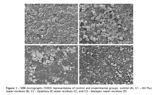
The 95% ethanol did not provide the complete removal of endodontic sealer residues on dentinal surface. The residues persistence was similar and there is no statistical difference among experimental groups (p > 0.05). In all specimens were observed a moderate amount of endodontic sealer residues coating the dentinal surface. Table I shows the frequency of scores assigned, mean and median scores in the experimental groups, regarding the presence of residues on dentin. SEM images representative of control group and experimental groups (G1, G2 and G3) are shown in figure 1. In control specimens, dentinal tubules were visible and dentinal surface without debris.


Discussion
Through SEM analysis, was possible to observe that 95% ethanol did not provide the complete removal of residues of epoxy-based (AH Plus), methacrylate-based (Epiphany SE) or calcium-based (Sealapex) sealers of dentinal surface. All specimens showed moderate amount of endodontic sealer residues on bovine dentine crown.
The method used to evaluate the presence of residues was through the analysis of dentine surface using scanning electron microscopy 9,15. Flat dentin fragments (0.5 cm x 0.5 cm) from bovine pulp chamber were used as substrate to avoid analysis in curved areas, which could adversely affect in interpretation of the results.
AH Plus sealer contains in its composition two non-polar resins: bisphenol-A and bisphenol-F epoxy resins 7. The efficacy of a solvent in dissolving a solute or softening a polymer may be explained by the concept that polar solvents are better at dissolving polar compounds 22. As ethanol is a polar solvent and resins of endodontic sealers are non-polar substances, it could be assumed that ethanol is incompletely miscible with these sealers, resulting in persistence of residues on dentinal surface. This date is in accordance with observed by Roberts et al. 22.
According to described by the manufacturer, to avoid a quick evaporation and enable an adequate time for use, the ethanol used in this study contains a low concentration of water in its composition. This may also have contributed to persistence of residues, because water is immiscible or incompletely miscible with some resin contained into the endodontic sealers 5,10,22. Although all specimens presented residues on dentinal surface, its characteristics were different, as shown in figure 2. To Epiphany SE, the dentinal tubules were totally obliterated by sealer, but on this layer, it had presence of residues that were not totally removed by 95% ethanol. Despite the ethanol is recommended to use in solubility test to methacrylate-based sealer, this substance did not provide adequate ability to removal residues 16. As Sealapex has a poorly formed matrix with low stability, high water absorption and reasonable degradation of this matrix occurs when in contact with water 3,18, showing diffuse residues on dentine and with smaller size than those of AH Plus.
Therefore, 95% ethanol used as endodontic sealer residues removal protocol was inefficient, maintaining residues that can have negative effects on the prognosis of endodontic treatment. This led to the conclusion that 95% ethanol was inefficiency to completely remove sealer residues on dentinal surface. Further studies should be undertaken in order to develop more efficacious cleaning protocols for the removal of endodontic sealers residues on dentinal surface, avoiding negative effects on bond strength of adhesive system or coronal discoloration of endodontically-treated teeth.
Conclusion
Through the methodology used in this study, it was possible to observe that 95% ethanol was inefficiency to provide the complete removal of residues of epoxy-based (AH Plus), methacrylate-based (Epiphany SE) or calcium-based (Sealapex) sealers on dentinal crown surface of bovine teeth.
References
1. Assmann E, Scarparo RK, Böttcher DE, Grecca FS. Dentin bond strength of two mineral trioxide aggregate-based and one epoxy resin-based sealers. J Endod. 2012 Feb;38(2):219-21. [ Links ]
2. Branstetter J, Von Fraunhofer JA. The physical properties and sealing action of endodontic sealer cements: a review of the literature. J Endod. 1982 Jul;8(7):312-6. [ Links ]
3. Caicedo R, Von Fraunhofer JA. The properties of endodontic sealer cements. J Endod. 1988 Nov;14(11):527-34. [ Links ]
4. Davis MC, Walton RE, Rivera EM. Sealer distribution in coronal dentin. J Endod. 2002 Jun;28(6):464-6. [ Links ]
5. Donnelly A, Sword J, Nishitani Y, Yoshiyama M, Agee K, Tay FR et al. Water sorption and solubility of methacrylate resin-based root canal sealers. J Endod. 2007 Aug;33(8):990-4. [ Links ]
6. Duarte MAH, Demarchi ACCO, Giaxa MH, Kuga MC, Fraga SC, Souza LCD. Evaluation of pH and calcium ion release of three root canal sealers. J Endod. 2000 Jul;26(7):389-90. [ Links ]
7. Goswami DN, Jha PC, Mahato K. Shellac as filler in sheet molding compound. Ind J Chem Tech. 2004 Jan;11(1):67-73. [ Links ]
8. Holland R, Souza V, Nery MJ, Bernabé PFE, Otoboni-Filho JA, Dezan-Junior E et al. Calcium salts deposition in rat connective tissue after the implantation of calcium hydroxide containing sealers. J Endod. 2002 Mar;28(3):173-6. [ Links ]
9. Hülsmann M, Rümmelin C, Schäfers F. Root canal cleanliness after preparation with different endodontic handpieces and hand instruments: a comparative SEM investigation. J Endod. 1997 May;23(5):301-6. [ Links ]
10. Kaplan AE, Goldberg F, Artaza LP, De Silvio A, Macchi RL. Disintegration of endodontic cements in water. J Endod. 1997 Jul;23(7):439-41. [ Links ]
11. Karapınar-KazandaĞ M, Bayrak OF, Yalvaç ME, Ersev H, Tanalp J, Sahin F et al. Cytotoxicity of 5 endodontic sealers on L929 cell line and human dental pulp cells. Int Endod J. 2011 Jul;44(7):626-34. [ Links ]
12. Kim YK, Grandini S, Ames JM, Gu LS, Kim SK, Pashley DH et al. Critical review on methacrylate resin-based root canal sealers. J Endod. 2010 Mar;36(3):383-99. [ Links ]
13. Kuga MC, Campos EA, Faria-Junior NB, Só MVR, Shinohara AL. Efficacy of NiTi rotary instruments in removing calcium hydroxide dressing residues from root canal walls. Braz Oral Res. 2012 Jan-Feb;26(1):19-23. [ Links ]
14. Kuga MC, Só MVR, Faria-Júnior NB, Keine KC, Faria G, Fabricio S et al. Persistence of resinous cement residues in dentin treated with different chemical removal protocols. Microsc Res Tech. 2012 Jul;75(7):982-5. [ Links ]
15. Kuga MC, Só MV, De Campos EA, Faria G, Keine KC, Dantas AA et al. Persistence of endodontic methacrylate-based cement residues on dentin adhesive surface treated with different chemical removal protocols. Microsc Res Tech. 2012 Oct;75(10):1432-6. [ Links ]
16. Moreira FCL, Antoniosi-Filho NR, Souza JB, Lopes LG. Sorption, solubility and residual monomers of a dental adhesive cured by different light-curing units. Braz Dent J. 2010 Nov-Dec;21(5):432-8. [ Links ]
17. Oliveira ACM, Lima LM, Pizzolitto AC, Santos-Pinto L. Evaluation of the smear layer and hybrid layer in noncarious and carious dentin prepared by air abrasion system and diamond tips. Microsc Res Tech. 2010 Jun;73(6):597-605. [ Links ]
18. Ørstavik D, Nordahl I, Tibballs JE. Dimensional change following setting of root canal sealer materials. Dent Mater. 2001 Nov;17(6):512-9. [ Links ]
19. Parsons JR, Walton RE, Ricks-Williamson L. In vitro longitudinal assessment of coronal discoloration from endodontic sealers. J Endod. 2001 Nov;27(11):699-702. [ Links ]
20. Plotino G, Buono L, Grande N, Pameijer CH, Somma F. Non vital bleaching: a review of the literature and clinical procedures. J Endod. 2007 Apr;34(4):394-407. [ Links ]
21. Resende LM, Rached-Junior FJ, Versiani MA, Souza-Gabriel AE, Miranda CE, Silva-Sousa YT et al. A comparative study of physicochemical properties of AH Plus, Epiphany, and Epiphany SE root canal sealers. Int Endod J. 2009 Sep;42(9):785-93. [ Links ]
22. Roberts S, Kim JR, Gu L, Kim YK, Mitchell QM, Pashley DH et al. The efficacy of different sealer removal protocols on bonding of self-etching adhesives to AH Plus-contaminated dentin. J Endod. 2009 Apr;35(4):563-7. [ Links ]
23. Saraç D, Bulucu B, Saraç S, Kulunk S. The effect of dentin-cleaning agents on resin cement bond strength to dentin. J Am Dent Assoc. 2008 Jun;139(6):751-8. [ Links ]
24. Silva DF, Stang EC, Campos EA, Kuga MC, Faria G, Kuga GK. Interference of the assessment method in pH values of an epoxy-based cement. RSBO. 2012 Apr-Jun;9(2):133-6. [ Links ]
 Corresponding author:
Milton Carlos Kuga
Avenida Saul Silveira, 5-01
CEP 17018-260 – Bauru – SP – Brasil
E-mail: kuga@foar.unesp.br
Received for publication: February 28, 2013
Accepted for publication: April 22, 2013


