Serviços Personalizados
Artigo
Links relacionados
Compartilhar
RSBO (Online)
versão On-line ISSN 1984-5685
RSBO (Online) vol.12 no.1 Joinville Jan./Mar. 2015
Case Report Article
Mesiodens surgery at deciduous and permanent dentition
Mariana DalledoneI; Paulo Afonso Tassi-JuniorII; Juliana Feltrin de SouzaI; Estela Maris LossoI
I Positivo University – Curitiba – PR – Brazil
II DDS
ABSTRACT
Introduction and Objective:To report a rare case of a patient who presented two mesiodens and the treatment performed at two moments. Case report: A 7 year-old male patient reported a supernumerary tooth extracted at age 4. The dental clinical exam revealed giroversion of permanent maxillary right central incisor. Cone-beam computed tomography (CBTC) revealed a presence of a mesiodens located at the buccal surface mesially to the permanent maxillary left central incisor and also indicated that the mesiodens was located close to the f loor of the nasal cavity. The surgery was performed with a conservative intervention and osteotomy by preserving the adjacent structure. The one-year following-up postoperative x-ray indicated new bone deposition and a more favorable eruption position of the right permanent maxillary lateral incisor. Conclusion: It can be concluded that an early diagnosis by CBTC allowed an adequate treatment planning, which avoid the formation of cysts and a prolonged retention of permanent tooth.
Keywords: supernumerary teeth; cone-bean computed tomography; pediatric dentistry.
Introduction
An alteration in the eruption of teeth or non-eruption is a frequent event in a pediatric pat ient, affect ing mainly the upper incisors group and representing an unpleasant aesthetical and functional factor. The phenomenon may be determined commonly by the presence of supernumerary tooth2,4.
Supernumerary tooth is an anomaly in number of tooth, where one or more teeth develop beyond the normal series2. Mesiodens can be defined as supernumerary teeth, located in between two upper central incisors, identified as having a coneshaped crown and short root. Most of mesiodens are located at the palatal area of permanent incisors5. This prevalence is low, from 0.15% to 1.9%, predominantly in males, with ratio of 2:15,6,8 .
Concerning to the etiology, the most accepted theory is the hyperactivity of the dental lamina2. Cone-shaped mesiodens occurs in isolated cases, and the incidence of multiples is a rare fact. If inverted positioned, it might be close to the floor of the nasal cavity. Supernumerary teeth in other locations may also cause failure of eruption of adjacent teeth, commonly, retention of the incisors. The problem is usually noticed with the eruption of the maxillary lateral incisors together with the failure of eruption of one or both central incisors. In these cases, the removal of the supernumerary tooth is recommended7.
With regard to pediatric dentistry practice, the dental procedures are related to pain, anesthesia, and parents' anxiety that may inf luence the children´s behavior, making the dental experience negative as well. In this field, the role of the pediatric dentist is to conduct the child using adequate care methods, minimizing the procedure duration, promoting conservative procedures, according to the age and psychology pattern of the child.
Cone-beam computed tomography (CBTC) is commonly applied to pediatric dentistry as complement of radiograph exam. It allowed the exact location of the supernumerary tooth and the relation with adjacent structure. It has been used to plan the surgery procedure, allowing a conservative surgery, more comfort to children and to ensure a rapid procedure execution3. Thus, the aim of this study was to report a case, in which the presence of two mesiodens was diagnosed using CBTC exam, and the surgical intervention was conducted at two different stages.
Material and methods
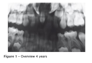
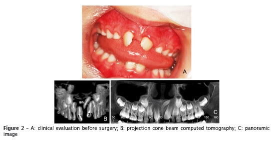
A 7-year-old male was referred to the Positivo University Dental Clinic with complain about misalignment of his teeth at the right permanent maxillary incisor. During the anamnesis, the parents stated that a supernumerary tooth had been extracted from the midline area at age 4 (figure 1). The child had an uneventful medical history with no reports on any type of allergies. Dental exam did not show any type of asymmetry and the patient was at mixed dentition stage. Orthodontic examination showed that the patient exhibited Class I malocclusion without detectable abnormalities on soft tissues. Concerning to the teeth, the patient showed the rotation of the right permanent maxillary central and lateral incisors (figure 2A).
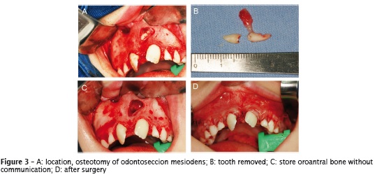
On cone-beam computed tomography (CBTC) and panoramic x-ray examinations, it was possible to observe the exact location of the supernumerary teeth, which was located buccal and mesially to the right permanent maxillary central incisor, without contact with the root of this tooth (figures 2B and C). Also, it could be observed a close proximity to the floor of the nasal cavity and the absence of bone between these structures, remaining only the nasal mucosa between the tooth and the nasal cavity. CBTC showed the exact location to perform the initial osteotomy, at the mesial surface of the root of the left permanent central incisor (figure 3A). The tooth was removed as well as a cystic lesion (figure 3B). No communication with the nasal cavity was detected (figure 3C) and the wound was sutured (figure 3D). The surgical procedure was performed under general anesthesia.
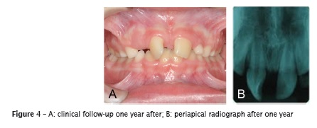
At one-year follow-up appointment, it could be observed a better positioning of the left maxillary incisor on tooth arch (figure 4A) and a new bone formation as shown by the x-ray (figure 4B).


Discussion
Dental literature states that mesiodens occurs solo at the palatal area, with the tendency of natural eruption in the oral cavity. In this case report, the incidence of two mesiodens makes it unusual by the fact that one had erupted at the oral cavity but the other at the labial area was not able to erupt.
CBTC was the elected complementary exam, allowing the proper surgical procedure, reducing undesirable trauma to hard and soft tissue1. Moreover, CBTC images allowed to evaluate the exact location of the supernumerary tooth with adjacent structures, the inverted position of the mesiodens was detected, without contact with the root of tooth #21, and proximity to the floor of the nasal cavity. Therefore, the CBTC images were able to perform a reliable surgery planning and a conservative osteotomy, which avoided an unexpected communication with the nasal cavity.
The adequate treatment planning is relevant by the fact that these inverted supernumerary teeth are generally apical located and retained, requiring a more complex surgical intervention. Because of this apical location, care should be taken in relation to the root development stage of adjacent teeth.
According to Rao and Chidzonga7, the removal of these teeth should be done only after the complete root closure of the adjacent permanent teeth. Moreover, the surgical intervention should be done with the child aging 8-10 years-old, when the roots of central and lateral incisors are developed and the child has a collaborative behavior.
On the other hand, it is agreed that the surgery moment depends on several factors, such as the type and position of the supernumerary tooth and its effect or potential effect on adjacent teeth. Thus, in this case, the surgical procedure was done at age 7 because of the unfavorable position of the tooth, located close to the floor of the nasal cavity and to the forming root of tooth #21. The indication of surgical treatment of unerupted mesiodens is a widely accepted proposal that can avoid or reduce orthodontic treatment time. This guideline was followed in this case.
Conclusion
The early diagnosis and planning of the surgical intervention either avoid or minimize the duration of orthodontic treatment, contributing to non-formation of cysts and longer impaction of permanent teeth.
The permanent maxillary left central incisor assumed a more favorable position on the arch and newly bone formation was observed at the mesiodens area.
References
1. American Association of Pediatric Dentistry. Guideline on management of the developing dentition and occlusion in pediatric dentistry. Pediatric Dentistry. 2008;30(7 Suppl):184-95. [ Links ]
2. Garvey MT, Barry HJ, Blake M. Supernumerary teeth – an overview of classification, diagnosis and management. J Can Dent Assoc. 1999;65(11): 612-6.
3. Gurgel CV, Costa AL, Kobayashi TY, Rios D, Silva SM, Machado MA et al. Cone beam computed tomography for diagnosis and treatment planning of supernumerary teeth. General Dentistry. 2012;60(3):e131-5.
4. Lawton H, Sandler PJ. Theapically repositioned flap in toothexposure. Dental Update. 1999;26: 236-8.
5. Meighani G, Pakdaman A. Diagnosis and management of supernumerary (mesiodens): a review of the literature. J Dent. 2010;7(1):41-9.
6. Rajab LD, Hamdan MA. Supernumerary teeth: review of the literature and a survey of 152 cases. International Journal of Paediatric Dentistry. 2002;12(4):244-54.
7. Rao PV, Chidzonga MM. Supernumerary teeth: literature review. The Central African Journal of Medicine. 2001;47(1):22-6.
8. Van Buggenhout G, Bailleul-Forestier I. Mesiodens. European Journal of Medical Genetics. 2008;51(2):178-81.
 Corresponding author:
Estela Maris Losso
Positivo University
Rua Pedro Viriato Parigot de Souza, n. 5.300 – Campo Comprido
CEP 81280-330 – Curitiba – PR – Brasil
E-mail: lossoem@gmail.com
Received for publication: March 13, 2014
Accepted for publication: July 12, 2014


