


Serviços Personalizados
Artigo
 pdf em Inglês
 Artigo em XML
 Referências do artigo
 Enviar este artigo por email
Links relacionados
Compartilhar

 Permalink
RPG. Revista de Pós-Graduação
versão impressa ISSN 0104-5695
RPG, Rev. pós-grad. vol.18 no.3 São Paulo Jul./Set. 2011
CASE REPORT
Expansive lesion of the anterior mandible in child: a case report of calcifying cystic odontogenic tumors
Lesão expansiva em região anterior de mandíbula em criança: relato de caso de tumor odontogênico cístico calcificante
Marco Antonio Trevizani MartinsI; Manoela Domingues MartinsI; Márcia Martins MarquesII; Sandra Kalil BussadoriIII; Cesar Angelo LascalaIV
IDepartment of Oral Pathology of School of Dentistry of Federal University of Rio Grande do Sul (UFRGS) – Porto Alegre/RS, Brazil
IIDepartment of Endodontics of School of Dentistry of São Paulo University (USP) – São Paulo/SP, Brazil
IIIRehabilitation Science Post Graduation Program of Nove de Julho University (UNINOVE) – São Paulo/SP, Brazil
IVDepartment of Radiology of School of Dentistry of São Paulo University – São Paulo/SP, Brazil
ABSTRACT
The calcifying cystic odontogenic tumor is an uncommon lesion that occurs in both jaws. This odontogenic neoplasm is generally asymptomatic, found either in routine radiographic examination or when causing bone expansion. The purpose of this report was to describe a case of calcifying cystic odontogenic tumor involving mandible in a 9-year-old girl, as well as discuss the importance of careful diagnostic procedures for the correct final diagnosis and establishment of the most appropriate treatment of this uncommon odontogenic lesion that can affect the patients of the pediatric group.
Descriptors: Odontogenic tumor. Odontogenic cysts. Diagnosis, Differential.
RESUMO
O tumor odontogênico cístico calcificante é uma lesão incomum que ocorre em ambos os maxilares. Esse tumor odontogênico é geralmente assintomático, sendo diagnosticado quando observado em exames radiográficos de rotina ou quando causa expansão óssea. O objetivo deste trabalho foi descrever um caso de tumor odontogênico cístico calcificante envolvendo mandíbula em uma menina de nove anos de idade, assim como realizar uma discussão sobre a importância de procedimentos diagnósticos cuidadosos para o correto diagnóstico final e o estabelecimento do tratamento mais apropriado para essa lesão odontegênica incomum que pode afetar paciente do grupo pediátrico.
Descritores: Tumores odontogênicos. Cistos odontogênicos. Diagnóstico diferencial.
INTRODUCTION
The calcifying cystic odontogenic tumor (CCOT) was formerly known as the calcifying odontogenic cyst or Gorlin cyst, but recently was reclassified according to the new World Health Organization (WHO) classification. This new terminology was due to the neoplastic nature of the lesion, which was previously classified as a cystic entity1. It is a rare odontogenic tumor constituting 0.37 to 2.1% of all odontogenic tumors5,12. Most of the lesions occur centrally within the jawbones, but peripheral (extraosseous) lesions of gingiva or edentulous alveolar ridge are reported in 12 to 20% of cases8.
The CCOT normally appears as a painless, slow-growing tumor8. Previous studies3,5,18 showed no predilection for either the maxilla or mandible; however, other studies indicated the mandible as a more affected area7,8. It tends to occur in the anterior portions of both jaws3,6. Patient age varies widely from infancy to elderly, with no gender predilection3,9. The mean age of patients with CCOT is 30.3 years, with most cases occurring between the second and third decades of life3. Most studies have recorded a pronounced peak frequency in the second decade, but others have reported a bimodal age distribution with a second peak in the sixth/seventh decade3,10.
Histologically, CCOT is a tumor characterized by the proliferation of fibrous connective tissue wall lined by an odontogenic epithelium of variable thickness with ghost cell masses that undergo calcification3,6,18.
Radiographic appearance is usually a unilocular radiolucency with possible radiopacities observed within the lesion3,6. The presence of calcification, ranging from tiny flecks to large masses, is an important radiographic feature detected in about half of all cases. Root resorption and root divergence are common. The tumor may be associated with impacted teeth in one-third of the cases. Patients experience alveolar bone expansion in a half of the cases4,14,16.
Treatment of CCOT involves enucleation. Recurrence is unusual and depends on the completeness of tumor removal4,14.
The purpose of this report was to describe a case involving the lateral incisor and canine of the right mandible in a 9-year-old girl. A discussion of the diagnosis, radiographic and microscopic findings, emphasizing its distinction from other bone lesions is presented.
CASE REPORT
A 9-year-old girl was referred to the Stomatology post-graduation clinic complaining of loose tooth and a painless swelling of the mandible, which has been present for one month. The patient medical history was non-contributory. The anamnesis revealed no other symptoms. Physical examination revealed a healthy condition of the patient. Extraoral examination showed
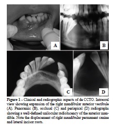
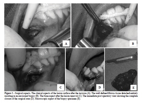
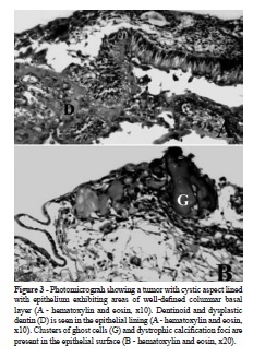
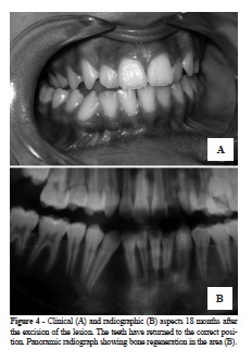
a mild face asymmetry in right anterior region of the mandible with no inflammation signs. Intraorally, there was a well-defined and firm swelling located at the right anterior mandible. The lesion extended from the right lateral incisor to right canine, expanding the lingual and buccal bone cortex (Figure 1A). The mucosa overlying the lesion was intact, non tender, but exhibited a purplish area. All teeth involved, although presenting little mobility, were vital. The lesion caused a separation of incisor and canine, with nonspecific rotation (Figure 1A). The radiographic examination comprised a dental panoramic (Figure 1B), mandibular occlusal (Figure 1C) and periapical (Figure 1D) radiographs. The images revealed a well-defined unilocular radiolucency delimited by radiopaque sclerotic border at the right mandible extending from lateral incisor to canine of the same side (Figure 1B-D). The adjacent teeth (incisor and canine) were divergent with no signs of root resorption. The clinical and radiographic differential diagnosis included central giant cell granuloma, keratocystic odontogenic tumor and ameloblastoma. Thus, an incisional biopsy was planned under local anesthesia; however, during the surgical approach, the lesion detached entirely resulting in an excisional biopsy (Figure 2). The postoperative course was uneventful. The biopsy material was fixed in a 10% formalin solution and submitted to histopathological analysis. Figure 3 shows the histopathological findings for the tumor. A cystic lesion with a fibrous capsule lined by odontogenic epithelium of varying thicknesses was observed (Figure 3). Columnar cells resembling ameloblasts (Figure 3A) and cuboidal cells forming a well-defined basal layer (Figure 3B) were present. In areas, the epithelium presented loosely arranged cells. Hyaline, eosinophilic and partially mineralized material (dysplastic dentine) were occasionally seen in the connective tissue adjacent to the epithelium (Figure 3A). Toward the lumen, epithelial cells without nucleus presented a round enlarged morphology slightly eosinophilic. These large ballooning cells, known as ghost cells, were a prominent feature. The ghost cells showed varying degrees of dystrophic calcification (Figure 3B). The lesion was diagnosed as a calcifying cystic odontogenic tumor. Further clinical (Figure 4A) and radiographic (Figure 4B) evaluation demonstrated that the teeth have returned to correct position. Radiographic examination showed a total bone formation (Figure 4B). No recurrence was detected in a clinical and radiographic follow-up of 18 months.
DISCUSSION
The CCTO is a mixed epithelial and mesenchymal odontogenic tumor that has a wide range of clinical and radiological appearance, which can simulate (or possibly coexist with) a different number of pathological conditions1. Based on these data, a detailed evaluation is necessary to establish a precise final diagnosis and an adequate treatment.
CCTO incidence varies in literature; however, all studies indicate that it is a rare lesion2,4. In fact, in a recent study, Fernandes et al.5 were able to detect only 12 cases (3.5%) of CCTO in 340 cases of odontogenic tumors in a Brazilian population.
The general clinical aspects (e.g. age, lesion localization and symptoms) of this case are in consonance with those previously described in the literature for CCTO2,3,7. Although the incidence of CCTO is mostly in the second decade of life, this lesion may occur at any age. In this case, it occurs in the first decade of life. The lesion appeared in the anterior portion of the mandible, and the literature indicates the anterior portions of both jaws as the most common sites for CCTO3-5,7-9,16.
In the present case, the CCTO presented a slow growing and a painless swelling that caused clinical teeth dislocation, common features of several benign intraosseous lesions. The radiographic examination demonstrated a solitary, well-circumscribed radiolucent area which displaced adjacent teeth. No calcifications were identified on plain radiographs. The absence of fever, lymphadenopathies and erythemas (intraoral and extraoral), associated with the presence of teeth vitality, excluded the diagnosis of inflammatory lesions. Due to the rarity of CCTO, the preoperative diagnosis is difficult to be done based only on clinical and radiographic features of the lesion. From a clinical and radiographic point of view, differential diagnosis included benign radiolucent lesions, such as central giant cell granuloma (CGCG), keratocystic odontogenic tumor (KCOT) and ameloblastoma11,13. Based on the fact that these lesions require different treatments, incisional biopsy and histopathologic analysis were necessary for the final diagnosis. In this case, incisional biopsy was planned under local anesthesia; however, when the lesion was accessed, it detached entirely resulting in an excisional biopsy. The diagnosis of CCTO was histologically determined.
Radiographically, CCTO generally appears as a unilocular lesion with well-defined margins in which calcifications may be observed. The radiographic aspects of the lesion described in this paper showed similar characteristics to those of the current literature3,4,6,14,16 although calcifications were not observed on plain film. The absence of evident mineralized tissue on radiographs was also reported in other studies in which calcified bodies were microscopically found. In fact, calcifications had been detected in about half of all CCTO3,4. Some reports have indicated that computed tomography (CT) may be more successful than plain film radiography in depicting such calcification, because such calcifications may be obscured in plain radiographs by superimposition of anatomic details14,16.
The CGCG is one of the most important lesions to be discussed in the differential diagnosis, because it has a predilection for women at any age, and most cases occur before the age of 30 years. Moreover, CGCG affects the anterior part of the jaw and 70% of the cases involve the mandible. Usually, this lesion may be present as a unilocular or multilocular radiolucency, often with poorly distinguished borders17.
Other important lesion to be considered in the differential diagnosis is the KCOT, that occurs in a wide age range, most commonly in the second and third decade, more frequently in men. Furthermore, this lesion affects mandible twice as often as the maxilla. It was previously known as the odontogenic keratocyst. The name was changed to reflect its neoplastic nature1,2,4. Ameloblastoma, specially the unicystic type, is seen in the second or third decades of life, and must be included in the differential diagnosis. The KCOT and ameloblastoma show slow growing; however, present local invasive behavior and high rate of recurrence if not treated with more aggressive procedures1. On the other hand, the CCTO and CGCG exhibit slow growing, but they are not invasive, thus the treatment generally involves only the enucleation of the lesion1.
The definitive diagnosis of CCTO is made histologically due to the lack of pathognomonic clinical characteristics and radiological features4. The most notable histological features of this pathologic entity include a cyst lining demonstrating clear cells known as ghost cells with a propensity to calcify and hyaline areas suggestive of immature or dysplastic dentine. The histological aspects described in this case are in accordance with literature1,4,6,8,10,14,18. The microscopic features confirm that calcified masses may not necessarily appear on radiographic examination even though they may be found microscopically.
The CCTO should be conservatively treated by surgical enucleation with curettage because recurrences are very uncommon. A follow-up period of at least 10 years is, therefore, recommended9,10,14,15. Most of the recurrent cases have occurred over five years following initial treatment. Maybe in this pediatric population the CCTO could be more aggressive, thus this case not only must be closely accompanied, but also has to be divulgated in the dental pediatric care field in order to aware the pediatricians of the possibility of occurrence of such pathological entity in children.
Fortunately, after a surgical conservative treatment, normal healing and absence of recurrence has been observed in a follow-up of 18 months. Although CCTO is an uncommon odontogenic lesion, it can affect the patients of the pediatric group. Careful diagnostic procedures and adequate interpretation of clinical, radiographic and histological findings are crucial for the correct final diagnosis and establishment of the most appropriate treatment.
REFERENCES
1. Barnes L, Eveson JW, Reichart PA, Sidransky D, editors. World Health Organization classification of tumors: pathology and genetics of tumors of the head and neck. Lyon: IARC; 2005. [ Links ]
2. Buchner A, Merrell PW, Carpenter WM. Relative frequency of central odontogenic tumors: a study of 1,088 cases from Northern California and comparison to studies from other parts of the world. J Oral Maxillofac Surg 2006;64(9): 1343-52. [ Links ]
3. Buchner A. The central (intraosseous) calcifying odontogenic cyst; an analysis of 215 cases. J Oral Maxillofac Surg 1991;49(4):330-9. [ Links ]
4. Erasmus JH, Thompson IO, van Rensburg LJ, van der Westhuijzen AJ. Central calcifying odontogenic cyst. A review of the literature and the role of advanced imaging techniques. Dentomaxillofac Radiol 1998;27(1):30-5. [ Links ]
5. Fernandes AM, Duarte EC, Pimenta FJ, Souza LN, Santos VR, Mesquita RA, et al. Odontogenic tumors: a study of 340 cases in a Brazilian population. J Oral Pathol Med 2005;34(10):583-7. [ Links ]
6. Hong SP, Ellis GL, Hartman KS. Calcifying odontogenic cyst. A review of ninety-two cases with reevaluation of their nature as cyst or neoplasms, the nature of ghost cells, and subclassification. Oral Surg Oral Med Oral Pathol 1991;72(1):56-64. [ Links ]
7. Iida S, Fukuda Y, Ueda T, Aikawa T, Arizpe JE, Okura M. Calcifying odontogenic cyst: radiologic findings in 11 cases. Oral Surg Oral Med Oral Pathol Oral Radiol Endod 2006;101(3):356-62. [ Links ]
8. Johnson A 3rd, Fletcher M, Gold L, Chen SY. Calcifying odontogenic cyst: a clinicopathologic study of 57 cases with immunohistochemical evaluation for cytokeratin. J Oral Maxillofac Surg 1997;55(7):679-83. [ Links ]
9. Jones AV, Craig GT, Franklin CD. Range and demographics of odontogenic cysts diagnosed in a UK population over a 30-year period. J Oral Pathol Med 2006;35(8):
500-7.
10. Li TJ, Yu SF. Clinicopathologic spectrum of the so-called calcifying odontogenic cysts: a study of 21 intraosseous cases with reconsideration of the terminology and classification. Am J Surg Pathol 2003;27(3):372-84. [ Links ]
11. McGowan RH, Browne RM. The calcifying odontogenic cyst: a problem of preoperative diagnosis. Br J Oral Surg 1982;20(3):203-12. [ Links ]
12. Shear M. Developmental odontogenic cysts. An update. J Oral Pathol Med 1994;23(1):1-11. [ Links ]
13. Sikes JW Jr, Ghali GE, Troulis MJ. Expansile intraosseous lesion of the maxilla. J Oral Maxillofac Surg 2000; 58(12):1395-400.
14. Slootweg P. Odontogenic tumors an update. Curr Diagnost Pathol 2006;12:54-65. [ Links ]
15. Souza LN, Souza AC, Gomes CC, Loyola AM, Durighetto AF Jr, Gomez RS, et al. Conservative treatment of calcifying odontogenic cyst: report of 3 cases. J Oral Maxillofac Surg 2007;65(11):2353-6.
16. Tanimoto K, Tomita S, Aoyama M, Furuki Y, Fujita M, Wada T. Radiographic characteristics of the calcifying odontogenic cyst. Int J Oral Maxillofac Surg 1988;17(1):29-32. [ Links ]
17. Walstad WR, Fields T, Schow SR, McKenna SJ. Expansile lesion of the anterior maxilla. J Oral Maxillofac Surg 1999;57(5):595-9. [ Links ]
18. Yoshida M, Kumamoto H, Ooya K, Mayanagi H. Histopathological and immunohistochemical analysis of calcifying odontogenic cysts. J Oral Pathol Med 2001;30(10):582-8. [ Links ]
 Corresponding address:
Rua Ramiro Barcelos, 2492, sala 503 – Santana
CEP 90035-003 – Porto Alegre/RS, Brazil
Phone: (55) 51 3308-5011
e-mail: manomartins@gmail.com
Received in: 21/1/11
Accepted in: 12/4/11
