


Serviços Personalizados
Artigo
 pdf em Inglês
 Artigo em XML
 Referências do artigo
 Enviar este artigo por email
Links relacionados
Compartilhar

 Permalink
Arquivos em Odontologia
versão impressa ISSN 1516-0939
Arq. Odontol. vol.46 no.3 Belo Horizonte Jul./Set. 2010
Radiographic analysis of traumatized primary teeth
Análise radiográfica da dentição decídua após traumatismo alveolodentário
Dione Dias TorrianiI; Elaine de Fatima Zanchin BaldisseiraII; Sílvia Aparecida Ximenes MouraIII; Renata da Luz FerroIV; Marília Leão GoettemsIII
IDepartment of Social and Preventive Dentistry, Federal University of Pelotas (UFPel), Pelotas, RS, Brazil
IIDepartment of Semiology and Clinical Studies, Federal University of Pelotas (UFPel), Pelotas, RS, Brazil
IIIDental Surgeon
IVGraduate Program in Dentistry, Federal University of Pelotas (UFPel), Pelotas, RS, Brazil
ABSTRACT
This study aimed to evaluate the frequency of radiographic findings in primary teeth, as well as their supporting tissues, following dental trauma, and the occurrence of sequelae according to the trauma type. One radiograph was taken soon after the trauma, and two follow-up radiographs, with approximately sixmonth intervals between each, were taken of 116 traumatized teeth from 65 patients at the Pelotas Dentistry School in Pelotas, Santa Catarina, Brazil. One hundred and ninety-five radiographs were analyzed to evaluate: tooth position, periodontal ligament space, root integrity, alveolar bone, and root canal aspects. Data were shown in the form of absolute and percentage frequencies. The Wilcoxon test (p<0.05) was used to detect differences in the images. The widening of the periodontal ligament and pathological root resorption, although with different evolutions, represented the most frequent findings. In the first radiograph, 35.6% of the teeth were diagnosed as presenting a widening of the periodontal ligament, as compared to 15.1% in the final radiograph. Pathological root resorption varied from 9.6% in the first radiograph to 35.8% in the final exam. Whereas alveolar bone resorption prevailed in intrusion cases and root canal obliteration in concussion/ subluxation cases, tooth displacement occurred most often in intrusion and lateral luxation/extrusion cases. Concussion, subluxation, and intrusion were the conditions which were most often associated with sequelae during this sample’s period of analysis. Radiographic sequelae resulting from alveolodental trauma in the primary dentition tend to vary. Identifying radiographic findings and relating them to the type of trauma can aid the professional in defining the prognosis and a proper treatment plan.
Uniterms: Pediatric dentistry. Tooth. Deciduous. Injuries. Radiography.
RESUMO
Este estudo visa identificar as sequelas radiográficas em dentes decíduos e seus tecidos de suporte, após traumatismo alveolodentário. Foram realizadas tomadas radiográficas logo após o traumatismo alveolodentário e duas radiografias subsequentes, executadas com intervalo de cerca de seis meses, em 116 dentes pertencentes a 65 pacientes, na Faculdade de Odontologia da Universidade Federal de Pelotas, Brasil. Nas cento e noventa e cinco radiografias foram analisadas a posição do dente, o espaço do ligamento periodontal, a integridade radicular, o osso alveolar e a luz do canal radicular. Os dados foram apresentados como freqüências simples e percentuais, sendo que o teste de Wilcoxon (p<0,05) foi utilizado para detectar diferenças entre as imagens radiográficas. A alteração do espaço do ligamento periodontal e a reabsorção radicular patológica foram os achados mais frequentes, apesar de apresentarem diferentes evoluções. O espessamento do espaço do ligamento periodontal, o qual foi diagnosticado em 35,6% das radiografias iniciais, foi visualizado em 15,1% das radiografias finais. A reabsorção radicular patológica variou de 9,6%, das tomadas radiográficas iniciais, para 35,8% das finais. Enquanto a reabsorção óssea alveolar predominou nos casos de intrusão, e a obliteração do conduto radicular nos casos de concussão e subluxação; a alteração da posição dentária ocorreu com maior frequência nas intrusões e luxações laterais/extrusões. Concussão, subluxação e intrusão foram os tipos de traumatismos alveolodentários mais frequentemente associados às sequelas durante o período de análise da amostra. A apresentação das sequelas radiográficas, decorrentes do traumatismo alveolodentário na dentição decídua, é diversificada. Identificar a correlação dos achados radiográficos com o tipo de traumatismo auxilia o profissional a estabelecer o prognóstico e plano de tratamento de acordo com cada tipo de trauma.
Descritores: Odontopediatria. Dente. Decíduo. Traumatismos. Radiografia.
INTRODUCTION
The epidemiologic relevance of dental trauma in primary teeth is well-known. Traumas include regular accidents involving children and often occur at an early age, showing an occurrence as high as 30%1,2 to 35%3,4. The highest incidence can be observed in children between 1 and 3 years of age, which points out the interdependence of the motor development and trauma occurrence1,5. In these cases, the upper incisors are the most commonly affected2,6,7.
Alterations, such as pulp necrosis8,10, fracture lines, dental mobility11, ankylosis, early loss of the primary tooth10, bone and root resorption9,10,12,13, dental discoloration, and pulp chamber obliteration10-12, are likely to represent dental trauma sequelae. Many of these conditions cannot be clinically viewed directly after the trauma. For this reason, a routine follow-up period is needed so as to detect and treat the traumas appropriately. Thus, radiographic assessment is essential, not only to diagnose, but also to deal with potential complications11, given that radiographs are able to identify otherwise temporarily asymptomatic, but no less injurious, sequelae.
The knowledge of trauma types and radiographic sequelae most likely related to the trauma is important. In this light, the aim of this descriptive study was to evaluate the frequency of radiographic findings in primary teeth and their supporting tissues following dental trauma, as well as the occurrence of sequelae according to the trauma type.
MATERIALS AND METHODS
The research protocol was submitted to by the Human Research Ethics Committee of the School of Dentistry, Federal University of Pelotas, and was approved under log #43/04. An Informed Consent Form was signed by the child’s parent/guardian so that the child could participate in this study. Data were obtained at the Center for the Study and Treatment of Dental Traumas in Primary Dentition at the School of Dentistry, Federal University of Pelotas. The protocol consists of filing a dental trauma record, including anamnesis, a clinical and radiographic exam of the affected area, as well as photographic documentation of the injuries. Regular follow-up is performed until the permanent successor has erupted in order to detect possible alterations.
Files from 259 patients attended to from May 2002 to May 2008 were analyzed. Those who presented insufficient clinical data or poor radiographs, showed extensive caries lesions in the traumatized tooth, or abandoned treatment sessions were excluded. Sixty-five patients with 116 traumatized teeth were included in the study. A total of 195 periapical or occlusal radiographs were taken upon first examination of each patient, from six months to one year following the trauma.
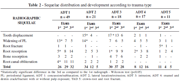
Dental trauma was classified according to Andreasen & Andreasen’s classification14 and divided into 5 groups: ADT 1 – concussion/ subluxation; ADT 2 – lateral luxation/extrusion; ADT 3 – intrusion; ADT 4 – enamel and dentin crack/ fracture with or without pulp exposure; ADT 5 – crown-root and root fracture.
Radiographic analysis was performed in a dark room in an X-ray viewing box with the aid of a 2X magnifying lens. The radiographs were arranged in chronological order to detect sequelae in the affected teeth and their evolution. An average of 30 radiographs was evaluated each day. Data were logged in a specific medical record.
The evaluation was carried out separately by two previously trained examiners (SAXM and EFZB), one of whom was a radiology specialist. The diagnoses of the two examiners were compared, and, in case of disagreement, a decision was reached by consensus.
The examiners received a dental record which included the child’s age at the time of the trauma and the affected tooth. The following preestablished criteria were used:
a) Position of the tooth: NORMAL or DISPLACED;
b) Periodontal ligament space: NORMAL or THICKENED;
c) Root integrity: NORMAL, with FRACTURE or with PATHOLOGICAL RESORPTION;
d) Bone tissue adjacent to affected tooth: NORMAL or with RESORPTION;
e) Root Canal Lumen: NORMAL or OBLITERATED.
Displacement was diagnosed in comparison with the homologous tooth. When two teeth, consecutive dental elements on a same arch, were displaced by dental trauma, they were diagnosed as displaced in relation to their neighboring teeth’s incisal edge or cusp.
Results were shown as simple and percentage frequencies. The Wilcoxon test (p<0.05) was used to detect differences on each traumatized tooth by comparing the final radiograph to the first. Statistical analysis was carried out using the Stata 9.1 software (2005).
RESULTS
Of the 116 evaluated teeth, 9 teeth were lost during follow-up due to early exfoliation or extraction: six of which belonged to the lateral/ extrusive luxation group, two to the intrusion group, and one to the crown-root/root fracture group.
Table 1 shows the distribution of sequelae found in the three radiographic examinations. The most frequent alterations found were the widening of the periodontal ligament space, which decreased in follow-up radiographs, and pathological root resorption, which increased during follow-up.
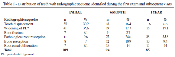
Table 2 presents sequelae development according to the trauma type. Assessing the position of the teeth, realignment occurred in the lateral/extrusive luxation (Z=-3.16; p<0.001) and in the intrusion groups (Z=-3.00; p<0.001). Periodontal ligament space thickening showed a significant decrease in concussion/subluxation (Z=2.13; p<0.001) and in lateral/extrusive luxation cases (Z=-3.16; p=0.02). A significant increase in root resorption in concussion/subluxation (Z=-2.67; p<0.001), intrusive luxation (Z=-2.53; p=0.01), and enamel and dentin crack/fracture (Z=-2.44; p=0.01) groups could be observed. Alveolar bone resorption occurring during the follow-up radiographic exams prevailed in intrusion cases. Obliteration of the root canal increased significantly in concussion/subluxation cases (Z=2.64; p<0.001).
DISCUSSION
Periodic radiographic evaluations associated with anamnesis and clinical examinations are crucial in cases of traumatized teeth. Their relevance must be made clear to parents/guardians by the professional so as to define the most appropriate moment to intervene, mainly because of the potential for late sequelae development.
Radiographs in primary dentitions are many times difficult to obtain due to the patient’s young age or because of his unwillingness to cooperate. According to the International Association of Dental Trauma, depending on the child’s ability to cope with the procedure and the type of injury suspected, several angles are recommended: a 90º horizontal angle, an occlusal view, and an extra-oral lateral view15. In this study, the occlusal technique by means of a size 2 film was preferred, as it is a simple procedure in which the film is easily fitted to the young child’s mouth and because it allows the dentist to assess tooth or root fractures, tooth displacement, and/or the widening of the periodontal ligament space11.
The difficulty of radiographic interpretation in primary teeth is also indisputable due to the overlapping of the permanent tooth follicle10.
According to Tyndall16, the professional must differentiate between artifact occurrence and anatomic accidents.
Upon evaluating the position of the tooth, in the lateral/extrusive and intrusion groups, statistical analysis showed a significant decrease in displacement between the first and third radiographs (Table 2). However, in the intrusion group, cases of displacement still occurred in the final radiograph. Borun & Andreasen10 also found a similar result upon evaluating primary incisors, which moved in a labial direction and presented a favorable prognosis, and found that 20% to 22% of the teeth do not totally re-erupt or re-erupt without perfect alignment. According to studies by Kenwood & Seow17, displaced teeth tend to develop necrosis over time, which reinforces the need for follow-up. In more severe displacement cases, compromising of the supporting structure may lead to early dental loss. Likewise, dental losses following lateral and/or extrusive luxation and intrusive luxation during follow-up, commonly caused by a premature exfoliation due to accelerated root resorption or the need for exodontics due to infection, could also be observed.
The widening of the periodontal ligament was observed in the concussion/subluxation group, although this is not a common characteristic finding of these trauma types15 (Table 2). This alteration may have been caused by periodontal fiber edema, which decreased during follow-up. Cases in which thickening persisted may well be compatible with pulp necrosis8,14. However, as the evaluations were solely based on radiographic aspects, the periodontal ligament condition may have been overestimated.
The decrease in root fractures in this study does not necessarily mean root healing had occurred (Table 2). Since most fractures occurred in teeth with crown-root or root fractures, either the extraction of the tooth or the removal of the coronal fragment was recommended, in turn maintaining the apical fragment to be resorbed15. According to a study by Kenwood & Seow17, out of eleven teeth with root fracture, 90% were normal upon first assessment whereas during in follow-up periods two teeth presented pulp necrosis. Both studies emphasize the importance of long-term follow-up.
An increase in pathological root resorption cases during follow-up could be observed in all cases except for the lateral/extrusive luxation group. Andreasen18 found a significant relationship between lateral luxation and intrusion and root resorption. Borum & Andreasen10 also found root resorption in intrusion and lateral luxation cases.
Pathological root resorption cases were detected in the first radiograph when taken soon after the trauma (Table 2). A history of previous traumas likely explains this finding. According to Holan & Fuks19, many primary tooth injuries are not perceived by parents, and radiographic signs of a previous injury were not supported by their personal accounts. In addition, Mortelliti & Needleman20 found that, out of 233 children with a record of vicious oral habits, traumas, and overjet, 14% presented radiographs which showed evidence of atypical root resorption. Moreover, when these risk factors were added, the total resorption values increased.
Obliteration of the root canal increase proved only to be statistically significant (p<0.001) in concussion/subluxation cases. Fried et al.11 reported that pulp calcification after subluxation increased in occurrence and severity over time. In contrast to this study, Kenwood’s & Seow17 and Ravn21 findings revealed pulp obliteration cases following crown fractures. In the present study, luxation revealed atresia signs during follow-up. Holan & Ram22 found pulp canal obliteration to be a common sequelae following intrusion of primary teeth and emphasized that the average age of children who had experienced some form of trauma was 3 years of age, at which time the apex may be open, favoring revascularization of the pulp and apposition of calcified deposits. Obliteration was observed both in diagnosis and follow-up radiographs. The same situation occurred in a study by Cardoso & Rocha23. It is important to mention that pulp obliteration is not an exclusive result of alveolodental trauma. It may occur by accelerating the physiological process, causing hard tissue apposition along the root canal walls.
The fact that sequelae, such as bone resorption, dental displacement, and pathological root resorption, are closely associated with intrusion shows the severity of this injury. Because it is strongly associated with secondary damage in permanent teeth24, the need for long term follow-up is emphasized.
It is evident, though, that a relation between radiographic findings and the clinical and symptomatological condition of the patient must be researched16. Often, the data from an image suggest a normal alteration which is not compatible with the clinical exam. In such cases, it is recommended to remain on alert, explaining the situation to parents/ guardians in search of signs which can aid in identifying the proper diagnosis.
All planned interventions for a young patient must necessarily take the future into account as part of a permanent healthcare program. Likewise, the treatment of dental trauma must fit this postulate. Thus, radiographic examinations are essential in this context.
CONCLUSION
Radiographic follow-up and the establishment of a connection between the type of alveolodental trauma and radiographic sequelae in primary dentitions can aid the dentist in defining the prognosis and the most appropriate treatment in an attempt to maintain the integrity of the deciduous tooth, the supporting tissues, and the permanent successor.
REFERENCES
1. Andreasen JO, Ravn JJ. Epidemiology of traumatic dental injuries to primary and permanent teeth in a Danish population sample. Int J Oral Surg. 1972; 1:235-9. [ Links ]
2. Ferguson FS, Ripa LW. Prevalence and type of traumatic injuries to the anterior teeth of preschool children. J Pedod. 1979; 4:3-8. [ Links ]
3. Garcia-Godoy F, Morban-Laucer F, Corominas LR. Traumatic dental injuries in preschoolchildren from Santo Domingo. Community Dent Oral Epidemiol. 1983; 11:127- 30. [ Links ]
4. Kramer PF, Zembrunsk C, Ferreira SH, Feldens CA. Traumatic dental injuries in brazilian preschool children. Dent Traumatol. 2003; 19:299-3. [ Links ]
5. Llarena del Rosario MEL, Alfaro VMA, Garcia- Godoy F. Traumatic injuries to primary teeth in México City children. Endod Dent Traumatol. 1992; 8:213-4. [ Links ]
6. Bijella MFTB, Yared FNF, Bijella VT, Lopes ES. Ocurrence of primary incisor traumatism in Brazilian children: a house-by-house survey. ASDC J Dent Child. 1990; 57:424-7. [ Links ]
7. Grimm S, Anntunes JLF, Castellanos RA, Narvai PC. Dental injury among Brasilian schoolchildren in the state of São Paulo. Dent Traumatol. 2004; 20:134-8. [ Links ]
8. Cunha RF, Pugliesi DMC, Percinoto C. Treatment of traumatized primary teeth: a conservative approach. Dent Traumatol. 2007; 23:360-3. [ Links ]
9. Gondim JO, Moreira Neto JJS. Evaluation of intruded primary incisors. Dent Traumatol. 2005; 21: 131-3. [ Links ]
10. Borum MK, Andreasen JO. Sequelae of trauma to primary maxillary incisors. I. Complication in the primary dentition. Endod Dent Traumatol. 1998; 14:31-44. [ Links ]
11. Fried I, Erickson P, Schwartz S, Keenan K. Subluxation injuries of maxillary primary anterior teeth: epidemiology and prognosis of 207 traumatized teeth. Pediatr Dent. 1996; 18:145- 51. [ Links ]
12. Jacobsen I, Sangnes G. Traumatized primary anterior teeth: prognosis related to calcific reations in the pulp cavity. Acta Odontol Scand. 1978; 36:199-204. [ Links ]
13. Cardoso M, Rocha MJC. Identification of factors associated with pathological root resorption in traumatized primary teeth. Dent Traumatol. 2008; 24:343-9. [ Links ]
14. Andreasen JO, Andreasen FM. Classification, etiology and epidemiologya. In: Text book and color atlas of traumatic injuries to the teeth. 3th ed. Copenhagen: Munksgaard; 1994. p.151-80. [ Links ]
15. Flores MT, Malmgren B, Andersson L, Andreasen JO, Bakland LK, Barnett F, et al. Guidelines for the management of traumatic dental injuries. III. Primary teeth. Dent Traumatol. 2007; 23:196-202. [ Links ]
16. Tyndal DA. The radiology of trauma. Dent Radiogr Photogr. 1985; 57:17-22. [ Links ]
17. Kenwood M, Seow WK. Sequelae of trauma to the primary dentition. J Pedod. 1989; 13:230-8. [ Links ]
18. Andreasen FM. Histological and bacteriological study of pulps extirpated after luxation injuries. Endod Dent Traumatol. 1988; 4:170-81. [ Links ]
19. Holam G, Fuks AB. The diagnostic value of coronal dark – gray discoloration in primary teeth following traumatic injuries. Pediatr Dent. 1996; 18:224-7. [ Links ]
20. Mortelliti GM, Needleman HL. Risk factors associated with atypical root resorption of the maxillary primary central incisors. Pediatr Dent. 1991; 13:273-7. [ Links ]
21. Ravn JJ. Sequelae of acute mechanical trauma in the primary dentition. ASDC J Dent Child. 1968; 84:281-9. [ Links ]
22. Holam G, Ram D. Sequelae and prognosis of intruded primary incisors: a retrospective study. Pediatr Dent. 1999; 21:247-7. [ Links ]
23. Cardoso M, Rocha MJC. Federal University of Santa Catarina follow-up management routine for traumatized primary teeth: part 1. Dent Traumatol. 2004; 20:307-13. [ Links ]
24. Sennhenn-Kirchner S, Jacobs H. Traumatic injuries to the primary dentition and effects on the permanent successors: a clinical follow-up study. Dent Traumatol. 2006; 22: 237-41. [ Links ]
 Author for correspondence:
Dione Dias Torriani
Rua Gonçalves Chaves 457, 4° andar - Centro
CEP: 96015560 - Pelotas- RS - Brasil
e-mail: dionedt@gmail.com
Received on 22/03/2010 – Accepted on 06/07/2010
Contato: dionedt@gmail.com, baldis@terra.com.br, silvia.axm@hotmail.com, renatalferro@ig.com.br, mariliagoettems@hotmail.com
