Serviços Personalizados
Artigo
Links relacionados
Compartilhar
Arquivos em Odontologia
versão impressa ISSN 1516-0939
Arq. Odontol. vol.49 no.1 Belo Horizonte Jan./Mar. 2013
Adjustment of cast metal post/cores modeled with different acrylic resins
Adaptação de pino/núcleos metálicos fundidos modelados com diferentes resina acrílicas
João Milton Rocha Gusmão I; Renato Piai Pereira I; Guilhermino Oliveira Alves II; Matheus Melo Pithon I; David Costa Moreira I
I Departamento de Saúde, Universidade Estadual do Sudoeste da Bahia (UESB), Vitória da Conquista, BA, Brasil
II Cirurgião dentista
Contact: joao.milton@ig.com.br, rppiai@uol.com.br, guild2@hotmail.com, matheuspithon@gmail.com, daviddcm@uol.com.br
ABSTRACT
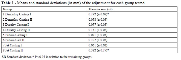
Aim: Evaluate the performance of four commercially available chemically-activated acrylic resins (CAARs) by measuring the level of displacement of the cores following casting. Materials and Methods: Two devices were constructed to model the cores based on a natural tooth. Forty post/cores were modeled, 10 in each of the following CAARs: Duralay (Reliance Dental, Illinois, USA), Pattern Resin (GC, Tokyo, Japan), Dencrilay (Dencril, Sao Paulo, Brazil), and Jet (Clássico, Sao Paulo, Brazil). Two casting rings were included, each of which contained 5 post/cores for each of the four CAARs tested, a total of 20 per ring. Following casting, the specimens were only sandblasted and separated from the feeding channels. The post/cores were placed in device 1 and, with the aid of an optical microscope, were attached to a digital camera. Images were then taken of the adjustment between the core and the remaining tooth on the labial surface. The images were processed using the Image Tool for Windows, version 3.0, measuring three fixed points in each specimen. Data were statistically analyzed using the ANOVA and Tukey test. Results: The 40 post/cores were divided into eight groups according to resin brand and casting, obtaining the following results for mean and standard deviation (in mm): Dencrilay 0.192 CI (± 0.08), CII 0.058 (± 0.03); Duralay CI 0.097 (± 0.03), CII 0.131 (± 0.06); Pattern CI 0.07 (± 0.03), CII 0.10 (± 0.05); and Jet CI 0.06 (± 0.02), CII 0.382 (± 0.17). Statistically significant differences could be identified when comparing the Dencrilay CI and the Jet CII with the remaining groups, which all proved to be unfavorable. Conclusion: The quality of cast metal post/core adjustment is not associated with the use of a specific acrylic resin.
Uniterms: Post and core techinique. Acrylic resins.
RESUMO
Objetivo: Avaliar o desempenho de 4 marcas comerciais de RAAQ medindo-se o nível de desadaptação dos núcleos pós fundição. Materiais e métodos: Foram construídos dois dispositivos, para modelagem dos núcleos a partir de um dente natural, foram modelados 40 pino/núcleos sendo 10 de cada uma das seguintes RAAQ: Duralay (Reliance Dental, Illinois, USA), Pattern Resin (GC, Tokio, Japan), Dencrilay (Dencril, São Paulo, Brasil) e Jet (Clássico, São Paulo, Brasil), foram incluído dois anéis para fundição, sendo que cada um continha 5 pino/núcleos de cada uma das 4 RAAQ testadas totalizando 20 por anel, após a fundição os espécimes foram apenas jateados e separados dos canais de alimentação. Os pino/núcleos foram recolocados no dispositivo 1 e com a ajuda de um microscópio ótico acoplado a uma camareira digital foram obtidas as imagens do gap entre o núcleo e o remanescente dentário na face vestibular, as imagem foram processada no programa Image Tool for Windows versão 3.0 medindo 3 pontos fixos em cada espécime. Os dados foram analisados estatisticamente com os testes ANOVA e Tukey. Resultados: Os 40 pinos foram divididos em 8 grupos segundo a marca e a fundição, obtendo as seguintes resultados de media (em mm) e desvio padrão; Dencrilay FI 0,192 (+-0,08), Dencrilay FII 0,058 (+-0,03), Duralay FI 0,097 (+-0,03), Duralay FII 0,131 (+-0,06), Pattern FI 0,07 (+-0,03), Pattern FII 0,10 (+- 0,05), Jet FI 0,06 (+-0,02) e Jet FII 0,382 (+-0,17) havendo diferença estatística apenas entre Dencrilay FI e Jet FII com os demais grupos sendo desfavoráveis para estes dois grupos mencionados. Conclusão: A qualidade de adaptação de pino/núcleos metálico fundidos não está associada ao uso de uma resina acrílica específica.
Descritores: Técnica para retentor intra-radicular. Resinas acrílicas.
INTRODUCTION
Cast metal post/core systems are still widely used in dentistry1,2. To obtain a cast, an impression of the root canal is made and the coronary portion is constructed with the specific anatomical features of each tooth, using a chemically-activated acrylic resin (CAAR) directly in the patient's mouth or on a plaster model that reproduces the prepared teeth with unobstructed root canals. The obtained acrylic structure (standard) is submitted to a laboratory casting process1,3 involving the use of one of several alloy alternatives: copper-aluminum (Al-Co), nickelchromium (Ni-Cr), and even noble alloys (platinumpalladium, Pt-Pd).
Although nonmetallic intracanal retainer systems, such as glass fiber, carbon fiber4, and ceramic posts, have grown in popularity in recent years, due to their superior aesthetic5 and biomechanical characteristics6-14, cast metal post/cores are indicated for posterior teeth and are generally the only option for inclined teeth or those that present torn roots, which require a change in the direction of the core in relation to the post. Cast metal post/cores are also used in anterior pillars of partial fixed dentures with no coronal remnant, since they present greater retention than nonmetallic posts, thus minimizing the risk of displacement9.
Despite the variety of commercially available alloys, dental health professionals usually choose one or two types of acrylic resin to model cast metal post/ cores. However, published studies confirming the best resin for this purpose are rare. Thus, the purpose of this study was to evaluate the adjustment of cast metal post/cores in vitro using a single alloy modeled with different chemically-activated acrylic resins.
MATERIALS AND METHODS
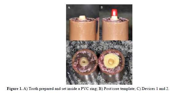
Initially, two devices were manufactured to model the post/cores, device 1 was constructed based on a natural tooth (central incisor) from the human teeth bank of the Dentistry course at Universidade Estadual do Sudoeste da Bahia (UESB). The manufacture of this device was performed by setting a tooth inside a 50-mm long and 20-mm thick PVC ring (Tiger, Ontario, Brazil) using a CAAR, maintaining 1 mm of the root exposed in the cervical region.
The tooth was endodontically treated and prepared for a full crown in such a way that the crown remnant was maintained vestibular to the surface plane (Figure 1A). Unblocking of the duct was perfomed using by drill bits of widths of 1, 2 and 3. Next, the post/core template was modeled (Figure 1B).
Next, device 2 was manufactured using a second PVC ring with the same dimensions, which was filled with a CAAR and fitted to device 1. To prevent bonding between the acrylic resin that filled the two parts, a layer of petroleum jelly was applied to the surface of the polymerized resin, while a layer of wax 7 (Clássico, São Paulo, Brazil) surrounded the core template. After the resin had been set, the wax 7 was removed, and the subsequent space was filled with dense condensation silicone (Heraeus Kulzer, São Paulo, Brazil). By fitting the two devices together, the material was molded by the core template, permitting its reproduction (Figure 1C).
After having manufactured the two devices, 10 post/cores were modeled from each of four different CAARs: Duralay (Reliance Dental, Illinois, USA), Pattern Resin (GC, Tokyo, Japan), Dencrilay (Dencril, São Paulo, Brazil), and Jet (Clássico, São Paulo, Brazil). At the same time, the casting molds were also created. The post/cores were obtained using the direct technique, in which the cores were modeled by the device, at which time the assembled piece was placed in a hydraulic press (VH, São Paulo, Brazil) at 15 kgf for 5 min. After having been manufactured, the post/cores were stored in containers of distilled water for 48 h until the time of inclusion.
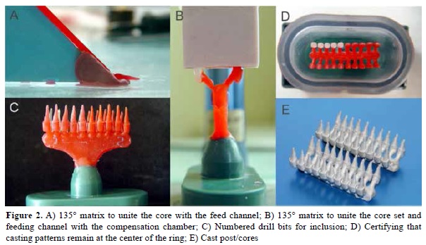
For the process of inclusion and casting, prefabricated wax thread feed conduits (Cerafix, São Paulo, Brazil), of 5 mm in length and 2 mm in diameter, were connected to the post/cores, forming an angle of 135° between their two long axes, secured by a matrix made from paper and dense silicone (Figure 2A). The post/core and conduit set was attached to the compensation chamber also at an angle of 135°, secured by another silicone matrix and with the aid of a delineator (Bioart, São Paulo, Brazil) (Fig. 2B).
The compensation chambers were made of utility wax (Clássico, São Paulo, Brazil) and each of the four extremities were numbered to identify the groups after casting (Figure 2C). Fixing the post/ cores at the center of the ring (Fig. 2D) was performed using the surface tension reducing agent, Surfacer (Polidental, Sao Paulo, Brazil), and Castorit Super C coating (Dentaurum GmbH & Co. KG, Ispringen, Germany), in accordance with the manufacturers' recommendations.


Two castings of 20 specimens were performed, five for each evaluated resin. A nickel-chromium alloy, Remanium CSe (Dentaurum GmbH & Co. KG, Ispringen, Germany), was used for this procedure. Following casting, the post/cores were submitted to a standard sandblasting procedure using aluminum oxide, separated from the feed channels using a silicon carbide disc (Carborundum, São Paulo, Brazil), and without any further processing or adjustment, were inserted in the prepared tooth. The test body was then attached to a base that maintained it at a set distance from the microscope objective (Alliance, São Paulo, Brazil), which was coupled to a digital camera used to obtain the images.
All of the images were evaluated considering three determined points in the space between the core and the vestibular coronal remnant, using the Image Tool for Windows software, version 3.0 (UTHSCSA, Texas, USA), aimed at measuring the displacement between the post and the remnant. After tabulating the data, the images were submitted to statistical analysis.
All statistical analyses were performed using the SPSS 15.0 software (SPSS Inc., Chicago, USA). Descriptive statistics, including mean and standard deviation, were calculated for both groups. The values of the displacement formed between the post and the tooth remnant were submitted to analysis of variance (ANOVA) to determine whether or not any statistical differences existed between the groups, followed by the Tukey test.

RESULTS
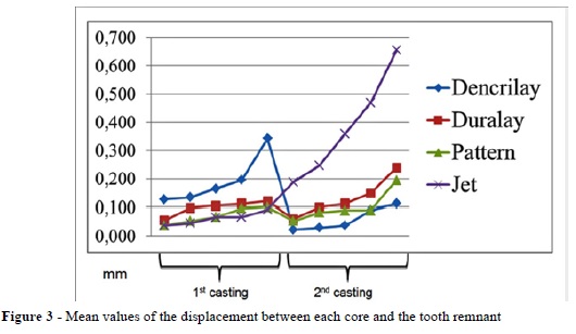
The mean values of the displacement between each core and the tooth remnant are presented in Figure 3, while the mean and standard deviation of each group are presented in Table 1. Comparisons between groups from the same casting revealed no statistical differences between the Dencrilay and Duralay (p.025), Dencrilay and Pattern Resin (p.001), and Dencrilay and Jet groups (p.000). In all cases, the largest displacement could be observed in the Dencrilay group. Statistically significant differences in displacement could be identified in the Dencrilay and Jet groups, which originated from the same CAAR produced in different castings. In the Dencrilay group, the first casting produced better adjusted posts (p.000), while in the Jet group, the second casting proved to be more well-adjusted (p.000). Finally, comparing the posts originating from different resins and different castings, the values obtained for first casting in the Dencrilay group and second casting in the Pattern group differed significantly (p.036) in favor of the Pattern group. By contrast, first casting in the Dencrilay group and second casting in the Jet group proved to be significantly different (p .000) in favor of the Dencrilay group. Second casting in the Jet group also showed statistically significant differences when compared with second casting in the Dencrilay group (p.000), first (p.000) and second casting in the Duralay group (p.000), and first (p.000) and second casting in the Pattern group (p.000).
DISCUSSION
CAARs are used in the modeling of root canals, either through direct or indirect techniques employed in the process of manufacturing cast metal post/cores16. The use of Duralay acrylic resin is preferred when performing this procedure, based on the argument that this material exhibits a greater dimensional stability after being pressed than do other commercial CAARs, despite the lack of studies supporting its superiority. Therefore, this study aimed to evaluate the performance of four different types of acrylic resin.
The majority of the specimens tested showed no statistically significant differences in displacement, regardless of the acrylic resin and casting, suggesting that the modeling capacity of the CAARs was not a determining factor as regards the variability in the observed values.
The present study opted to use an endodontically treated natural tooth, in contrast to the study by Rennó and Contim3, who used a metal structure to simulate the root and coronal remnant, since the use of a natural tooth more clearly simulates clinical practice17-20.
The nickel-chromium alloy used in this study was appropriate to obtain cast metal post/cores, and although it has a high modulus of elasticity, it has a low degree of oxidation15 when compared to similarly suitable alloys, such as copper-aluminum. To standardize the experiment, only one commercial batch of the alloy was used. The casting procedure was performed using two rings on which five specimens of each of the four evaluated resins were placed. Two castings were performed for each resin group, and the same coating was used on both rings.
The Image Tool software (UTHSCSA, Texas, USA) is an appropriate tool to perform the proposed evaluation technique, which in association with an optical microscope, allowed for the measurement of the displacement between the cast metal post/core and the tooth remnant at three points2, from which a mean value was calculated for the studied specimens.
Considering the values obtained from the same CAAR and different castings, statistically significant differences were verified for Dencrilay and Jet, with Dencrilay presenting better results in the second casting and Jet in the first casting. This finding suggests that even when following a rigorous standardized procedure, sequential castings can be associated with a variable quality of cast metal post/ core adjustment to tooth remnants.
Considering the values obtained for all test groups, the least displacement was observed for the second casting in the Dencrilay group (0.058 ± 0.03 mm), followed by the first casting in the Jet group (0.06 ± 0.02 mm), showing that different materials in different castings showed favorable adjustment results. By contrast, the first casting in the Dencrilay group (0.192 ± 0.08 mm) and the second casting in the Jet group (0.382 ± 0.17 mm) presented unfavorable values that were statistically significant when compared with the groups tested. This finding shows that the results could be obtained regardless of the materials and castings used.
Given these findings and considering the limitations of this study, the final analysis verified that cast metal post/core adjustment is not associated with a specific acrylic resin. When applied to clinical practice, this suggests that the preferential use of a particular resin is more strongly related to personal choice, considering the ease of handling, setting time, availability21-23, and cost, than with the qualities of the resin regarding its function in the manufacturing of casting molds.
CONCLUSION
Based on the present study's results, it can be concluded that the quality of cast metal post/core adjustment is not associated with the use of a specific acrylic resin.
REFERENCES
1. Louro RL, Vieira IM, Firme CT. Uso do núcleo metálico fundido na reconstrução de dentes tratados endodonticamente: relato de caso clínico. UFES Rev Odontol. 2008; 10:69-75. [ Links ]
2. Walton TR. An up to 15-year longitudinal study of 515 metal-ceramic FPDs: part 2. Modes of failure and influence of various clinical characteristics. Int J Prosthodont. 2003;16:177–82.
3. Rennó DG, Contin I. Avaliação da adaptação de retentores intrarradiculares fundidos obtidos por modelagem direta com RAAQ- estudo in vitro. RPG Rev Pos Grad. 2006; 13:25-30.
4. Ferrari M, Vichi A, Garcia-Godoy F. Clinical evaluation of fiber-reinforced epóxi resin posts and cast post and core. Am J Dent. 2000; 13:15-8.
5. Ferrari M, Vichi A, Mannocci F, Mason PN. Retrospective study of the clinical performance of fiber posts. Am J Dent. 2000; 13:9-13.
6. Schwartz RS, Robbins JW. Post Placement and restoration of endodontically treated teeth: a literature review. J Endod. 2004; 30:289-301.
7. Hansen PA, LeBlanc M, Cook NB, Williams K. The quality of posts and cores made using a reduce-time casting technique. Oper Dent. 2009; 34:709-15.
8. Asmussen E, Peutzfeld A, Heitmann T. Stifiness elastic limit, and strenght of types of endodontic posts. J Dent. 1999; 27:275-8.
9. Giovani AR, Vansan LP, Sousa Neto MD, Paulino SM. In vitro fracture resistance of glassfiber and cast metal posts with different lengths. J Prosthet Dent. 2009;101:183-8.
10. Akkayan B, Gulmez T. Resistance to fracture of endodontically treated teeth restored with different post systems. J Prosthet Dent. 2002; 87: 431-7.
11. Muniz L, Mathias P, Teixeira ML, Muhana M, Costa L, Ticianeli MG, et al. Reabilitação estética em dentes tratados endodonticamente. Pinos de fibra e possibilidades clínicas conservadoras. São Paulo: Santos; 2010.
12. Santos AF, Meira JB, Tanaka CB, Xavier TA, Ballester RY, Lima RG, et al. Can fiber posts increase root stresses and reduce fracture. J Dent Res. 2010; 89: 587-91.
13. Naumann M, Blankenstein F, Dietrich T. Survival of glass fibre reinforced composite post restorations after 2 years - an observational clinical study. J Dent. 2005; 33:305-12.
14. Malferrari S, Monaco C, Scotti R. Clinical evaluation of teeth restored with quartz fiber-reinforced epoxi resin posts. Int J Prosthod. 2003; 16:39-44.
15. Gadbut J, Wenschhot DE, Properties of níquel alloys, Int J Prosthodont In: Metals handbook, vol3- Properties and Selection. Stainless steel, tool Materials and special Purpose Metals, ASM Metal Park. 1980; 3: 128-70.
16. Sabbak SA. Indirect fabrication of multiple postand- cores patterns with a vinil polissiloxane matrix. J Prosthet Dent. 2002; 88: 555-7.
17. Mendoza, DB, Eakle S, Kahl EA, Ho R. Root reinforcement with a resin bonded preformed post. J Prosthet Dent. 1997; 78: 10-4.
18. Sidoli GE, King PA, Setchell DJ. An in vitro evaluation of a carbon fiber-based post and core system. J Prostet Dent. 1997; 78: 5-9.
19. Martinez Inssua A, Silva L, Rilo B, Santana U. Comparison of the fracture resistances of pulpless teeth restore with a cast post and core or carbon fiber post with composite core. J Prosthet Dent. 1998; 80: 527-32.
20. Newman MP, Yaman P, Dennison J, Rafter M, Billy E. Fracture resistance of endodontically treated teeth restored with composite posts. J Prosthet Dent. 2003; 89: 360-7.
21. Saito T, Campos TN, Fernandes IM, Tortamano P. Moldagem de preparação dental com anel de cobre e godiva. Bras Odontol. 2002; 45: 555-7.
22. Rosentiel SF, Land MF, Fujimoto J. Contemporary fixed prosthodontics. St Louis: Mosby; 1995.
23. Kleine A, Nobilo MAA, Henriques GEP, Mesquita MF. Influência de materiais de moldagem e de técnicas de transferência em implantes osseointegrados na precisão dimensional linear de modelos de gesso. RPG Ver Pos Grad. 2002; 9: 349-57.


