


Serviços Personalizados
Artigo
 pdf em Inglês
 Artigo em XML
 Referências do artigo
 Enviar este artigo por email
Links relacionados
Compartilhar

 Permalink
Stomatos
versão impressa ISSN 1519-4442
Stomatos vol.20 no.38 Canoas Jan./Jun. 2014
Adenomatoid hyperplasia of minor salivary glands: a report of two cases
Hiperplasia adenomatoide de glândulas salivares menores: relato de dois casos
Cleverson Patussi I; Regiane Benez Bixofis I; Fernando Luiz Zanferri II; Roberta Targa S. Zanicotti III; Laurindo Moacir Sassi IV; Juliana Lucena Schussel II
I DDSs and residents in Oral and Maxillofacial Surgery at the Department of Oral and Maxillofacial Surgery, Erasto Gaertner Hospital, Curitiba, PR, Brazil
II DDSs and hold a PhD degree in Stomatology; they are part of the clinical staff of the Department of Oral and Maxillofacial Surgery, Erasto Gaertner Hospital, Curitiba, PR, Brazil
III DDS and holds a MSc degree in Stomatology; she is part of the clinical staff of the Department of Oral and Maxillofacial Surgery, Erasto Gaertner Hospital, Curitiba, PR, Brazil
IV DDS and holds a PhD degree in Head and Neck Surgery; he is chief of the Department of Oral and Maxillofacial Surgery, Erasto Gaertner Hospital, Curitiba, PR, Brazil
The authors have no conflicts of interest to declare concerning the publication of this manuscript.
ABSTRACT
Introduction: Adenomatoid hyperplasia of minor salivary glands is a rare benign lesion that can be mistakenly diagnosed as other types of salivary gland neoplasms. It presents as a small firm nodule or as an exophytic mass, usually painless with normal mucosa, and slightly red or blue. Histologically, aggregates of relatively normal acinar lobule mucosa can be seen in larger amounts than expected, causing increased volume and sometimes pain. Case report: This article describes two cases of adenomatoid hyperplasia of minor salivary glands, the first in the left buccal mucosa of a 12 year-old boy and the second in the labial mucosa of a 44 year-old woman, and a review of previous reports of this pathology in English literature. The clinical appearance of the tumor is indistinguishable from salivary gland neoplasms and pathological examination is therefore essential for definitive diagnosis of this pathology. In both cases the treatment chosen after diagnosis was follow up and no changes were observed over 2 years from the first appointment.
Keywords: Minor Salivary Glands, Salivary Gland Diseases, Salivary Gland Neoplasms.
RESUMO
Introdução: A hiperplasia adenomatoide de glândulas salivares menores é uma lesão benigna rara que pode ser confundida com outras neoplasias de glândulas salivares. Apresenta-se como um pequeno nódulo firme ou como massa exofítica, geralmente indolor, com mucosa de aparência normal, levemente avermelhada ou azul. Histologicamente, observa-se a presença de agregados acinares relativamente normais e lóbulos da mucosa em maiores quantidades, causando aumento do volume e, por vezes, dor. Relato do caso: São relatatos dois casos de hiperplasia adenomatoide de glândula salivar menor, sendo um em menino de 12 anos de idade, em mucosa bucal esquerda, e o outro em mulher de 44 anos de idade em mucosa labial; uma revisão dos relatos anteriores já descritos na literatura também é apresentada. O aparecimento clínico do tumor é indistinguível em comparação com neoplasia da glândula salivar; assim, o exame patológico é essencial para o diagnóstico definitivo desta patologia. Nos dois casos descritos, o tratamento escolhido foi o acompanhamento clínico após diagnóstico; não foram observadas alterações no período de 2 anos desde a primeira consulta.
Palavras-chave: Glândulas Salivares, Doenças das Glândulas Salivares, Neoplasias das Glândulas Salivares.
INTRODUCTION
Adenomatoid hyperplasia of minor salivary glands was first described in 1971 by Giansanti et al. 1 as a benign lesion of the oral cavity frequently located in the hard palate, but also being seen in the soft palate, retromolar area, buccal mucosa, lips and tongue.
This is a rare lesion, and its true nature has not yet been clarified, with doubt remaining on whether it is a hyperplasic or hamartomatous condition. Clinically, it presents as a small firm nodular, exophytic mass, usually painless and with normal mucosa, slightly red or blue. Histological examination is essential for correct diagnosis, to achieve differentiation from other salivary gland neoplasms 2,3.These lesions can occur at any age, usually during the fourth and fifth decades of life, and have a slight predilection for men. There are no reports in the literature of adenomatoid hyperplasia associated with salivary gland neoplasms 4. They have no known etiology, although it is speculated that local trauma may be an etiological factor for development of a hyperplasic lesion 5. They have also been classified as hamartomas, and are considered as a developmental variation by some authors 4.When they are considered to be hyperplasia secondary to irritation, cigarette smoking and wearing prostheses can be important factors in development of this tumor type 5.
The objective of this article is to describe two cases of this lesion and also to present a revision of cases previously described in the English-language literature in the form of case reports, highlighting the rarity of this manifestation.
CASE REPORT
Case 1
A 12-year-old Caucasian boy complaining of a lesion in the left buccal mucosa was referred by his orthodontist to the Department of Maxillofacial Surgery at Erasto Gaertner Hospital for evaluation.
No relevant medical history was reported other than medication for attention deficit disorder. There was no history of a traumatic episode affecting the area or any event that could be associated with the appearance of the lesion. The patient had good hygiene and was undergoing orthodontic treatment.
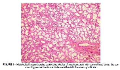
The lesion presented as an asymptomatic, whitish colored, increase in volume of approximately 5 mm in diameter on the left buccal mucosa. An excisional biopsy was performed and sent for histopathological examination. Microscopically, the lesion was well circumscribed showing coalescing lobules of mucinous acini with some dilated ducts and the surrounding connective tissue was dense with mild inflammatory infiltrate. The final diagnosis was adenomatoid hyperplasia of minor salivary glands. No signs of relapse were observed over a two-year follow-up period (Figures 1 and 2).
Case 2
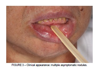
A 44 year-old, afro-descendent woman, was referred to the Department of Maxillofacial Surgery at Erasto Gaertner Hospital, complaining of an asymptomatic lesion of oral mucosa detected by her dentist during routine examination. Arterial hypertension was the only relevant element of her medical history and she reported hyposalivation. Clinical examination showed multiple asymptomatic nodular lesions on lip mucosa with an average size of 3 mm and normal coloration (Figure 3). Incisional biopsy revealed adenomatoid hyperplasia of minor salivary glands after histopathological examination. After two year's follow-up, the patient has no further complaints and the lesions are stable.
DISCUSSION
Adenomatoid hyperplasia of minor salivary glands (AHMSG) is an uncommon clinicopathologic lesion characterized by a clinically evident, nodular, non-painful swelling, usually mimicking a salivary gland neoplasm or a fibroma, but histologically comprised of normal-appearing minor salivary gland lobules 4.
Seifert et al.6 believe that one etiological factor of adenomatoid hyperplasia may be sialadenitis, considering that this major salivary gland disease can affect and influence the minor salivary glands, leading to the occurrence of this condition. However, Brannon et al. 2 do not believe there is any evidence to support this statement as true. In the cases described here, there was no manifestation of this condition before the onset of AHMSG, and so no relationship was detected with sialadenitis. In fact, no oral findings or systemic disorders were present that the authors could have considered possible etiologic factors in these lesions.
Since the first description by Giansanti et al. 1 in 1971, a total of 82 cases have been reported in the English-language scientific literature (Table 1).
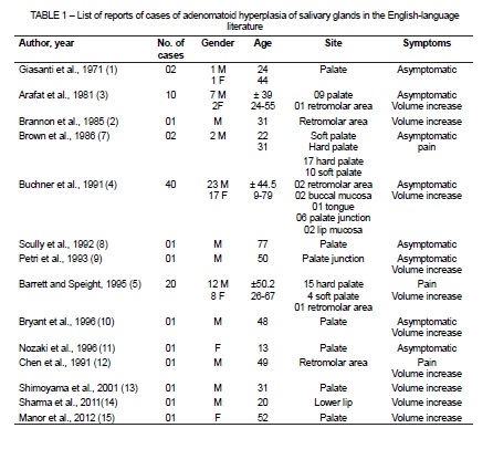
Age at occurrence can vary from 9 to 79 years, as shown by Buchner et al. 4, with a slight preference for men, the most common sites being the hard or soft palate, but AHSMG is also frequently found in the retromolar area. A correct diagnosis of AHSMG will exclude malignant salivary neoplasm and improve patient care.
Adenomatoid hyperplasia of the minor salivary gland is considered a benign tumor. However, it is not currently known whether the chromosomal aberrations associated with it are related to its malignant potential. Since the precise mechanisms involved in its development and progression are still not clear, any additional data that might bring us closer to full understanding are important 15.
This lesion can sometimes be confused with mucocele, especially if it appears on the lower lip 14. Bhavna et al. 16 state that the lower lip is the most common site for mucocele and that they generally have a bluish surface, but not always. As such, mucocele is on the list of differential diagnoses for AHSMG, when found on the lower lip, as was seen in one of cases reported in this article.
The interest in this entity arises from the fact that although it is a benign pseudotumoral lesion, it can be clinically confused with benign or malignant tumors and even, if fine needle aspiration cytology is used, with low grade mucoepidermoid tumors 17. If due care is not taken, mistaken diagnosis could therefore result in inappropriate treatment 14.
Our cases occurred at unusual sites and only two other cases have been reported in the buccal mucosa and three in lip mucosa 4-14.Although systemic conditions and some medications may cause enlargement of major salivary glands, there is no report showing this is associated with minor salivary gland hyperplasia. The patient in case 1 takes medication for attention deficit disorder, but there has been no link made between central nervous system stimulants and AHMSG to date. However, the patient is hyperactive, which could lead to traumas in the mouth area.
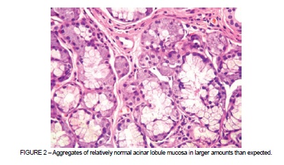
Histologically, the presence of aggregates of relatively normal acinar lobule mucosa can be seen in larger amounts than expected. Sometimes glands may be enlarged and will be located near the mucosal surface. Occasionally there is localized chronic inflammation 4. Furthermore, there is no report in the literature that relates adenomatoid hyperplasia with benign or malignant gland neoplasm.
Infiltration by fat of the stroma may occasionally be seen, such as adipose tissue that can sometimes be observed in the periphery of salivary gland tissue. In the elderly, alcoholics, diabetics and the malnourished, adipose tissue shows this peculiarity in the interacinar gap, forming a true fatty infiltration of the stroma 18.
When a lesion is confirmed to be an adenomatoid hyperplasia of minor salivary glands on excisional biopsy, no further treatment is required because these lesions do not recur. The present case is consistent with all of these findings 12.
In conclusion, the literature is clear that these lesions may be associated with local trauma and that their histological characteristics are benign. The clinical appearance of these tumors is indistinguishable from salivary gland neoplasms and so pathological examination is essential for definitive diagnosis of this pathology.
REFERENCES
1. Giansanti JS, Baker GO, Waldron CA. Intraoral mucinous minor salivary gland lesions presenting clinically as tumors. Oral Surg Oral Med Oral Pathol. 1971;32(6):918-22. [ Links ]
2. Brannon RB, Houston GD, Meader CL. Adenomatoid hyperplasia of mucous salivary glands: a case involving the retromolar area. Oral Surg Oral Med Oral Pathol. 1985;60(2):188-90.
3. Arafat A, Brannon RB, Ellis GL. Adenomatoid hyperplasia of mucous salivary glands. Oral Surg Oral Med Oral Pathol. 1981;52(1):51-5.
4. Buchner A, Merrell PW, Carpenter WM, Leider AS. Adenomatoid hyperplasia of minor salivary glands. Oral Surg Oral Med Oral Pathol. 1991;71(5):583-7.
 Correspondence:
Cleverson Patussi
Department of Oral and Maxillofacial Surgery
Erasto Gaertner Hospital
Rua Dr. Ovande do Amaral, 20
CEP 81520-060, Curitiba, PR, Brazil
E-mail: cleversonpatussi@hotmail.com
