Stomatos
ISSN 1519-4442
Stomatos vol.20 no.38 Canoas ene./jun. 2014
Unusual fusion of a distomolar with a third molar assessed by cone-beam computed tomography
Fusão incomum de distomolar com terceiro molar avaliado por tomografia computadorizada de feixe cônico
Debora Duarte Moreira I; Sergio Lins de-Azevedo-Vaz I; Saulo Leonardo Sousa Melo I; Deborah Queiroz de Freitas I
I Department of Oral Diagnosis, Division of Oral Radiology, Faculdade de Odontologia de Piracicaba, Universidade Estadual de Campinas (UNICAMP), Piracicaba, SP, Brazil
The authors have no conflicts of interest to declare concerning the publication of this manuscript.
ABSTRACT
Supernumerary teeth are teeth that occur in addition to the normal series. They can be observed in all quadrants of the jaw, with highest incidence in the maxilla. When a supernumerary tooth is distal to the most posterior molar, it is called a distomolar. Distomolars are more common unilaterally, in the maxilla and in black people and affect 2.2% of the population. In contrast, fusion is the result of the union of two separate tooth germs, forming a single tooth joined by dentin and/ or enamel, and fusion of a permanent tooth with a supernumerary accounts for fewer than 0.1% of cases, usually involving anterior maxillary teeth. Periapical radiographs are routinely used for endodontic diagnosis and preoperative planning, for transoperative guidance and for postoperative follow-up. However, the two-dimensional nature of this imaging technique can impose limitations on the ability to determine the anatomy of root canals in teeth with anatomical variations. The objective of this case report is to describe a rare case of fusion of a distomolar with a third molar, assessed using cone-beam computed tomography (CBCT).
Keywords: Third Molar, Supernumerary Tooth, Tooth Abnormalities, Cone-Beam Computed Tomography.
RESUMO
Os dentes supranumerários são uma anomalia e podem ser vistos em todos os quadrantes dos maxilares, com maior incidência na maxila. Quando os dentes supranumerários ocorrem distalmente ao terceiro molar, eles são denominados de dentes distomolares. Os distomolares ocorrem mais comumente unilateralmente na maxila de pessoas negras e afetam 2.2% da população. Por outro lado, a fusão ocorre pela união de dois germes dentários separados, desenvolvendo um único dente unido pela dentina e/ou pelo esmalte. A frequência de fusão de dentes permanentes e supranumerários é menor do que 0.1%, e normalmente envolve dentes anteriores da maxila. Radiografias periapicais são rotineiramente utilizadas em endodontia para o diagnóstico e planejamento pré-operatório, bem como durante o trans e pós-operatório. Entretanto, limitações relacionadas à bidimensionalidade dessa modalidade de imagens podem impedir a visualização adequada da anatomia dos canais radiculares dos dentes com variações anatômicas. O objetivo do presente relato foi descrever um caso raro de fusão por meio da tomografia computadorizada de feixe cônico.
Palavras-chave: Terceiro Molar, Dente Supranumerário, Anomalias Dentárias, Tomografia Computadorizada de Feixe Cônico.
INTRODUCTION
Supernumerary teeth are teeth that occur in addition to the normal series and they have been observed in all quadrants of the jaw, with highest incidence in the maxilla 1,2. When the supernumerary tooth occurs in a position distal to the most posterior molar, it is known as a distomolar, while if it erupts buccally or lingually to any molar, it is called a paramolar 1-3. Distomolars are more common unilaterally in the maxilla of black people and affect 2.2% of the population 3,4. It is important to be aware of the existence of a distomolar when a third molar is to be extracted, since the surgeon responsible for work-up must decide whether these teeth should be extracted (simultaneously with the third molar or independent of the third molar) or left alone 3.
In contrast, fusion is the result of the union of two separate tooth germs, forming a single tooth joined by dentin and/or enamel 5,6. Depending on the stage of development of the tooth and the moment of union, the union can be defined as complete or incomplete 5. A four-category classification has been suggested for these anomalous teeth: i) concrescence of teeth, two teeth fused by coalescence of their cementum; ii) fused teeth, teeth joined by dentine in their developmental stage; iii) geminated teeth, when the tooth bud of a single tooth attempts to divide; and iv) dens invaginatus 7-9. When fusion occurs between a normal tooth and a supernumerary tooth it can be difficult, if not impossible, to differentiate from gemination 5-10. The exact etiology of fusion is unknown, but it is believed to be related to physical forces or pressure that provokes close contact between developing teeth 5-11. The frequency of fusion of a permanent tooth with a supernumerary is less than 0.1% of cases and usually involves anterior maxillary teeth 9-12. These fused teeth tend to cause spacing and alignment problems, increasing predisposition to caries and periodontal diseases 5-12. Management of fused teeth with pulpal involvement can be considered an endodontic dilemma 11.
In endodontics, conventional intraoral periapical radiographs are an important diagnostic tool for assessing the root canal configuration. However, they only produce two-dimensional images of three-dimensional objects, resulting in superimposition of structures. For this reason, they are sometimes not capable of revealing complicated root canal system morphology, as in the case of fused teeth. It is possible that such problems can be overcome by using advanced diagnostic methods, such as cone-beam computed tomography (CBCT) 10-13.
The objective of this case report is to describe a rare case of fusion of a distomolar with a third molar, assessed using CBCT.
CASE REPORT
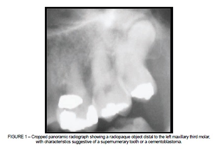
A 47-year-old black woman was referred to our oral radiology center (Faculdade de Odontologia de Piracicaba, Universidade Estadual de Campinas, Piracicaba, Brazil) for panoramic radiographic imaging, as the first step in planning for dental implant treatment. When analyzing the image, we noted a radiopaque object, distal to the left maxillary third molar, with characteristics suggestive of a supernumerary tooth or a cementoblastoma (Figure 1).

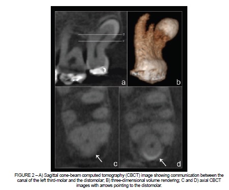
However, it was not possible to determine the boundary between tooth and the incidental finding. On the periapical image, acquired using a standard parallel technique, the radiopaque area was not entirely recorded because of its superior location. Therefore, in order to arrive at a differential diagnosis and determine the relationship between the incidental finding and the tooth, a CBCT scan was performed (Picasso Trio, E-WOO Technology, Giheung-gu, Republic of Korea) using a 5x5 cm field of view (FOV) and a 0.2 mm voxel size, in high-quality mode and with the metal artifact algorithm activated. The scan clearly showed a distomolar in an inverted position, atypically fused to the third molar as a disto-palatal root, with the canal of the third molar ending at the pulp chamber of the distomolar (Figure 2).

DISCUSSION
Periapical radiographs are routinely used for endodontic diagnosis and preoperative planning, for transoperative guidance and for postoperative follow-up. However, the two-dimensional nature of this imaging technique can impose limitations on its ability to reveal the anatomy of root canals in teeth with anatomical variations. In the case described here, CBCT was of paramount importance to clinical management of the patient, since it enabled differential diagnosis between a cementoblastoma and a supernumerary tooth, which require different treatment approaches. The first is a benign tumor that requires complete removal with extraction or root amputation and endodontic treatment of the tooth involved 6. In contrast, a supernumerary tooth with no associated disease process does not require surgical removal 6.
Differential diagnosis between fusion and germination is difficult and sometimes these entities cannot be distinguished from each other. We assumed that we were faced with a case of fused teeth in view of the union between the crown of the supernumerary tooth (in an inverted position) and the root of 28. The higher prevalence of unilateral distomolars in the maxilla of black people was also confirmatory. From this perspective, this case supports the theory of physical forces provoking close contact between developing teeth, bearing in mind the union between the pulp chambers of the supernumerary and the third molar.
Detection of fusion of teeth has clinical significance because treatment varies depending on severity. For asymptomatic cases, routine review and careful maintenance are required. In the event of future endodontic treatment of any of the teeth involved, a different approach to instrumentation and obturation would be required. In the present case, for example, there was no apical foramen to be debrided. Similar situations have been described by Ballalet al. 1, Rudagi et al. 8 and Song et al. 13, in which CT scans helped in planning endodontic treatment. Moreover, Indra et al. 11 and Sachdeva et al. 9 performed endodontic treatment on fused teeth without the aid of CT, but only because they were treating anterior teeth, in which there is less overlapping of structures and radiographic examination suffices.
Multidisciplinary treatment may be needed in cases requiring esthetic and/or functional improvement. Combined treatments comprise tooth extraction, endodontic treatment, reduction of mesiodistal tooth dimensions, followed by orthodontic treatment, tooth hemisection, and/or intentional replantation 14,15. Steinbock et al. 16 described a case of a F"large central incisor" (i.e. a central incisor fused with a supernumerary tooth) that was causing tooth crowding and esthetic problems. The case was treated by section and extraction of the fused supernumerary teeth; direct pulp capping of the remain incisor using mineral trioxide aggregate (MTA); orthodontic treatment to improve tooth alignment; and composite restoration of the crown of the central incisors. Similar treatment was applied in the case described by Guler et al. 14. Communication between the pulp of fused teeth should taken into consideration when the treatment planned involves sectioning the malformed tooth. Although root canal treatment is routinely considered in these cases, it can be avoided by performing pulp capping using MTA, if the exposed site is not infected and is limited in size.
CONCLUSION
This report presented a case of fusion of a distomolar with a third molar that mimicked a cementoblastoma on the periapical radiograph, but was correctly diagnosed using CBCT images. Since the patient was asymptomatic, routine review and careful maintenance were prescribed.
REFERENCES
1. Ballal S, Sachdeva GS, Kandaswamy D. Endodontic management of a fused mandibular second molar and paramolar with the aid of spiral computed tomography: a case report. J Endod. 2007;33:1247-51. [ Links ]
2. Ferreira-Junior O, Ávila LD, Sampieri MBS, Dias-Ribeiro E, Chen WL, Fan S. Impacted lower third molar fused with a supernumerary tooth: diagnosis and treatment planning using cone-beam computed tomography. Int J Oral Sci. 2009;1:224-8.
3. Shahzad KM, Roth LE. Prevalence and management of fourth molars: a retrospective study and literature review. J Oral Maxillofac Surg. 2012;70:272-5.
4. Kokten G, Balcioglu H, Buyukertan M. Supernumerary fourth and fifth molars: a report of two cases. J Contemp Dent Pract. 2003;15:67-76.
5. Shafer WG, Hine MK, Levy BM. Developmental disturbances of oral and paraoral structures. In: Shafer WG, Hine MK, Levy BM, editors. A text book of oral pathology. 4th ed. Philadelphia: WB Saunders; 1993. p. 38-9.
6. Neville BW, Damm DD, Allen CM, Bouquet JE. Abnormalities of teeth. In: Neville BW, Damm DD, Allen CM, Bouquet JE, editors. Oral and maxillofacial pathology. 2nd ed. Oxford: WB Saunders; 2002. p. 69-77.
7. Tsesis I, Steinbock N, Rosenberg E, Kaufman AY. Endodontic treatment of developmental anomalies in posterior teeth: treatment of geminated/fused teeth – report of two cases. Int Endod J. 2003;36:372-9.
8. Rudagi K, Rudagi BM, Metgud S, Wagle R. Endodontic management of mandibular second molar fused to a supernumerary tooth, using spiral computed tomography as a diagnostic aid: a case report. Case Rep Dent. 2012:614129.
9. Sachdeva GS, Malhotra D, Sachdeva LT, Sharma N, Negi A. Endodontic management of mandibular central incisor fused to a supernumerary tooth associated with a talon cusp: a case report. Int Endod J. 2012;45:590-6.
10. Rani K, Metgud S, Yakub SS, Pai U, Toshniwal NG, Bawaskar N. Endodontic and esthetic management of maxillary lateral incisor fused to a supernumerary tooth associated with a talon cusp by using spiral computed tomography as a diagnostic aid: a case report. J Endod. 2010;36:345-9.
11. Indra R, Srinivasan MR, Farzana H, Karthikeyan BDS. Endodontic management of a fused maxillary lateral incisor with a supernumerary tooth: a case report. J Endod. 2006;32:1217-9.
12. Nunes E, de Moraes IG, Novaes PMO, de Sousa SMG. Bilateral fusion of mandibular second molar with supernumerary teeth: case report. Braz Dent J. 2002;12:137-41.
13. Song CK, Chang HS, Min KS. Endodontic management of supernumerary tooth fused with maxillary first molar by using cone-beam computed tomography. J Endod. 2010;36:1901-4.
14. Guler DD, Tunc ES, Arici N, Ozkan N. Multidisciplinary management of a fused tooth: a case report. Case Rep Dent. 2013;634052.
15. Brunet-Llobet L, Miranda-Rius J, Lahor-Soler E, Cahuana A. A fused maxillary central incisor and its multidisciplinary treatment: an 18-year follow-up. Case Rep Dent. 2014;503478.
16. Steinbock N, Wigler R, Kaufman AY, Lin S, Naaj IAE, Aizenbud D. Fusion of central incisors with supernumerary teeth: a 10-year follow-up of multidisciplinary treatment. J Endod. 2014;40:1020-4.
 Correspondence:
Debora Duarte Moreira
Department of Oral Diagnosis
Faculdade de Odontologia de Piracicaba, Universidade Estadual de Campinas (UNICAMP)
Av. Limeira, 901, Caixa Postal 52
CEP 13414-903, Piracicaba, SP, Brazil
E-mail: dededm@hotmail.com


