


 Inglés


 Permalink
Stomatos
ISSN 1519-4442
Stomatos vol.20 no.39 Canoas jul./dic. 2014
Do different dentin tubular patterns of primary molars affect the bond strength of total-etching and self-etching adhesive systems?
Diferentes padrões tubulares dentinários de molares decíduos interferem na força de união de adesivos baseados no condicionamento ácido total e autocondicionantes?
Henrique Castilhos Ruschel I; Míriam Lacalle Turbino II; Antonio C. Guedes-Pinto III
I Board-certified in Cell Biology, Histology, and Microscopic Anatomy from Universidade Luterana do Brasil (ULBRA), Canoas, RS, Brazil; is an MSc in Pediatric Dentistry from Universidade Federal do Rio de Janeiro (UFRJ), Rio de Janeiro, RJ, Brazil; is a PhD in Pediatric Dentistry from Universidade de São Paulo (USP), São Paulo, SP, Brazil; and is a professor at the School of Dentistry at ULBRA, Canoas, RS, Brazil
II MSc and a PhD in Restorative Dentistry from USP, São Paulo, SP, Brazil; and a professor of the Department of Restorative Dentistry at USP, São Paulo, SP, Brazil
III MSc and a PhD in Pediatric Dentistry from USP, São Paulo, SP, Brazil; and a professor of the Department of Pediatric Dentistry at USP, São Paulo, SP, Brazil
The authors have no conflicts of interest to declare concerning the publication of this manuscript.
ABSTRACT
Aims: To evaluate the tensile bond strength of self-etching and total-etching adhesive systems to the different dentin surfaces of primary molars and to analyze the resin-dentin interface. Methodology: In this in vitro study, dentin samples 35 to 65% distant from the pulp (intermediate dentin) were obtained from buccal and lingual surfaces at the middle third of the crown of first and second primary molars. Dentin surfaces were prepared with 400 and 600-grit silicon carbide paper. Three adhesive systems (Prime & Bond NT, AdheSE and Clearfil SE Bond) were tested on the first and second primary molar surfaces (n=15); inverted truncated cones of resin composite with a 2.0 mm bonding diameter were built. After 24 hour storage in distilled water at 37°C, the specimens were submitted to the tensile bond strength test. To analyze the resin-dentin interface under scanning electron microscopy, samples were prepared with the same three adhesive systems (n=5). Results: No differences between first and second primary molar dentin substrates could be observed in mean bond strength values (ANOVA; p>0.05). The following mean bond strength values (MPa) were obtained: 15.65±3.70 (Prime & Bond NT), 19.47±7.09 (AdheSE) and 17.14 ±5.35 (Clearfil SE Bond). There were no statistically significant differences between the self-etching adhesive systems. The presence of hybrid a layer and tags were observed in all groups. Conclusions: Contemporary adhesive systems showed similar behaviors on both dentin tubular surfaces of primary molars. Follow-up studies of the clinical performance of these materials are needed.
Keywords: Dentin-Bonding Agents; Dentin; Tooth, Deciduous.
RESUMO
Objetivos: Avaliar a resistência de união, pelo ensaio de tração, de sistemas adesivos autocondicionantes e baseados no condicionamento ácido total em diferentes superfícies dentinárias de molares decíduos e analisar a interface de união adesiva. Metodologia: Neste estudo in vitro, amostras de dentina de 35 a 65% de distância pulpar (dentina intermediária) foram obtidas do terço médio das faces vestibular e lingual/palatina de primeiros e segundos molares decíduos. As superfícies dentinárias foram polidas com lixas de carboneto de silício de granulação 400 e 600. Três sistemas adesivos (Prime & Bond NT, AdheSE e Clearfil SE Bond) foram empregados nas amostras dos primeiros e segundos molares (n=15); corpos de prova foram confeccionados em resina composta com uma área de adesão de 2 mm de diâmetro. Após 24 horas de armazenagem em água destilada a 37ºC, fez-se o ensaio de tração. Para a análise da interface de união adesiva entre sistema adesivo e dentina usando microscopia eletrônica, as amostras foram preparadas com os mesmos três sistemas adesivos (n=5). Resultados: Não foram observadas diferenças estatisticamente significantes nos valores de adesão entre primeiros e segundos molares (ANOVA; p>0,05). Os seguintes valores médios de adesão em MPa foram obtidos: 15,65±3,70 (Prime & Bond NT), 19,47±7,09 (AdheSE) e 17,14±5,35 (Clearfil SE Bond). Não foram observadas diferenças estatisticamente significantes entre os adesivos autocondicionantes. A formação de camada híbrida e tags foi observada em todos os grupos. Conclusões: Os sistemas adesivos contemporâneos apresentaram comportamentos similares em ambos os tipos de superfícies tubulares dentinárias dos molares decíduos. Estudos de seguimento são necessários para o acompanhamento do desempenho clínico desses materiais.
Palavras-chave: Agentes de Adesão Dentinária; Dentina; Dente Decíduo.
INTRODUCTION
Dentistry has evolved considerably throughout the years, not only concerning techniques, but especially in the development of adhesive materials. In vitro tests, such as bond strength tests (tensile, shear, microtensile and microshear tests), have been developed with the purpose of analyzing the performance of adhesive systems on dental tissues and predict their clinical performance 1.
The bonding protocol of conventional adhesive systems involves dentin acidetching, followed by application of the primer and the adhesive. Some of these functions have been combined in the form of single-bottle adhesive systems. Adhesive bonding to the dentin is obtained by the formation of resin tags within tubules and also by the hybrid layer resulting from impregnation of the adhesive system into demineralized dentin 2-4.
Self-etching adhesive systems were developed with the purpose of eliminating the surgical steps of acid etching, washing and drying, which can be critical and interfere with the final quality of adhesion, due to the instability on the demineralized dentin matrix 3,5. The presence of a hybrid layer and tags in primary teeth has been observed with the use of total-etching and self-etching adhesive systems 1,3,6-12.
The study of the structural characteristics of the dentin tissue and a better understanding of the structure of mineralized tissues of primary teeth are extremely important to understand bonding mechanisms and interactions on this tissue 13. The dentin structure is formed by several tubules that do not have a uniform pattern throughout their extension. Few studies have evaluated dentin tubular pattern and its variations in primary teeth 14,15. In a previous study of the tubular pattern in first and second primary molars, we compared the dentin of the middle third of the crown of these teeth at a depth of 35 to 65% from the pulp 15. A comparison between the values obtained in the two types of dentin substrate showed statistically significant differences for tubule diameter and density in these teeth, with higher values on second molars when compared with first molars.
Correlations between dentin tubular pattern and dentin bond strength values have been reported in permanent teeth 16-19. Thus, it could be questioned if the different dentin tubular patterns found in primary molars described in our previous study 15 would not interfere with bond strength values of adhesive systems.
Therefore, the present study aimed to evaluate tensile bond strength of totaletching and self-etching adhesive systems to the dentin of first and second primary human molars. Additionally, the adhesive interface (resin-dentin) was analyzed by scanning electron microscopy (SEM).
METHODOLOGY
Tooth selection
Sixty-five primary human molars (33 first molars and 32 second molars) obtained at the Human Tooth Bank of the School of Dentistry of University of São Paulo were used. All teeth were healthy and showed complete root resorption. Selected teeth should not show clinically visible resorption on the pulp-chamber internal walls. Teeth were stored in distilled water cooled at 4ºC, which was changed weekly until the experiment was performed.
The present study was approved by the Research Ethics Committee of FOUSP (protocol no. 76/03).
Dentin sample collection
Selected primary molars were divided into two groups (first and second primary molars) and fixed with their occlusal surfaces facing the upper central portion of acrylic resin blocks. Teeth were fixed with the same acrylic resin used in the manufacturing of the blocks; molar occlusal surfaces were previously etched with 37% phosphoric acid gel (Condicionador Dental Gel; Dentsply, Petrópolis, RJ, Brazil) for 60 seconds.
Dentin samples were obtained according to the methodology employed in our previous study 15 and described below. The dentin surfaces on which adhesive procedures were performed were the same in all specimens and had similar tubular patterns as previously described by the authors 15. Based on that study, the crowns of the primary molars were divided into three equal parts in occlusal-cervical direction, corresponding to the cervical, middle and occlusal thirds of the crown. Then the cervical third of the crown was separated from the remainder of the crown by sectioning with a double-sided diamond disc (KG Sorensen, Cotia, São Paulo, Brazil), under cooling conditions (Figure 1a).
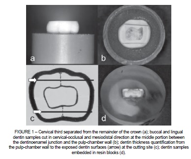
Dentin thickness at the buccal and lingual surfaces of the middle third of the crown of primary molars was quantified using a micrometer (Microdurometer HMV- 2000, Shimadzu, Kyoto, Japan). Measurements were taken at the central portion of each surface, considering a straight line extending from the dentinoenamel junction to the pulp-chamber wall.
Subsequently, each surface was cut in cervical-occlusal and mesiodistal direction with a diamond disc, at the middle portion between the dentinoenamel junction and the pulp-chamber wall, under cooling conditions (Figure 1b). Dentin thickness from the pulp-chamber wall to the exposed dentin surfaces at the cutting site was measured using a micrometer. Measurements followed the same procedure as that used for determining total dentin thickness at each surface (Figure 1c).
Based on total dentin thickness and on the distance between the exposed dentin surface and the pulp-chamber wall, the percentage of the distance from these surfaces to the pulp was determined. Only surfaces that were 35 to 65% distant from the pulp remained in the study, with total dentin thickness corresponding to 100%. Thus, it was possible to standardize the depth of the dentin samples on the tensile bond strength tests carried out.
Next, the sequence of cuts was completed to generate dentin samples for subsequent use in tensile bond strength tests and bonding interface analysis.
After these procedures, 77 and 81 dentin samples were obtained from first and second primary molars, respectively, all 35 to 65% distant from the pulp. Of this total sample, 45 were used in tensile tests and 15 in SEM analysis for each dentin substrate, i.e., first and second primary molars. The remaining samples were stored in case there was the need of repeating some of the experiments due to failure.
Preparation and distribution of dentin samples into groups
Dentin samples were embedded in resin blocks (Figure 1d), and the surfaces of the blocks were ground to expose the dentin surface using 400 and 600-grit silicon carbide papers (EXTEC Corp, Enfield, Connecticut , USA) on a polisher machine (Ecomet 3, Buehler Ltd., Lake Bluff, Illinois, USA) for about 10 seconds for each grinding under 400 rpm. This procedure exposed the dentin surface embedded in the block and standardized the smear layer formed.
Samples were distributed into three groups, according the adhesive system used: a single-bottle adhesive system, based on the total-etching technique, and two self-etching adhesive systems. Additionally, Filtek Z250 composite resin was used to build the specimens (Table 1). Each group was formed by two subgroups, corresponding to the two tooth types (first and second primary molars). Fifteen dentin samples were used in each subgroup for each adhesive system (total of 90 samples). Dentin surfaces were treated according to the adhesive system used, following the recommendation provided by the manufacturer.
Building of specimens for the tensile bond strength test
Following application of the adhesive system corresponding to each group, the resin block/dentin fragment was adjusted and fixed to a metallic clamping device together with a bipartite cylinder matrix measuring 3 mm in height. The two juxtaposed parts of the matrix formed a cone-shaped central cavity, with its vertex facing the dentin fragment. Filtek Z250 composite resin was inserted into the central cavity of the matrix in three increments, with each increment light-cured for 20 seconds. Hence, the set obtained comprised the resin block with the dentin fragment embedded and, attached to the latter, a specimen in the shape of a conical trunk with the smaller base in contact with the dentin. This base had 2.0 mm of diameter, with a bonding area of 0.0314 cm2.
Tensile bond strength evaluation
After 24-hour storage in distilled water at 37°C, specimens were submitted to the tensile bond strength test using a universal testing machine (Mini-Instron – Model 4442, Instron Corp., Norwood, Massachusetts, USA) running at a crosshead speed of 0.5 mm/ min. Tensile strength values were recorded in Newton (N) and then converted into Mega Pascal (MPa), considering the already described bonding area.
Mean values for the tensile bond strength test were compared by analysis of variance (ANOVA) and Tukey's test. The GMC software, version 2002, was used to analyze the results.
Preparation of samples for SEM analysis
The 30 dentin samples (15 primary first molars and 15 primary second molars) were divided into the same three groups according to the adhesive system employed. Samples were fixed with wax onto a glass plate, with the dentin surface facing up. Adhesive systems were applied and Filtek Z250 composite resin was placed in two increments, each of them measuring 2 mm and light-cured for 20 seconds. Samples were then stored in distilled water at 37 oC for 24 hours. Subsequently, dentin/resin samples were split in two, and one of the halves was used for SEM analysis of the resin-dentin interface.
Samples were immersed in HCl 2N for 2 minutes, then washed in distilled water with ultrasound for 10 minutes and immersed in 10% sodium hypochlorite solution for 5 minutes at room temperature. Next, samples were washed again in distilled water with ultrasound for 10 minutes and dehydrated in increasing concentrations of ethanol – 25% (20 minutes), 50% (20 minutes), 70% (20 minutes), 95% (30 minutes) and 100% (1 hour). Afterwards, samples were immersed in hexamethyldisilazane (HMDS) for 10 minutes and let dry on absorbent paper for 2 hours.
Samples were fixed in stubs with the fractured surface facing up and covered with a 20 nm layer of gold-palladium (BALTEC MED 020 – Coating System, Balzers, Liechtenstein). The entire extension of the bonding area between dentin and resin was analyzed with SEM (Philips XL 20, Amsterdam, The Netherlands), using a voltage of 20 Kv.
RESULTS
The values obtained showed a normal, homogeneous distribution. Therefore, mean values between the groups were compared using ANOVA and Tukey's test. The comparison between the different types of adhesive systems tested showed a statistically significant difference at 5% (ANOVA; p=0.003). However, when comparing mean bond strength values according to dentin substrate, i.e., dentin samples of first and second primary molars, no statistically significant differences were found (ANOVA; p=2987) (Table 2).
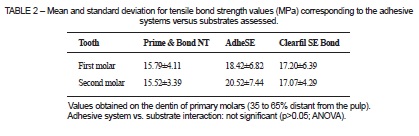
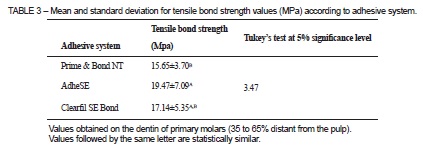
When analyzing the different adhesive systems tested, AdheSE presented higher mean bond strength values (19.47±7.09 MPa) than the other groups, at a statistically significant difference with regard to Prime Bond NT. Clearfil SE Bond, with a mean bond strength value of 17.14±5.35 MPa, did not show statistically significant differences in relation to AdheSE or Prime Bond NT (Table 3).
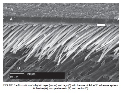
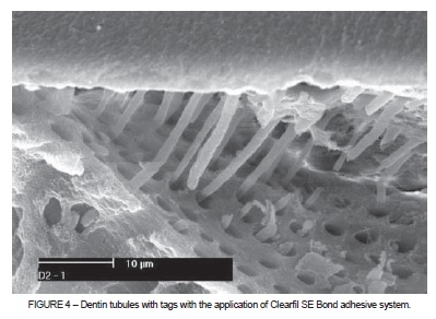
A descriptive analysis of the resin-dentin interface revealed the presence of a hybrid layer and resin tags within dentinal tubules in all adhesive systems. This aspect was evidenced in both types of dentin substrates, and no differences were observed between them. Hybrid layer thickness was not uniform throughout the adhesive interface, with values ranging from 1 to 3 μm. The AdheSE adhesive system showed a higher number and length of resin tags (Figures 2 to 4).

DISCUSSION
The lack of standardization in the methods used to test bond strength and the high number of variables assessed hindered the comparison between our results and those of other studies. In this sense, some aspects of the methodology adopted should be discussed.
The teeth used in this study were obtained at the same Human Tooth Bank used in the study by Bengtson et al. 1. According to those authors, due to difficulties obtaining teeth, many studies on bond strength in primary teeth include specimens with different times of exfoliation and storage. Nevertheless, these variables do not seem to interfere with tensile dentin bond strength values 1. The first and second molars used in this study were in the same range of the dental biological cycle, with all showing complete tooth resorption.
According to Pashley et al. 5, several variables can infl uence bond strength tests, and one of such factors is the size of the area to be tested. This is because the higher the area the greater the possibility of defects in the dentin-resin interface 5. In the present investigation, a 2.0 mm-diameter matrix was built, because dentin samples, mainly those from first primary molars, had a very reduced size. The use of a 2.0 mm-diameter matrix favors a smaller bonding area (0.0314 cm2), reducing the risk of adhesive interface failures 1,20. Studies with a 2.0 mm-diameter bonding area, similar to that used in the present investigation, did not quantify the depth between dentin and pulp at the bonding site 1,20,21. These aspects make it difficult to compare the results from these studies.
Most studies obtain dentin samples by superficial grinding of a dental surface until exposing an area sufficient for adhesion 1,20-22. However, this methodology does not allow to analyze the actual depth of the dentin surface at the bonding site. In this study, due to the sample collection method employed 15, it was possible to standardize this variable and analyze only the intermediate dentin (35 to 65% distant from pulp). Some previous studies have already described bond strength tests performed in primary teeth and focusing on the intermediate dentin 10,12. However, in those studies, this location was estimated rather than quantified, as sample preparation did not allow for such quantification.
Microtensile and microshear bond strength tests yield smaller bonding areas 23,24. In the present study, because the main purpose was to standardize dentin depth at the bonding site, the methodology employed to obtain dentin samples would not allow for the microtensile test to be performed.
The use of buccal and lingual dentin allowed us to obtain a fl at dentin area sufficiently large for adhesion. Additionally, the majority of tubules were disposed perpendicularly to the surface, as also observed in our previous study 15. Other studies have demonstrated the interference of tubular orientation on bond strength values 25,26.
Ruschel and Chevitarese 15 found higher tubule density and diameter in second molars compared to first molars. Because some studies associate bond strength values and 38 tubular pattern at the bonding site in permanent teeth 16-19, we hypothesized whether these differences between primary molars would interfere with bonding interaction with these substrates. Therefore, in this study, dentin samples were obtained following the same methodology of our previous study 15. The results showed that the values did not differ between the two types of dentin substrate. This is highly important for standardizing the methodology employed in the collection of dentin samples for in vitro bond strength tests.
Some studies have reported differences in adhesion due to variations in tubular pattern in permanent teeth (16,19), while other studies did not confirm this finding 17,18. Considering the lack of information on this aspect in primary teeth, this study contributes to demonstrate that the different tubular patterns of primary molars 15 do not interfere with the bond strength of adhesive systems to these teeth. Thus, it could be suggested that the composition and the action mechanism of the adhesive system employed is more important in the final product of the bonding process than the tubular pattern at the bonding site. The components of adhesive systems act differently on the smear layer and the underlying dentin. Therefore, the interaction between the adhesive system and the dentin may vary depending on the pH of the etching agent and on the capacity of the bonding agent to penetrate into demineralized dentin, especially in primary teeth 11,20.
This investigation found a hybrid layer and tags within dentinal tubules regardless of the action mechanism of the adhesive system employed. This was also observed in other studies assessing the dentin of primary teeth after the use of total-etching and self-etching systems 1,6-12. There may be a co-participation of both (hybrid layer and tags) in the bonding mechanism 27-29. Perhaps the smaller diameter and tubule density observed in primary first molars would result in a more solid dentin structure, in which the bonding process would more easily induce the formation of a hybrid layer in intertubular dentin. Conversely, in second molars, which show a greater number and diameter of dentinal tubules, it is possible that the tags could contribute to the bonding process.
Marquezan et al. 30 also failed to find differences in bond strength in primary teeth with the use of AdheSE vs. Clearfil SE Bond. In permanent teeth, Sensi et al. 31 found higher bond strength values for AdheSE, but again not statistically different from those associated with Clearfil SE Bond. The higher bond strength values obtained for AdheSE could be partially explained by the higher number of tags associated with this adhesive system, as the hybrid layer was present in all groups in this study, and the variation in its thickness has not been associated with bond strength values in primary dentin 7-10.
The present analysis of bond strength values led us to conclude that new selfetching systems show good bonding performance in vitro in the dentin of primary teeth. In fact, in primary teeth, some self-etching adhesives have shown higher bond strength values when compared to previous acid-etching adhesives 10,21, whereas others have shown lower values 11,12. Similar bond strength values have also been reported in primary and permanent teeth 4,7,9,20,30,32.
In pediatric dentistry, the development of adhesives requiring lower operative time is extremely important. With the purpose of reducing the number of surgical steps, single-bottle adhesive systems, based on total-etching, have been introduced into the market. In addition, self-etching systems have considerably reduced the time required for the clinical procedure. Finally, their indication is reinforced by the results of this study. However, etch-and-rise bonding systems are often preferred when large areas of enamel are still present 3. Thus, it is important evaluate the clinical performance of these self-etching systems on primary teeth, especially over time.
CONCLUSIONS
The present study showed that the different tubular patterns found in first and second primary molars did not interfere with bond strength values, and that self-etching adhesive systems shoed good bonding to the dentin of primary teeth. More follow-up studies of the clinical performance of these materials are needed.
REFERENCES
1. Bengtson CR, Bengtson AL, Bengtson NG, Turbino ML. Do the origins of primary teeth affect the bond strength of a self-etching adhesive system to dentin? Braz Oral Res. 2010;24:355-60. [ Links ]
2. Nakabayashi N, Nakamura M, Yasuda N. Hybrid layer as a dentin-bonding mechanism. J Esthet Dent. 1991;3:133-8.
3. Ozer F, Blatz MB. Self-etch and etch-and-rinse adhesive systems in clinical dentistry. Compend Contin Educ Dent. 2013;34:12-14.
4. Reis A, Loguercio AD, Manso AP, Grande RH, Schiltz-Taing M, Suh B, et al. Microtensile bond strengths for six 2-step and two 1-step self-etch adhesive systems to enamel and dentin. Am J Dent. 2013;26:44-50.
5. Pashley DH, Sano H, Ciucchi B, Yoshiyama M, Carvalho RM. Adhesion testing of dentin bonding agents: a review. Dent Mat. 1995;11:117-25.
6. Telles PDS, Aparecida M, Machado M, Nor JE. SEM study of a self-etching primer adhesive system used for dentin bonding in primary and permanent teeth. Pediatr Dent. 2001;23:315-20.
7. Burrow MF, Nopnakeepong U, Phrukkanon S. A comparison of microtensile bond strengths of several dentin bonding systems to primary and permanent dentin. Dent Mater. 2002;18:239-45.
8. Kaaden C, Schmalz G, Powers JM. Morphological characterization of the resin-dentin interface in primary teeth. Clin Oral Investig. 2003;7:235-40.
9. Casagrande L, De Hipolito V, De Goes MF, de Araujo FB. Bond strength and interfacial morphology of two adhesive systems to deciduous dentin: in vitro study. J Clin Pediatr Dent. 2005;29:317-22.
10. Nakornchai S, Harnirattisai C, Surarit R, Thiradilok S. Microtensile bond strength of a total-etching versus self-etching adhesive to caries-affected and intact dentin in primary teeth. J Am Dent Assoc. 2005;136:477-83.
11. Sardella TN, de Castro FL, Sanabe ME, Hebling J. Shortening of primary dentin etching time and its implication on bond strength. J Dent. 2005;33:355-62.
12. Sacramento PA, De Carvalho FG, Pascon FM, Borges AF, Alves MC, Hosoya Y, et al. Infl uence of NaOCl irrigation and water storage on the degradation and microstructure of the resin/primary dentin interface. J Adhes Dent. 2011;13:213-20.
13. Ruschel HC, Ligocki GD, Flaminghi DL, Fossati AC. Microstructure of mineralized tissues in human primary teeth. J Clin Pediatr Dent. 2011;35:295-300.
14. Koutsi V, Noomam RG, Horner JA, Simpson MD, Matthews WG, Pashley DH. The effect of dentin depth on the permeability and ultrastructure of primary molars. Pediatr Dent. 1994;16:29-35.
15. Ruschel HC, Chevitarese O. Density and diameter of dentinal tubules of first and second primary human molars – comparative scanning electron microscopy study. J Clin Pediatr Dent. 2002;26:297-304.
16. Öilo G, Olsson S. Tensile bond strength of dentin adhesives: a comparison of materials and methods. Dent Mater. 1990;6:138-44.
17. Shimada Y, Iwamoto N, Kawashima M, Burrow MF, Tagami J. Shear bond strength of current adhesive systems to enamel, dentin and dentin-enamel junction region. Oper Dent. 2003;28:585-90.
18. Toledano M, Osorio R, Ceballos L, Fuentes MV, Fernandes CA, Tay FR, et al. Microtensile bond strength of several adhesive systems to different dentin depths. Am J Dent. 2003;16:292-8.
19. Sattabanasuk V, Shimada Y, Tagami J. The bond of resin to different dentin surface characteristics. Oper Dent. 2004;29:333-41.
20. Stalin A, Varma BR, Jayanthi. Comparative evaluation of tensile-bond strength, fracture mode and microleakage of fifth, and sixth generation adhesive systems in primary dentition. J Indian Soc Pedod Prev Dent. 2005;23:83-8.
21. Agostini FG, Kaaden C, Powers JM. Bond strength of self-etching primers to enamel and dentin of primary teeth. Pediatr Dent. 2001;23:481-6.
22. Torres CP, Chinelatti MA, Gomes-Silva JM, Borsatto MC, Palma-Dibb RG. Tensile bond strength to primary dentin after different etching times. J Dent Child. 2007;74:113-7.
23. Pashley DH, Carvalho RM, Sano H, Nakajima M, Yoshiyama M, Shono Y, et al. The microtensile bond test: a review. J Adhes Dent. 1999;1:299-309.
24. Sano H, Shono T, Sonoda H, Takatsu T, Ciucchi B, Carvalho R, et al. Relationship between surface area for adhesion and tensile bond strength-evaluation of a micro-tensile bond test. Dent Mater. 1994;10:236-40.
25. Phrukkanon S, Burrow MF, Tyas MJ. The effect of dentine location and tubule orientation on the bond strengths between resin and dentine. J Dent. 1999;27:265-74.
26. Çehreli ZC, Akca T. Effect of dentinal tubule orientation on the microtensile bond strength to primary dentin. J Dent Child. 2003;70:139-44.
27. Carvalho RM, Ciuchi B, Sano H, Yoshiyama M, Pashley DH. Resin diffusion through demineralized dentin matrix. Rev Odontol USP 1999;13:417-24.
28. Vashisth P, Mittal M, Goswami M, Chaudhary S, Dwivedi S. Bond strength and interfacial morphology of different dentin adhesives in primary teeth. J Dent (Tehran). 2014;11:179-87.
29. Mithiborwala S, Chaugule V, Munshi AK, Patil V. A comparison of the resin tag penetration of the total etch and the self-etch dentin bonding systems in the primary teeth: An in vitro study. Contemp Clin Dent. 2012;3:158-63.
30. Marquezan M, da Silveira BL, Burnett LH Jr, Rodrigues CR, Kramer PF. Microtensile bond strength of contemporary adhesives to primary enamel and dentin. J Clin Pediatr Dent. 2008;32:127-32.
31. Sensi LG, Lopes GC, Monteiro Jr S, Baratieri LN, Vieira LC. Dentin bond strength of self-etching primers/adhesives. Oper Dent. 2005;30:63-8.
32. Gonzalez G, Rich AP, Finkelman MD, Defuria C. Shear bond strength of seventh generation bonding agents on dentin of primary teeth--an in vitro study. Gen Dent. 2012;60:46-50.
 Correspondence:
Henrique Castilhos Ruschel
Rua da República, 338/806
CEP 90050-320, Porto Alegre, RS, Brasil
E-mail: henrirus@gmail.com
