


Serviços Personalizados
Artigo
 pdf em Inglês
 Artigo em XML
 Referências do artigo
 Enviar este artigo por email
Links relacionados
Compartilhar

 Permalink
Stomatos
versão impressa ISSN 1519-4442
Stomatos vol.22 no.42 Canoas Jan./Jun. 2016
Factors associated with mandibular movement and vertical dimension of occlusion in elderly complete-denture wearers
Fatores associados com movimentos mandibulares e dimensão vertical de oclusão em idosos usuários de prótese total
Luiz Otávio Behrensdorf Reis I; Rita de Cássia Costa Ribeiro de Almeida I; Thiago Britto Ribeiro II; Angélica Gonzatti III; Noéli Boscato IV
I DDS, MSc, and PhD students at Graduate Program in Dentistry (Prosthodontics), School of Dentistry, Universidade Federal de Pelotas (UFPel), Pelotas, RS, Brazil
II DDS (private practice), Pelotas, RS, Brazil
III DDS (private practice), Palmitos, SC, Brazil
IV DDS, MSc, and PhD; and associate professor at Graduate Program in Dentistry, School of Dentistry (Prosthodontics), UFPel, Pelotas, RS, Brazil
The authors have no conflicts of interest to declare concerning the publication of this manuscript.
ABSTRACT
Objective: This study evaluated the effect of age, duration of edentulism, and duration of current complete-denture use on the extent of mandibular movements and on reestablishment of vertical dimension of occlusion in elderly complete-denture wearers. Methods: Thirty volunteers were selected based on predetermined inclusion criteria. Extent of mandibular movements was assessed using intraoral Gothic arch tracings while the study participants were still wearing their old complete dentures (T0). Change in vertical dimension of occlusion was determined from measurements taken using frontal images, acquired before substituting the old dentures (T0) and seven days after the last appointment for adjusting the new complete dentures (T1). Statistical analyses were performed using Pearson correlation coefficient and one-way analysis of variance with a post hoc Bonferroni test (α = 0.05). Results: Pearson correlation analysis found statistically significant negative correlation between vertical dimension of occlusion and mandibular movement measurements and age and between vertical dimension of occlusion and mandibular movement measurements and duration of denture use. The Bonferroni test detected differences for all parameters evaluated, except between duration of edentulism and extent of mandibular movement. Conclusion: The study findings show that older age and long-term use of dentures were linked to limited reestablishment of vertical dimension of occlusion and range of mandibular motion. However, the effect of duration of edentulism was not clear.
Keywords: Mouth, edentulous; Elderly; Denture, complete; Dental occlusion.
RESUMO
Objetivo: Este estudo avaliou o efeito de idade, tempo de edentulismo e tempo de uso da atual prótese total na extensão dos movimentos mandibulares e restabelecimento da dimensão vertical de oclusão em idosos usuários de prótese total. Metodologia: Trinta voluntários foram selecionados a partir de critérios de inclusão predeterminados. A extensão de movimentos mandibulares foi avaliada usando o registro intraoral do arco gótico de Gysi enquanto os participantes do estudo ainda usavam suas antigas próteses totais (T0). Alteração na dimensão vertical de oclusão foi determinada a partir de medidas realizadas em imagens frontais, obtidas antes da troca da prótese antiga (T0) e sete dias após o último ajuste da nova prótese (T1). Análise estatística foi realizada usando coeficiente de correlação de Pearson e análise de variância de uma via seguida do teste de Bonferroni (α = 0,05). Resultados: A análise de correlação de Pearson encontrou uma correlação negativa estatisticamente significativa entre dimensão vertical de oclusão e movimentos mandibulares e idade, e entre dimensão vertical de oclusão e movimentos mandibulares e tempo de uso da dentadura. O teste de Bonferroni encontrou diferenças estatísticas para todos os parâmetros avaliados, exceto entre tempo de edentulismo e amplitude de movimentos mandibulares. Conclusão: Os achados mostram que idade avançada e longo tempo de uso das próteses totais originaram limitado restabelecimento da dimensão vertical de oclusão e amplitude de movimentos mandibulares. Entretanto, o efeito da duração do edentulismo não foi claro.
Palavras-chave: Arcada edêntula; Idoso; Prótese total; Oclusão dentária.
INTRODUCTION
The literature reports a high prevalence of tooth loss among older people1,2. Dental implants have become the treatment of choice for edentulous patients3, but conventional complete dentures are still widely used4.
Previously published research shows that long-term complete-denture wearers often present with loss of vertical dimension of occlusion (VDO) and unstable dentures that are associated with wearing artificial teeth and progressive residual ridge resorption on both the maxilla and mandible5-7. Studies conducted with these individuals suggest that these aging-associated factors can culminate in the collapse of masticatory muscles, thereby jeopardizing basic functions such as mandibular movements (MM)8-10. In turn, compromised MM are associated with systemic diseases11 such as senile dementia, stress-related disorders, and cognitive dysfunctions, which may be due to nutritional deficiencies12-15.
This study evaluated the effect of age in years, duration of edentulism, and duration of current complete-denture use on the extent of MM and on reestablishment of VDO in complete-denture wearers. The null hypothesis in this study was that there are no relationships between increasing age, duration of edentulism, or duration of current complete-denture use and compromised MM and VDO.
METHODOLOGY
Experimental design
This observational, cross-sectional (for MM), and longitudinal (for changes in VDO) study was approved by the Local Human Research Ethics Committee (protocol no. 63/2013) and conducted in accordance with the Strengthening the Reporting of Observational Studies in Epidemiology (STROBE) guidelines16.
Participants
All elderly complete-denture wearers who came to a social center for seniors between March and November 2014 were invited to participate in the study. Those who met the following inclusion criteria were enrolled: complete-denture wearers (aged 60 to 90 years) who had used the same set of complete dentures for at least five years and needed to have them replaced; had experienced loss of VDO and had loose and unstable dentures due to changes in the orientation of occlusal plane; were free from temporomandibular disorders (TMDs), from limitations affecting opening the mouth, and from pain; and were able to comply with the experimental protocol (i.e., come to the dental clinic to be photographed and to have MM-Gysi arch tracing)17.
Initially, 131 elderly people were invited to participate; 97 of them did not meet the inclusion criteria and 4 declined to participate because of difficulties traveling to the clinic or work-related duties. After signing a written informed consent form based on the Declaration of Helsinki, the subjects enrolled were evaluated for VDO and extent of MM, at two and one time points respectively, and a questionnaire was administered to collect data on current age, duration of edentulism, and length of time using their most recent set of complete dentures.
The sample size (n = 8) needed to compare age, duration of edentulism, and duration of current complete-denture use across tertiles in years was calculated assuming that statistical tests would be used with a study power of 80% (α = 0.05)9. A total of 30 volunteers were enrolled, to account for potential losses during the experiment. The study participants were sequentially assigned to tertiles by years (n = 10 per tertile), according to their age, duration of edentulism, and duration of current complete-denture use.
Clinical procedures
After the initial interview, all participants were fitted with new conventionally manufactured complete dentures. Participants' maxillomandibular relationships were evaluated by obtaining their VDO from interocclusal wax records, using the metric, esthetic and phonetic methods. Mandibular and maxillary wax records were adjusted, and the wax was built up or removed to obtain the optimum VDO. The Dawson bimanual technique was used to achieve centric relation18.
After their new dentures had been fitted, participants were requested to return to the clinic for possible adjustments after 24 hours and 7, 14, and 21 days and then, finally, a week after their last adjustment. All clinical procedures followed the same standardized procedures. All materials were used in accordance with their manufacturers' instructions.
MM and VDO measurements
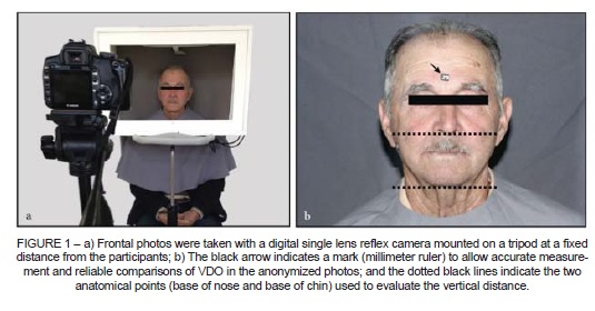
Frontal photos were taken using a digital single lens refl ex camera (Canon EOS Rebel XTi, Tokyo, Japan) mounted on a tripod19. The camera lens was set up in line with the Frankfort horizontal plane to ensure that each VDO photograph was standardized20,21. The vertical distance from the base of the nose to the base of the chin was measured twice to determine VDO (Figure 1), once before the new dentures were fitted (T0), and then again seven days after the last appointment for adjusting the new complete dentures (T1). The measurements at T1 were subtracted from those at T0 to calculate the difference in VDO, with a 6-week interval between evaluations.
To determine MM, each participant was instructed to perform anteroposterior and laterolateral mandibular movements for 10 minutes on an intraoral record inserted into his or her mouth22. Digital images provided intraoral Gothic arch tracings and anteroposterior (x-axis) and laterolateral (y-axis) MM measurements (Figure 2). Mandibular movement measurements were conducted once, at T0, while the participants were still wearing their old complete dentures. Afterwards, all subjects were provided with new complete dentures.
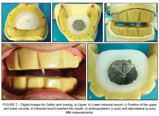
Each digital image included a millimeter ruler to allow accurate measurement and reliable comparisons of VDO and MM in the anonymized photos21. The images were imported into image-processing software (Image Tool version 3.0, University of Texas Health Science Center, San Antonio, TX, US)18 and VDO and MM measurements were taken by one calibrated and blinded examiner.
Statistical analysis
Data were recorded and analyzed using STATA version 13.0 (StataCorp LP, College Station, TX) to a significance level of 5%. Differences in reestablishment of VDO and extent of MM according to age, duration of current complete-denture use, and duration of edentulism were assessed using one-way analysis of variance with a post hoc Bonferroni test to compare outcomes across tertiles in years. Correlations between variables and MM and VDO outcomes were tested using Pearson correlation analysis.
RESULTS
The sample recruited for this study comprised 24 women and 6 men with a mean (standard deviation) age of 70.4 (10.1) years. They had been wearing their current sets of complete dentures for a mean (SD) of 17 (2.2) years and had been edentulous for a mean (SD) of 30.8 (18.5) years.
Pearson correlation analysis detected negative correlations (P < 0.05) between duration of current complete-denture use and VDO and MM measurements and between age and VDO and MM measurements (Table 1).
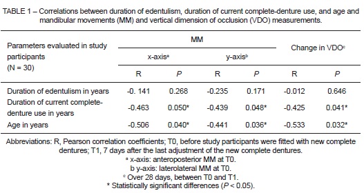
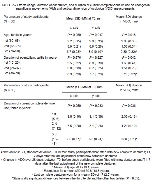
Table 2 shows the effects that the parameters investigated had on reestablishment of MM and VDO. Bonferroni testing detected statistically significant differences between the third tertiles for age and duration of complete-denture use (oldest ages and longest periods wearing current dentures, respectively) and the other tertiles (P < 0.05) for MM and for changes in VDO, but not for duration of edentulism and MM.
DISCUSSION
To the best of the researchers' knowledge, this is the first study to evaluate the effects of increasing age, duration of edentulism, and duration of current complete-denture use on the extent of MM and on reestablishment of VDO in elderly complete-denture wearers.
The null hypothesis tested was partially rejected as both age and duration of current complete-denture use were negatively correlated with optimized MM and VDO. In addition, increase in the length (in years) of any parameter resulted in lower VDO and MM measurements, the only exception being duration of edentulism and MM measurements.
Although the supporting tissue and associated facial structures deteriorate progressively as edentulous patients age6, these results show that the duration of edentulism was the only parameter that did not have a significant negative correlation with the outcomes evaluated. Likewise, the strong negative correlations between the outcomes investigated and duration of closed-denture use and age indicate that age8,9 and long-term use of complete dentures compromise MM 9,10,22 and VDO20.
These research findings corroborate studies that have reported that age affects the function of the stomatognathic system and masticatory muscles8-15,23. The lower reestablishment of VDO achieved for older patients could be linked to this20. Study participants aged 76 to 90 years had reduced Gothic arch tracing. This impaired mobility is probably because of reduced VDO and residual ridge resorption over the years compromising the translational capacity of the temporomandibular joints, as the effective working length of the masticatory muscles changes7. Notwithstanding, the higher proportion of older participants with adhesions or disk displacements without reduction might be another explanation and since the prevalence of TMD pain is reduced in elderly populations, anamnestic indices are not helpful for excluding painless TMDs5. Thus, ageing appeared to have an important effect on the parameters evaluated.
However, these findings also indicate that long-term complete-denture wearers should change their dentures regularly to maintain the health of the stomatognathic system because the duration of closed-denture use has a greater effect on the outcome than does duration of edentulism8. These results agree with studies showing that optimum prosthodontic treatment for edentulism enables proper masticatory muscle activity24,25 and could consequently improve MM and VDO patterns. Furthermore, this is an important issue because reduced MM can result in impaired mastication; an epidemiological risk factor for the development of cognitive dysfunction12-13.
One of this study's strong points is the methods used to measure MM and VDO, using standardized VDO frontal photographs and Gothic arch tracing records. Additionally, using Image Tool software and including the millimeter ruler in each photographic record ensured that measurements were accurate and reliable21.
Our findings should be interpreted with caution because to our knowledge this is the first study that has evaluated both MM and VDO under the infl uence of all outcomes investigated. One of the limitations of this study was the short-term VDO measurement, since long-term effects are more relevant clinically. Mandibular movements were only assessed once because the aim was to determine the magnitude of MM before any clinical and prosthodontic treatment, to avoid infl uence on MM measurements from the new prostheses. Further research should be conducted to evaluate the functional parameters of edentulous patients since the factors that predispose people to oral and/or systemic diseases—and their frequency and severity—can change over time.
CONCLUSION
The study findings show that older age and long-term use of dentures were linked with limited reestablishment of vertical dimension of occlusion and range of mandibular motion. However, the effect of duration of edentulism was not clear.
ACKNOWLEDGEMENTS
The authors would like to thank the seniors at the Centro de Extensão em Atenção à Terceira Idade (CETRES) for volunteering to take part in this study and are grateful for financial support provided by Programa de Extensão Universitária, Ministério da Educação e Cultura, Secretaria de Educação Superior (MEC/SESu).
REFERENCES
1. Colussi CF, Freitas SF. Epidemiological aspects of oral health among the elderly in Brazil. Cad Saude Publica. 2002;18:1313-20. [ Links ]
2. Peres MA, Barbato PR, Reis SC, Freitas CH, Antunes JL. Tooth loss in Brazil: analysis of the 2010 Brazilian Oral Health Survey. Rev Saude Publica. 2013;47:1-11.
3. Sivaramakrishnan G, Sridharan K. Comparison of implant supported mandibular overdentures and conventional dentures on quality of life: A systematic review and meta-analysis of randomized controlled studies. Aust Dent J. 2016 Feb 2. doi: 10.1111/ adj.12416. [Epub ahead of print]
4. Carlsson GE, Omar R. The future of complete dentures in oral rehabilitation. A critical review. J Oral Rehabil. 2010;37:143-56.
5. Grunert I, Grubwieser GJ, Ulmer H. Bilateral investigation of the temporomandibular joint. An autopsy study of edentulous individuals. J Oral Rehabil. 2000;27:671-81.
6. Zarb GA. A panacea for the edentulous predicament? Int J Prosthodont. 2013;26:405-6.
7. Tallgren A. The continuing reduction of the residual alveolar ridges in complete denture wearers: a mixed-longitudinal study covering 25 years. J Prosthet Dent. 2003;89:427-35.
8. Karlsson S, Persson M, Carlsson GE. Mandibular movement and velocity in relation to state of dentition and age. J Oral Rehabil. 1991;18:1-8.
9. Almeida RC, Rosa WL, Boscato N. The effect of occlusal splint pretreatment on mandibular movements and vertical dimension of occlusion in long-term complete denture wearers. Int J Prosthodont. 2016;29:287-9.
10. Gonçalves TM, Vilanova LS, Gonçalves LM, Garcia RC. Kinesiographic study of masticatory movements in denture wearers with normal and resorbed denture-bearing areas. J Prosthet Dent. 2014;112:1343-8.
11. Nakata M. Masticatory function and its effects on general health. Int Dent J. 1998;48:540-8.
12. Ono Y, Yamamoto T, Kubo KY, Onozuka M. Occlusion and brain function: mastication as a prevention of cognitive dysfunction. J Oral Rehabil. 2010;37:624-40.
13. Kim JM, Stewart R, Prince M, Kim SW, Yang SJ, Shin IS, et al. Dental health, nutritional status and recent-onset dementia in a Korean community population. Int J Geriatr Psychiatry. 2007;22:850-5.
14. Delmonico MJ, Harris TB, Visser M, Park SW, Conroy MB, Velasquez-Mieyer, et al. Longitudinal study of muscle strength, quality, and adipose tissue infiltration. Am J Clin Nutr. 2009;1:1579-85.
15. Manini TM, Hong SL, Clark BC. Aging and muscle: a neuron's perspective. Curr Opin Clin Nutr Metab Care. 2013;6:21-6.
16. Von Elm E, Altman DG, Egger M, Pocock SJ, Gotzsche PC, Vandenbroucke JP. The Strengthening the Reporting of Observational Studies in Epidemiology (STROBE) statement: guidelines for reporting observational studies. Lancet. 2007;370:1453-7.
17. Schiffman E, Ohrbach R, Truelove, Look J, Anderson G, Goulet JP, et al. Diagnostic Criteria for Temporomandibular Disorders (DC/TMD) for clinical and research applications: recommendations of the International RDC/TMD Consortium Network and Orofacial Pain Special Interest Group. J Oral Facial Pain Headache 2014;28:6-27.
18. Zarb GA, Bolarder CL, Hickey JC, Carlsson GE, eds. Boucher's prosthodontic treatment for edentulous patients. 10th ed. St Louis: Mosby; 1997.
19. Chou JC, Thompson GA, Aggarwal HA, Bosio JA, Irelan JP. Effect of occlusal vertical dimension on lip positions at smile. J Prosthet Dent. 2014;112:533-9.
20. Desai S, Upadhyay M, Nanda R. Dynamic smile analysis: changes with age. Am J Orthod Dentofacial Orthop. 2009;136:310.e1-10; discussion 310-1.
21. Baba K, Tsukiyama Y, Clark GT. Reliability, validity, and utility of various occlusal measurement methods and techniques. J Prosthet Dent. 2000;83:83-9.
22. Casselli H, Landulpho AB, Silva WA, Silva FA. Electrognathographic evaluations of rehabilitated edentulous patients. Braz Oral Res. 2007;21:355-61.
23. Mays KA. Reestablishing occlusal vertical dimension using a diagnostic treatment prosthesis in the edentulous patient: a clinical report. J Prosthodont. 2003;12:30-6.
24. Zuccolotto MC, Vitti M, Nobilo KA, Regalo SC, Siessere S, Bataglion C. Electromyographic evaluation of masseter and anterior temporalis muscles in rest position of edentulous patients with temporomandibular disorders, before and after using complete dentures with sliding plates. Gerodontology. 2007;24:105-10.
25. Trulsson U, Engstrand P, Berggren U, Nannmark U, Branemark PI. Edentulousness and oral rehabilitation: experiences from the patients' perspective. Eur J Oral Sci. 2002;110:417-24.
 Correspondence:
Noéli Boscato
Graduate Program in Dentistry
School of Dentistry, UFPel
Rua Gonçalves Chaves, 457, 2º andar
CEP 96015-560, Pelotas, RS, Brazil
E-mail: noeliboscato@gmail.com
