


Serviços Personalizados
Artigo
 pdf em Inglês
 Artigo em XML
 Referências do artigo
 Enviar este artigo por email
Links relacionados
Compartilhar

 Permalink
Brazilian Journal of Oral Sciences
versão On-line ISSN 1677-3225
Braz. J. Oral Sci. vol.10 no.3 Piracicaba Jul./Set. 2011
ORIGINAL ARTICLE
Use of dental dimensions estimated from personal portraits in human identification
Rachel Lima Ribeiro TinocoI; Laíse Nascimento Correia LimaI; Mário Marques FernandesII; Luiz Francesquini JuniorIII; Eduardo Daruge JuniorIV
I DDS, MSc, Department of Forensic Dentistry, Piracicaba Dental School, University of Campinas, Brazil
II DDS, MSc, Biomedical Service of Prosecute Council of Rio Grande do Sul State, Brazil
III DDS, MSc, PhD, Department of Forensic Dentistry, Piracicaba Dental School, University of Campinas, Brazil
IV DDS, MSc, PhD, Assistant Professor, Department of Forensic Dentistry, Piracicaba Dental School, University of Campinas, Brazil
ABSTRACT
Many cases of human identification in which traditional methods are not applicable challenge the experts' capability and versatility. In the absence of ante-mortem records, superimposition of skull images over photographs of a possible victim arises as a possible alternative. Aim: The present study was a pilot work willing to validate a new method of sizing images of the face by the use of proportionality principles, taking as reference a few predetermined accessories: a pair of sunglasses, a hat and a necklace. Methods: Twenty-one volunteers were photographed using each one of the accessories mentioned above. Pictures of the dental arches were also taken, with millimeter scale adjacent. The images with accessories were examined by a single operator, who estimated the mesiodistal width of the upper central incisor, for later comparison with the real measures. Results: The accuracy of the method was evaluated by the Student's t-test, which showed that the estimated measures were statistically greater than the real ones. Conclusions: The analysis of the data collected showed that the use of the accessories as a dimensional scale did not generate reliable results.
Keywords: forensic dentistry, victims' identification, tooth.
Introduction
Forensic dentistry is an important science for human identification, especially when conventional methods cannot be applied, usually due to advanced decomposition, carbonization or fragmentation of the body. In such cases, and in the absence of antemortem dental data to be compared, craniofacial image superimposition can be an accurate and reliable technique for human identification1-3. However, before superimposing images of the skull over photographs of a possible victim, it is mandatory that they both are properly sized in the same scale4-5.
Authors that apply computer-assisted craniofacial superimposition size reach the scale through craniometric and cephalometric landmarks easily identifiable in the face and the skull6-7, even thought this assessment is influenced by the thickness of local soft tissue - amount and disposition of fat - which can vary among different populations8.
Considering that the teeth are naturally visible throughout life, not being influenced by soft tissue, and commonly shown in smiling pictures, they are reliable size reference for comparing images of skull and personal pictures9. Although technical advances allow the three-dimensional facial reconstruction10-12, this is not an identification method, but rather a form to disseminate a facial approximation of that subject, willing to achieve someone who knew him/her, and can provide reliable antemortem records for human identification.
Human identification through craniofacial image superimposition has been tested and used by different researchers and experts. However, the proper scaling of the photographs to be compared is a precondition for an accurate evaluation. The aim of this pilot study was to test the feasibility of obtaining real size of teeth from personal photographs, using common accessories as size reference, in order to use them for the proper scaling required for image superimposition.
Material and methods
Sampling
Pictures of 21 volunteers (8 males and 13 females) were taken by a single photographer. All the subjects were aged between 21 and 60 years, and agreed to participate of the study by signing an appropriate document. Subjects with absence or anomalies in the upper central incisors were excluded, as well as those with notorious systemic pathology.
Methods
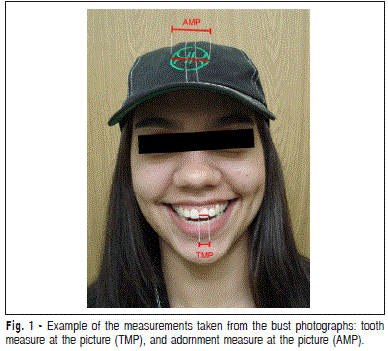
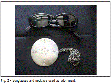
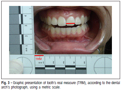
From each participant the following photographs were taken: The images obtained were stored in a computer managed by Windows Vista™, and analyzed by the software Adobe Photoshop™. From the bust photographs, the mesiodistal dimensions of the upper central incisors were measured, which were called TMP (tooth measure at the picture). From the same picture, using the same zoom, the size of the adornment (sunglass, a rounded detail of the cap, or necklace) were measured, to obtain what were called AMP (adornment measure at the picture), as shown in Figure 1, with the cap. Figure 2 shows the sunglasses and the necklace. All the measurements were stored in an Excel™ file. Then, the life-size of the adornments worn by the subjects were taken, with a digital caliper (Digimess, São Paulo, Brazil), which was called adornment's real measure (ARM). From the measures taken, tooth's real measure (TRM) was estimated, according to algebra fundamental rules of proportion, translated by the following equation: Once the estimations were concluded and stored, the pictures of the dental arches with metric scale were analyzed, and the mesiodistal dimensions of the upper central incisor were taken with a manual caliper, according to the scale at the picture, as graphically represented in Figure 3. The values obtained from this measuring procedure were then compared to the estimations. All measurements and estimations were made by a single examiner. Statistical analysis
• dental arch, in frontal view, with lip spacer, and scale number 2 from American Board of Forensic Dentistry placed at the same visual plane of the upper central incisors;
• bust photograph, wearing a pair of sun glasses, and smiling;
• bust photograph, wearing a cap, and smiling;
• bust photograph wearing a round pendant necklace, and smiling – only the female subjects. 
Results
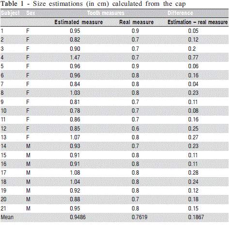
The dental dimensions estimated from the size of the cap were overestimated in 100% of the cases, with a mean difference of 0.1867 mm, as can be seen in Table 1. According to the Student's t-test, the estimated dental width was statistically higher than the real width (p = 0.0000169), at a significance level of 5%.
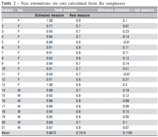
When estimating the dental width from sunglass dimensions, overestimated values were found again, except for two cases of underestimation (9.52% of the sample). The mean difference between estimated and real measurements was equal to 0.1190cm, as shown on Table 2. The values estimated from measurements of the sunglasses were considered statistically higher than the actual measurements (p = 0.0000018), with a significance level of 5%.
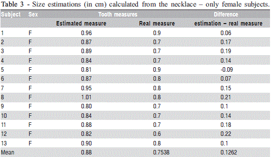
The estimated tooth measures from the dimensional relation with the necklace were overestimated in most cases (92.31%). It is noteworthy that this type of calculation, using the necklace as a reference point, was performed only in female participants, since they have virtual exclusivity in the use of this type of adornment. The mean difference between estimated and actual measurements was equivalent to 0.1262 cm, as shown in Table 3. Tooth measures estimated from the necklace were significantly higher (p = 0.0001307) than the actual measurements, with a significance level of 5%
Discussion
Since the early twentieth century, researchers have analyzed craniofacial correlations and the relationship between the face and the underlying bone morphology. On this line, are noteworthy the studies about the shape of the nose and the nasal bone, as well as variations in nose shapes of different ethnic groups13-14. These studies underlie the current principles of facial reconstruction15, which became the main target for this type of research16.
As occurs in the nose, eyes also show great relationship between soft tissue and bone structure. The position of ectocanthus was studied by Whitnall in 2,000 skulls, and can be located in the so called tubercle of Whitnall. This anatomical point constitutes the major reference for sizing and positioning the image of the skull in relation to the face in superimposition methods15.
The objects used as a dimensional reference in this study were selected because they frequently appear in personal pictures, in the same spatial plane of the teeth. However, the facial sizing from the teeth dimensions naturally requires the recovery of these teeth, which does not always occur, especially considering the frequency of post-mortem loss of anterior teeth.
The proposed method overestimated the mesiodistal width of the central incisor, in its best result – by measuring sunglasses - 1.19 mm on average (15.62% of the real average). The worst performance was achieved by having the cap as size reference, overestimating the dental width in 1.867 mm on average. The less satisfactory result of the cap in relation to other items may be related to the position of the cap slightly oblique in relation to the plane of the central incisors. Further studies are suggested.
The analysis of a personal photograph for human identification purposes requires excellent image resolution for detailed observation and accurate superimposition with the cranial image. The analysis of teeth in photographs only becomes possible under optimal conditions, close and high resolution images. Still, the use of tooth width as a size reference, given its small dimensions, creates a greater risk of failure, since fractions of a millimeter can represent important distances in the final result. Thus, when sizing images using anatomical landmarks, the larger the reference, the greater the chance of success.
Although previous studies have been engaged in creating protocols and methods of sizing and positioning skull and face images for superimposition, the correct size and position are more easily achieved through successive attempts, and movement of the images3,6,1,17-19. The method evaluated in the present study is therefore inaccurate for sizing facial images for superimposition with human identification purposes.
References
1. Dorion RBJ. Photographic superimposition. J Forensic Sci. 1983; 28: 724-34. [ Links ]
2. Chai DS, Lan YW, Tao C, Gui RJ, Mu YC, Feng JH et al. A study on the standard for forensic anthropologic identification of skull-image superimposition. J Forensic Sci. 1989; 34: 1343-56.
3. Austin-Smith D, Maples W. The reliability of skull/photograph superimposition in individual identification. J Forensic Sci. 1994; 39: 446-55.
4. Iscan MY, Helmer RP. Forensic analysis of the skull: craniofacial analysis, reconstruction, and identification. New York: Wiley-Liss; 1993.
5. Jayaprakash PT, Srinivasan GJ, Amravaneswaran MG. Cranio-facial morphhoanalysis: a new method for enhancing reliability while identifying skulls by photo superimposition. Forensic Sci Int. 2001; 117: 121-43.
6. Yoshino M, Imaizumi K, Miyasaka S, Seta S. Evaluation of anatomical consistency in cranio-facial superimposition images. Forensic Sci Int. 1995; 74: 125-34.
7. Yoshino M, Matsuda H, Kubota S, Imaizumi K, Miyasaka S, Seta S. Computer-assisted skull identification system using video superimposition. Forensic Sci Int. 1997; 90: 231-44.
8. Starbuck JM, Ward RE. The affect of tissue depth variation on craniofacial reconstructions. Forensic Sci Int. 2007; 172: 130-6.
9. Marks M, Bennett JL, Wilson L. Digital video image capture in establishing positive identification. J Forensic Sci. 1997; 42: 492-5.
10. De Greef S, Willems G. Three-dimensional cranio-facial reconstruction in forensic identification: latest progress and new tendencies in the 21st century. J Forensic Sci. 2005; 50: 12-7.
11. Ricci A, Marella GL, Apostol MA. A new experimental approach to computer-aided face/skull identification in forensic anthropology. Am J Forensic Med Pathol. 2006; 27: 46-9.
12. Quatrehomme G, Balaguer T, Staccini P, Alunni-Perret V. Assessment of the accuracy of three-dimensional manual craniofacial reconstruction: a series of 25 controlled cases. Int J Legal Med. 2007; 121: 469-75.
13. Glanville EV. Nasal shape, prognathism and adaptation in man. Am J Phys Anthropol. 1969; 30: 29-37. 14. Macho GA. An appraisal of plastic reconstruction of the external nose. J Forensic Sci. 1986; 31: 1391-403.
15. Wilkinson C. Forensic facial reconstruction. Cambridge: Cambridge University Press; 2004. 16. Prokopec M, Ubelaker DH. Reconstructing the shape of the nose according to the skull. Forensic Sci Commun. 2002; 4: 1-4.
17. Pesce Delfino V, Colonna M, Vacca E, Potente F, Introna FJ. Computeraided skull/face superimposition. Am J Forensic Med Pathol. 1986; 7: 201-2.
18. Gruner O. Identification of skulls: a historical review and practical applications. In: Iscan MY, Helmer RP. Forensic analysis of the skull: craniofacial analysis, reconstruction, and identification. New York: Wiley- Liss; 1993.
19. Pesce Delfino V, Vacca E, Potente F, Lettini T, Colonna M. Shape analytical morphometry in computer-aided skull identification via video superimposition. In: Iscan MY, Helmer RP. Forensic analysis of the skull: craniofacial analysis, reconstruction, and identification. New York: Wiley-Liss; 1993.
 Correspondence:
Rachel Lima Ribeiro Tinoco
Piracicaba Dental School - University of Campinas
Department of Forensic Dentistry
Av. Limeira, 901 - Caixa Postal 52
Piracicaba - SP - CEP 13414-903
Phone: (55) 19 2106 5200 / (55) 21 9963 4751
E-mail: racheltinoco@live.com
Received for publication: December 07, 2010
Accepted: June 21, 2011
