


Serviços Personalizados
Artigo
 pdf em Inglês
 Artigo em XML
 Referências do artigo
 Enviar este artigo por email
Links relacionados
Compartilhar

 Permalink
Brazilian Journal of Oral Sciences
versão On-line ISSN 1677-3225
Braz. J. Oral Sci. vol.10 no.3 Piracicaba Jul./Set. 2011
ORIGINAL ARTICLE
Comparative clinical evaluation of chemomechanical caries removal agent Papacarie® with conventional method among rural population in India - in vivo study
Sanjeet SinghI;Deepti Jawa SinghII; Shipra JaidkaIII; Rani SomaniIV
I Senior lecturer, Department of Oral and Maxillofacial Pathology, D J College of Dental Sciences and Research, Modinagar (U.P) India
II Senior lecturer, Department of Paedodontics and Preventive Dentistry, D J College of Dental Sciences and Research, Modinagar (U.P) India
III Reader, Department of Paedodontics and Preventive Dentistry, D J College of Dental Sciences and Research, Modinagar (U.P) India
IV Professor, Department of Paedodontics and Preventive Dentistry, D J College of Dental Sciences and Research, Modinagar (U.P) India
ABSTRACT
The use of minimally invasive procedures and attention to patient comfort are of great importance, especially for dental treatment in young children. This has led to the development of chemomechanical methods for caries removal. Aim: To evaluate and compare the antimicrobial efficacy, efficacy in terms of time consumption and pain perception of chemomechanical caries removal agent Papacarie® and conventional method of caries removal. Methods: Subjects for this study were chosen from children admitted to dental clinic for restorative procedures. Forty children (age 4-8 years) with early childhood caries were included in this study. Two primary teeth with comparable degrees of carious destruction were chosen in each child for caries removal with either Papacarie® or rotary instruments. The time taken for caries removal was measured using stopwatch. Pain response during caries removal was evaluated using the Wong Baker Face Pain Scale. Dentin samples of both groups were taken prior to, and after caries removal for microbiological analysis. Results The time taken for caries removal in chemomechanical caries removal method was three times longer than the conventional method. Pain score during chemomechanical method of caries removal was 1.525 as compared with 6.65 when conventional method was used. The antimicrobial efficacy of chemomechanical caries removal was significantly similar to conventional method. Conclusions: Papacarie® can be an effective clinical alternative treatment for the removal of occlusal dentinal caries in cavitated primary molars.
Keywords: chemomechanical caries removal, Papacarie®, dental caries.
Introduction
Painless dentistry and minimal intervention providing comfort, relief, solace and instillation of positive attitude towards dental treatments are some of the factors justifying the specialty of pediatric dentistry. Despite the decline of its prevalence, caries continues to affect a significant portion of world population and treatment of the decay is still a challenge for researchers. In children, especially those with dental anxiety, caries removal by means of conventional instruments is considered an unpleasant step of the restorative process mainly because of pain, drilling and noise1. Furthermore, drilling results in rapid and excessive removal of tooth structure and may cause harmful thermal and pressure effects to the pulp2. These disadvantages of conventional method had led to a more gentle, comfortable and conservative caries excavation method aimed at providing minimal thermal changes, less vibration and less pain, and removal of infected dentine only.
With an advancing era of science, much superior technique of removing dental caries by means of chemomechanical agents was first introduced in 1975 by Habib et al.3 by using 5% sodium hypochlorite, which is a non specific proteolytic agent. As sodium hypochlorite was found eventually too corrosive to be used in healthy tissue, Goldman et al.4 made an attempt to minimize the problem by introducing GK-101 for removal of dental caries in 19764. It was FDA approved for use in USA in 1984 and was marketed in 1985 by the name of Caridex system5. Despite its effectiveness, Caridex had certain limitations like long working time, short shelf life and requirement of large volume of solutions along with a special pump6. Rolf Bornstein et al in mid 1990’s introduced Carisolv as a successor to Caridex7-8.Carisolv was quite a success in the field of dentistry but with its long use certain drawbacks of the system has been reported which includes requirement of customized instruments that increased the cost of solution.
In 2003, a research project in Brazil conducted by Bassadori et al.9 led to the development of a new formula to universalize the use of the chemomechanical method for caries removal, which was launched for use in public health in 2005 under the brand name Papacarie7. Papain gel, the basic component of this product, is responsible for its bactericidal, bacteriostatic and inflammatory characteristics.
Various in vivo and in vitro studies have been done using different chemomechanical caries removal agents namely 5% sodium hypochlorite, GK101, Caridex, Carisolv, but literature on the efficacy of chemomechanical caries removal using Papain gel is scanty3-8,10. Thus the need to evaluate the recent material especially in young children arose.
The aims of the present in vivo study were to compare the time taken in caries removal by Papacarie® as a chemomechanical caries removal agent with conventional method; to compare the pain response associated with the chemomechanical and conventional methods during caries removal; to evaluate and compare the antimicrobial efficacy of both methods.
Material and methods
The present in vivo study was carried out at the Department of Pedodontics and Preventive Dentistry at D.J. College of Dental Sciences and Research, Modinagar (Distt. Gaziabad, State Uttar Pradesh, India), involving 40 healthy children aged 4-8 years. Two contralateral cavitated primary molars, with occlusal caries having approximately equal-sized cavity openings (diameter 1.5-2 mm measured with a metallic caliper) with brown and softened dentine and having defects with comparable depths (less than 1.5-2 mm measured with a WHO Periodontal probe) were chosen for the study. The patients selected were fully cooperative as judged by the Frankle Shiere and Fogel Four Point behavior rating scale11. Parents/guardians responsible for each child were fully informed of the details of the study, and asked to sign a consent form authorizing their child’s participation in the study in agreement with the ethical principles of the DJ Dental IEC declaration with reference number DJD/IEC/05. Thus, the 80 contralateral primary molars from the 40 children were divided equally for the conventional and chemomechanical methods of caries removal. In each child, one tooth was randomly selected to be treated with either Papacarie® or the other conventional method. Since both molars in each child were exposed to a similar oral environment, hence this study was more suitable to compare the two treatment modalities.
Both chemomechanical and conventional methods of caries removal were carried out under rubber dam isolation ( Figure 1) in order to obtain moisture control and avoid microbial contamination.
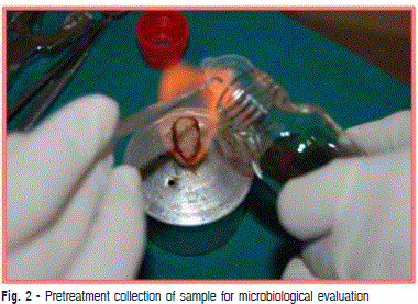
TThe first sample from the superficial carious lesion from both cavities was removed with the help of a sterile spoon excavator and transferred to a sterile vial containing 20 mL of BHI broth for microbiological evaluation ( Figure 2).
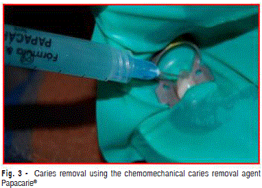
Caries removal by chemomechanical method (Figure 3)
According to the manufacturer’s instructions, the syringe containing Papacarie® was removed from the refrigerator 30 min before treatment. Papacarie® was applied with the help of an applicator tip into the cavity and left for 30-40 s. The softened dentine tissue was removed using the excavator in a pendulum motion in a pressureless manner. The remaining gel was removed with cotton pellet soaked in saline. This procedure was repeated as many times as necessary, until the darkish color of the gel was revealed. The cavity was not washed or rinsed between the gel applications. The cavities were considered caries-free when there was no change in the color of Papacarie® gel.

Caries removal by the conventional method
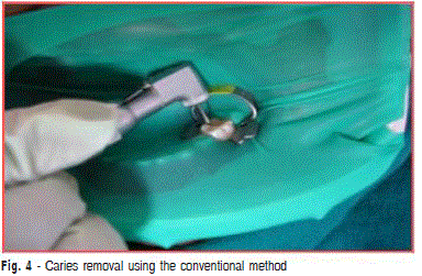
Conventional caries removal was carried out using a sterile No. 16 straight bur on a contra-angle micromotor handpiece at slow and intermittent speed, without water spray ( Figure 4). After caries removal, dentine was considered cariesfree, using established Ericksons clinical (optical and tactile) criteria7.The second sample from both cavities was then taken from the cavity floor with a sterile spoon excavator and transferred to another sterile vial containing 20 mL of BHI broth for microbiological eval-uation.
The preparation time for each caries-removal technique Fig. 4 - Caries removal using the conventional method was determined using a stopwatch. The time taken for the experimental group was calculated from the beginning of gel application until the end of the caries removal procedure.
After the removal of caries was completed, the Wong Baker Faces Pain Scale12 was used to evaluate whether the child felt any pain during the procedure and accordingly the pain scores was given to them for both the methods used for caries removal separately.
Microbial cultivation and evaluation
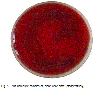
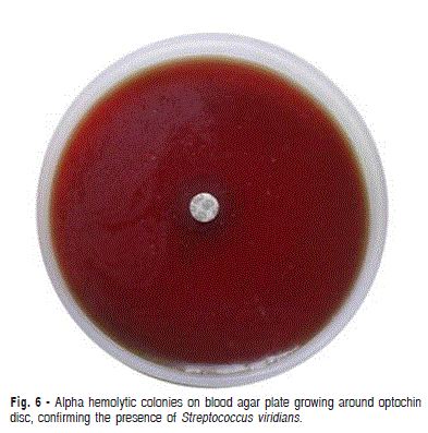
The dentin samples of both groups were processed in the microbiological laboratory within 1 hour of collection. Each sample was vortexed for about 30 s in order to dislodge the bacteria from the dentin. A sterile loop full of sample was collected and was cultured with aseptic technique onto 10% blood agar plates by streaking method. The plates were incubated at 37ºC in 5% CO2 atmosphere in candle jar for 48 h. Thereafter, the plates were observed for the growth of alpha hemolytic streptococci ( Figure 5). The alpha hemolytic green colored colonies from the primary plates were picked up with a sterile loop and were subcultured onto another blood agar plate for examination of colony characteristics and identification of viridans streptococci. During isolation, a disc of optochin was placed in the primary inoculum to exclude S. pneumoniae ( Figure 6). These plates were also incubated at 37ºC in 5%Co2 atmosphere in candle jar. After overnight incubation, the plates were observed for colony characteristics using magnifying lens. The plates were further evaluated using Compound Light microscope. The colonies showing convex appearance were identified as viridans streptococci. Further Gram staining was done to identify the streptococci in chains and the plates were divided into four quadrants and bacterial count was done using a magnifying lens. The total viable bacterial count was determined and expressed as number of bacteria per mL of medium. Because of the wide range of total numbers, 5 classes were defined for the total viable count: 0: no growth; 1: < 103; 2: 1001-104; 3: 10001-105(discrete growth); 4: uncountable (confluent growth).
Data were analyzed statistically using the Z-test for time and pain assessment at 1% level of significance and paired t test for microbiological evaluation at 1% level of significance.
Results
In the present study, 80 primary molars obtained from 40 children aged 4 to 8 years were evaluated in terms of time spent for caries removal, pain response and microbiological assessment.
Time consumption and pain perception
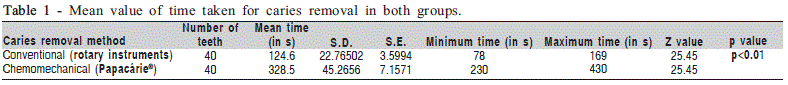
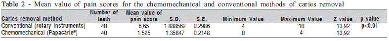
Table 1 shows that the mean time required for caries removal with the chemomechanical method (328.5 ± 45.26 s) was 193.9 s longer than the time spent with the conventional method (124.6 ± 22.76 s) (p<0.01). Table 2 shows that the mean value of pain score using the conventional method was significantly higher (6.65 ± 1.888) compared with the chemomechanical method (1.525 ± 1.35847) (p<0.01).
Microbial count
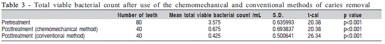
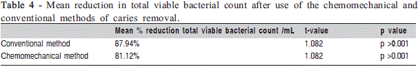
Table 3 shows the total viable bacterial count before and after caries removal by both methods. The mean total viable count of S. viridians was 3.575 bacteria/mL before caries removal. The mean total viable Streptococcus count was reduced to 0.675 after caries removal with the chemomechanical agent and 0.425 with conventional method (Table 3). The difference between the pretreatment sample score and posttreatment sample score was found to be statistically significant in both groups (p<0.01). This corresponds to a mean reduction of total viable count of 87.94% and 81.12% for the conventional and chemomechanical methods, respectively ( Table 4). The results also indicated that both methods of caries removal had an almost similar percentage of reduction of bacterial count when analyzed statistically.
Discussion
Dental caries is still one of the most common diseases affecting the human population. It is one of the main etiological factors of dental pain, so its treatment is not only required, but also demanded since the inception of mankind.
The development of caries removal techniques in restorative dentistry is progressing towards a more biological and conservative direction. Chemomechanical caries removal became an area of interest in dental research due to its concept of sound tissue preservation. As only carious dentin is removed, the painful removal of sound dentin is avoided and hence, the need for local anesthesia is minimized. The latest production of chemomechanical caries removal Papacarie® has been developed in Brazil in order to overcome the clinical limitations of other products. The present study was performed to assess the efficacy of this new product to remove carious lesion in primary teeth. As the microflora is one of the main etiological factors in caries occurrence, it is essential to reduce the microbial counts in carious lesions. With regard to oral microflora, evidence has shown that acidogenic species such as viridians streptococci (S. mutans and S. sobrinus) are strictly associated with the onset and presence of dental caries13. Mutans streptococci are mainly implicated with the initiation of enamel caries and gradually increase with the completion of the primary dentition and presence of proximal contacts between primary molars. Hence, in the present study, the efficacy of the chemomechanical method was assessed by evaluating its antimicrobial efficacy.
Papain, the main ingredient of Papacarie®, is an enzyme similar to human pepsin, used in food technology and pharmaceutical and cosmetic industries. Guzman and Guzman14 performed clinical studies on patients with skin lesions caused by burns, observing that the enzymatic action of papain was considered excellent in areas with necrotic and purulent processes. Udok and Storojuk15 also verified that papain aided cleansing necrotic tissue and secretions, shortening the period of tissue repair.
Flindt16 demonstrated that papain acts only in infected tissues because infected tissues lack plasmatic anti protease called Al anti trypsin, this is only present in sound tissues and it inhibit protein digestion16. The absence of Al anti trypsin enzyme in infected tissues allows Papain to break the partially degraded molecules. Dawkins et al.17 showed that Papain has bactericidal and bacteriostatic properties which inhibit the growth of gram positive and gram negative organism.
In addition to papain, Papacarie® also contains chloramines, toluidine blue, salts and thickening vehicle. Chloramine has bactericidal and disinfectant properties. The antiseptic properties of chloramine were recently documented in an in vitro study18. These are broadly used to chemically soften the carious dentine. According to Maragakis et al.19, the partially degraded collagen in carious dentine was chlorinated by chemomechanical caries removal solutions. The chlorination affects the secondary or quaternary structure of collagen, by disrupting hydrogen bonding and thus facilitating the carious tissue removal.
An in vitro evaluation of Papacarie® cytotoxicity using different concentrations of papain (2, 4, 6, 8, and 10%) on fibroblast culture found not cytotoxic effects, suggesting that Papacarie® is safe to use in pediatric patients19.
In the present study, the efficacy of Papacarie® in terms of time taken for caries and pain perception along with its comparison with the conventional method was evaluated. It was observed that chemomechanical caries removal was approximately 3.25 min longer in removing caries, which was statistically highly significant. This result is consistent with those of Jawa D et al. and Bassadori et al.9 it has been stated that Papacarie® requires more than one application for its action to work20. Carrillo et al.21 reported that the chemomechanical removal of carious tissue using Papacarie® had treatment duration of 8 min per tooth. The longer treatment time in that study was due to the evaluation done on special needs children.
In the present study, the analysis of pain perception during caries removal according to the overall rating by patients revealed a higher comfort level with the chemomechanical method compared with the conventional method. These findings are in accordance with those of Silva et al.15, who demonstrated that caries removal using Papacarie® is significantly less painful in comparison with the conventional method. The wide difference in the pain scores between Papacarie® and conventional method could be because Papacarie® acts only on the dead infected cells and does not damage the healthy tissues. Anusavice and Kincheloe22 demonstrated that removing carious dentin generally elicits little or no painful sensation, while removing sound dentin often results in some level of pain.
Clinically, in general practice, soft and presumably irreversibly destroyed dentine is removed prior to restoring the cavity. The clinical criteria for complete caries removal differ around the world. Over the years, several investigators have defined caries-free dentin and the number of microorganisms that can be left in the cavity that will not promote further disease progressl1. When excavating a lesion, the bulk of microorganisms are removed along with most of the necrotic dentin. This does not render the prepared cavity bacteria-free, and the rationale behind removal of carious dentin is still uncertain and based on rather blunt clinical criteria. Banerjee et al.1 reviewed this problem and concluded that `it is not possible to remove all infected dentin.
In the present study, the mean total viable count after caries removal was reduced to 0.675 score per mL from 3.525 using chemomechanical caries removal (Papacarie®) and 0.425 score per mL with conventional drilling which mean less than 103 CFU /mL .These reductions were highly significant.
Kneist and Heinrich-Wetzien23, Azrak et al.24 and Subramaniam et al.25showed that the mean total viable count was significantly reduced after caries removal using a chemomechanical caries removal agent (Carisolv™) and conventional drilling, which is in accordance with the findings of the present study.
Although complete caries removal was achieved by both methods, Jawa et al.20 observed under light microscopy that there was less marked destruction of dentinal tubules when caries was removed with Papacarie®. In a scanning electron microscopic study, Bassadori et al.9 observed conventional removal of carious tissue using diamond and/or stainless steel burs in permanent teeth with dentin caries left a residual smear layer, whereas the use of Papacarie® resulted in more preservation of dentin structure and bacterial removal9,20.
The results of this study indicated that Papacarie® is efficient in caries removal from open and accessible occlusal lesions. This finding is in agreement with those of Bussadori et al.9, Kotb et al.26. Papacarie®, a virtually painless, noninvasive technique of caries removal was proven efficient, easy to perform, inexpensive and comfortable to the patient. Thus, its use can be recommended for caries removal in patients seeking an alternative to conventional treatment.
References
1. Banerjee A, Watson TF, Kidd EAM. Dentine caries excavation: a review of current clinical techniques. Br Dent J. 2000; 188: 476-82. [ Links ]
2. Willershausen B, Azrak B, Wilms S. Fear of dental treatment and its possible effects on oral health. Eur J Med Res. 1999; 4: 72-7.
3. Habib CM, Kronman J, Goldman M. A chemical evaluation of collagen and hydroxyproline after treatment with GK-101 (N-chloroglycine). Pharmacol Ther Dent. 1975; 2: 209-15.
4. Goldman M,kronam JH .A preliminary report on a chemomechanical means of caries removal. J Am Dent Assoc. 1976; 93: 1149-53.
5. Schutzbank SG, Galaini J, Kronman JH, Goldman M, Clarke REA. A comparative in vitro study of the effect of GK-101 and GK-101E in caries removal. J Dent Res. 1978; 57: 861-4.
6. Beeley JA, Yip HK, Stevenson AG. Chemomechanical caries removal: a review of techniques and latest developments. Br Dent J. 2000; 188: 427-30
7. Ericson D, Zimmerman M, Raber H, Götrick B, Bornstein R, Thorell J. Clinical evaluation of efficacy and safety of a new method for chemo-mechanical removal of caries. A multicentre study. Caries Res. 1999; 33: 171-7.
8. Bornstein R, Wedenberg C. Pulpal reactions in rat incisors to Caridex. Aust Dent J. 1990; 35: 505-8.
9. Bussadori SK, Castro. LC, Galvao AC. Papain gel: a new chemo¬mechanical caries removal agent. J Clin Pediatr Dent. 2005; 30: 115-19.
10. Burke F M, Lynch E. The effect of chemomechanical caries removal on the bond strength of glass polyalkenoate cement to dentine. J Dent. 1994: 22: 283-91.
11. Frankle SN, Shiere FR, Fogel HR. Should the parent remain with the child in the dental operatory. J Dent Child. 1962; 29: 150-4.
12. Wong DL, Hockenberry-Eaton M, Wilson D, Winkelstein Ml, Schwartz P. Wong’s Essentials Of Pediatric Nursing. 6 ed. Saint Louis: Mosby; 2001.
13. Roeters FJ, Van der Hoeven JS, Burgersdijk RC, Schaeken MJ. Lactobacilli, mutans streptococci and dental caries: a longitudi¬nal study in 2 year old children up to the age of 5 years. Caries Res. 1995; 29: 272-9.
14. Guzman AY, Guzman MGS. The enzymatic debridement of suppura¬tions, necrotic lesions and burns with papain. J Int Coil Surg, 1953, 20: 695-702.
15. . Udok VM, Storojuk VT. Use of papain in trating suppurative postoper¬ative soft tissue complications and daises. Khirurgiia.1981; 19: 99-101.
16. Flindt M. Health and safety aspects of working with enzymes. Process Biochem. 1979; 13: 3-7.
17. Dawkins G ,Hewitt H,Wint Y Obiefuna P H,Wint B. Antimicrobial effects of Carica papaya fruit on common wound organisms. West Indian Med J. 2003; 52: 290-2.
18. Fuursted K, Hjort A, Knudsen L. Evaluation of bactericidal activity and lag of regrowth (post antibiotic effect) of five antiseptics on nine bacterial pathogens Antimicrob Chemother. 1997; 40: 221-6.
19. Margakis GM, Hahn P, Hellwig E. Clinical evaluation by patients of chemomechanical varies removal in primary molars and its acceptance by patients. Caries Res. 2001; 35: 205-10.
20. Jawa D, Singh S, Somani R, Jaidka S, Sirkar K, Jaidka R Comparative evaluation of efficacy of chemomechanical caries removal agent Papacarie and conventional method of caries removal: an in vitro Study. J Ind Soc Pedod Prev Dent. 2010; 28: 73-7.
21. Carrillo CM, Tanaka MH, Cesar MF, Camargo MA, JulianoY, Novo NF. Use of papain gel in disabled patients. J Dent Child. 2008; 75: 222-8.
22. Anusavice KJ, Kincheloe JE Comparison of pain associated with mechanical and chemomechanical removal of caries J Dent Res. 1987; 66: 1680-3.
23. Kneist S, Heinrich-Weltzien R: Antibacterial action of Carisolv. In: Albrektsson T, Bratthall D, Glantz P, Lindhe J, editors). Tissue preservation in caries treatment. London: Quintessence; 2001. p.205-19.
24. Azrak B, Callaway A, Grundheber A, Stender E Comparison of the efficacy of chemomechanical caries removal (Carisolv) with that of conventional excavation in reducing the cariogenic flora. Int J Paediatr Dent. 2004; 14: 182-91.
25. Subramaniam P, Babu KL, Neeraja G. Comparison of the antimicrobial efficacy of chemomechanical caries removal (Carisolv) with that of conventional drilling in reducing cariogenic flora. J Clin Pediatr Dent. 2008; 32: 215-9.
26. Kotb RM, Abdella AA, El Kateb MA, Ahmed AM. Clinical evaluation of Papacarie in primary teeth. J Clin Pediatr Dent. 2009; 34: 117-23.
 Correspondence:
Deepti Jawa Singh
A-28 ,Defence Colony, Mawana Road, Meerut
250001, Uttar Pradesh , India
Phone: 09720038877, 0121-2622435
E-mail: jawadeepti@rediffmail.com
Received for publication: September 23, 2010
Accepted: June 16, 2011
