Serviços Personalizados
Artigo
Links relacionados
Compartilhar
Brazilian Journal of Oral Sciences
versão On-line ISSN 1677-3225
Braz. J. Oral Sci. vol.10 no.3 Piracicaba Jul./Set. 2011
ORIGINAL ARTICLE
Do self-ligating brackets favor greater bacterial aggregation?
Matheus M. PithonI;Rogério L. dos SantosII; Leonard Euller NascimentoII; Amanda Osorio AyresII; Daniela AlvianoIII; Ana Maria BologneseIV
I Professor of Orthodontics, University of Southwestern Bahia (UESB), Bahia, Brazil
II PhD student in Orthodontics; Federal University of Rio de Janeiro, Rio de Janeiro, RJ, Brazil
III Adjunct Professor of Microbiology, Federal University of Rio de Janeiro, Brazil
IV Full Professor of Orthodontics, Federal University of Rio de Janeiro, Brazil
ABSTRACT
Aim: To verify the hypothesis that self-ligating brackets favor greater aggregation of microorganisms when compared with conventional brackets. Methods: Four types of self-ligating metal brackets were evaluated. Initially, 50 brackets were divided into five groups (n=10): Morelli Conventional, GAC (In-Ovation R, Dentsply Caulk), Aditek (Easy Clip), Ormco (Damon System) and 3M Unitek (Smart Clip). An in vivo evaluation was carried out in which the brackets were bonded to the mandibular teeth of five healthy individuals who had not undergone previous orthodontic treatment. The right hemiarch brackets were used for bacterial plaque collection and those on the left side were examined by scanning electron microscopy (SEM). Before bracket bonding, the bacterial plaque material aggregated to the tooth surfaces was collected, with the areas of choice being the cervical-buccal and mesial and distal interproximal regions. After 21 days had elapsed since bonding, the plaque adhered to the winglet, channel and cervical regions of the bracket bases was collected. The materials collected were diluted and seeded on Petri dishes onto Mitis salivarius medium specific for S. mutans and non-specified BHI culture medium. Colony forming unit (CFU) counts were performed visually after 24, 48 and 72 h of incubation. Results: Greater bacterial accumulation was observed on the winglets of 3M brackets, with statistical statistically significant differences from the other types (p<0.05). As regards the channel regions, most microorganisms accumulated in the Ormco Group (p<0.05), and in the cervical region of Aditek brackets. In all evaluated regions, those with least bacterial accumulation were the conventional brackets. Conclusions: The hypothesis was confirmed, as the self-ligating brackets were shown to have greater bacterial accumulation when compared with the conventional brackets.
Keywords: orthodontic brackets, microbiology, dental plaque, biofilms.
Introduction
Self-ligating brackets have been introduced to Orthodontics several decades ago. Harradine1 mentioned that the concept of self-ligation is as old as that of the edgewise appliance. Nevertheless, over the last two decades, there has been increasing production and dissemination of these accessories with active and passive modes of ligation2-4.
One of the most favorable aspects with the use of self-ligating brackets, according to the literature, would be elimination of the elastomers and steel ligature wires5-6. Two basic advantages are achieved with this procedure5-6: eradication of cross contamination that may occur accidentally during ligature placement, and improvement in oral hygiene by the patients. The latter advantage would be attributed to the fact that without the ligature, the bracket surface would be freer for cleaning purposes5-6.
There is a rich ecosystem in the oral cavity, with a countless number of microorganisms. Although both periodontal disease and dental caries are considered multifactorial diseases, the bacteria in dental plaque are the main factor in their onset and progression. However, there are situations that comprise so-called "ecological stress", with reference to the displacement of microbiological equilibrium, creating conditions that favor the growth and appearance of caries and/or periodontopathogenic bacteria7,8.
The different accessory components of fixed orthodontic appliances contribute to the change in the equilibrium of oral ecology9. The presence of brackets and ligatures has been shown to be related to in gingival inflammation and increased risk of tooth surface decalcification, which ultimately results in the appearance of white spots and caries10,11.
In spite of the literature guiding the thought that the self-ligating bracket system favors less aggregation of mutans group microorganisms, up to now, there are no consistent clinical evaluations in this sense4,12. Based on this premise, the aim of this study was to investigate the hypothesis that selfligating brackets favor greater aggregation of S. mutans and other microorganisms when compared with conventional brackets.
Material and methods
Brackets
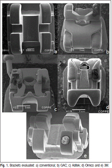
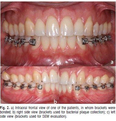
Fifty brackets were evaluated, being 40 of the selfligating type of four different commercial brands and 10 of the conventional type (n=10). The brackets were divided into five groups, described as follows: conventional (Morelli, Sorocaba, SP, Brazil), GAC (In-Ovation R, Dentsply Caulk, Milford, DE, USA), Aditek (Easy Clip, Cravinhos, SP, Brazil), Ormco (Damon System, Orange, CA, USA), and 3M Unitek (Smart Clip, Monrovia, CA, USA) ( Figure 1). The bracket channels (passive type) were filled with segments of round 0.020" wire (TP Ortho, Tokyo, Japan). The wire received a gray colored elastic ligature (Morelli, Sorocaba, SP, Brazil) on the conventional brackets and on the others, it was fixed by the specific system of each type of bracket (Figure 2).
Subjects
The sample was constituted by 5 male individuals with mean age of 25.3 years, with complete permanent dentition, normal occlusion and no previous orthodontic treatment. The brackets were bonded in the mandibular arch, the right side being used for microbial count and the left for scanning electron microscopy (SEM) analysis ( Figure 2). This study was approved by the Ethics Committee (Protocol 125/2008).


Methods
Initially, the individuals had their oral hygiene calibrated, according to the modified Bass-type technique, and also received oral hygiene kits containing a toothbrush (Procter & Gamble/Oral B, São Paulo, Brazil), dental floss "Super Floss" (Procter & Gamble/Oral B, São Paulo, Brazil) and toothpaste (Colgate-Palmolive, São Paulo, Brazil).
One week after receiving oral hygiene instructions, the bacterial plaque material aggregated to the tooth surfaces was collected with No. 20 absorbent paper cones (Dentsply Ind. e Com. Ltda., Petrópolis, RJ, Brazil), obtained from the colonized sites on the canines, mandibular first and second premolars and mandibular first and second molars, of which the supragingival areas of choice were as follows: a) cervical-buccal; b) mesial interproximal and c) distal interproximal areas.
Immediately after the initial collection of microorganisms, the brackets were bonded to the tooth surfaces according to the bonding technique proposed by the orthodontic adhesive manufacturer (Transbond XT, Monrovia, CA, USA).
After 21 days had elapsed since bonding, the second bacterial plaque collection was performed on the brackets, in the cervical-distal regions of the winglets, channel and in the cervical region of the bracket base.
After plaque collection, initial dilution and homogeniza-tion were performed in a mechanical vibrator, using 1 ml of sterile saline solution composed of 0.85% sodium chloride and 1% sodium thioglycollate as reducing agent for each 1mg of plaque collected. After this 0.1 mL aliquots of each dilution were seeded in Petri dishes containing the culture media Mitis Salivarius (MS) specific for of S. mutans and BHI broth, non-specific for bacteria and fungi. The dishes were incubated in an oven for bacterial growth for periods of 24, 48 and 72 h. Colonies formed in all the culture media were macroscopically identified and counted by visual inspection. The valuation periods were 24, 48 and 72 h.
Statistical Analysis
Data were analyzed with the SPSS 13.0 program (SPSS Inc., Chicago, IL, USA). The number of colonies found in MS and BHI media was analyzed statistically by application of the Wilcoxon test (p-value <0.05). Descriptive statistics included the means of the evaluated groups.
SEM Analysis
After removal from the left side of the arches, the brackets were fixed in an ascending ethanol series and thereafter any residual water was eliminated by critical point drying (CPD- 030 critical point dryer; Bal-Tec AG, Balzers, Liechtenstein). The dehydrated parts were fixed on stubs with a silver-based adhesive, sputter-coated with gold, placed on acrylic well plates and examined with a scanning electron microscope (Scanning Microscope JEOL-SM 5310, Tokyo, Japan).
Results
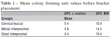
The CFU counts obtained from the plaque collected from tooth surfaces before bracket placement are shown in Table 1. The quantitative results of the number of bacterial colonies formed in the plaque adhered to the winglets of brackets revealed that Group 3M provided adherence of the largest number of colonies (p<0.05) followed by Aditek, GAC, Ormco and the conventional group. When the collection site was the channels, the brackets showing the greatest accumulation were the Ormco brand (p<0.05) and those that formed the fewest colonies were the conventional type. In turn, in the cervical region the Aditek brackets were those that formed most colonies, followed by GAC, as shown in Table 2. It is worth pointing out that the values found were similar for S. mutans and for the non-specific microorganisms.
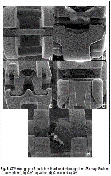
The analysis of the brackets removed from the oral cavity under SEM revealed larger plaque accumulation on the winglets of 3M brackets, followed by Aditek, GAC, Ormco and the conventional type. The channels of Ormco brackets were those that accumulated greater amount of plaque, followed by GAC, Aditek and 3M. In the cervical region the largest plaque accumulation could be seen on the Aditek brackets, followed by the GAC, 3M, and Ormco types ( Figure 3). In all the evaluated regions, the conventional brackets had the least plaque accumulation.


Discussion
In the present study, the proposal was to make a quantitative evaluation of the number of CFU present on conventional and self-ligating brackets of different designs. The evaluation was made in vivo in a model that individuals who had never undergone orthodontic treatment were used as hosts to the brackets. Initially, the oral hygiene of all patients was standardized to prevent individual variations from compromising the results. The use of any medications during the experimental period was not allowed in order to avoid alteration of the microbiota.
Bacterial plaque was collected before bracket bonding. The purpose of this procedure was to quantify the number of CFU present on the tooth surface. The regions of choice were the cervical-buccal, mesial and distal interproximal regions, as these areas are the most difficult to clean from an anatomical point of view.
The obtained results showed that in spite of the tooth surfaces being free for correct cleaning, they housed bacteria. For S. mutans counts the cervical-buccal region (5.4 CFU) was where microorganisms most concentrated, followed by the mesial (3.6 CFU) and distal (3.4 CFU) interproximal regions. As regards the non-specific bacterial counts, the interproximal regions were those with more CFU (19.4 CFU in the distal and 14.2 CFU in the mesial) followed by the cervical-buccal (10.4 CFU) regions.
The culture media used for seeding Petri dishes were MS, specific for growing S. mutans, while BHI broth, a nonspecific medium for bacteria and fungi. The choice for MS was made because of the well known role of S. mutans in dental caries development. The literature has stated that one of the major problems resulting from the use of an orthodontic appliance is the greater susceptibility to the development of dental caries13-15. The non-specific BHI medium was used to evaluate the presence of other microorganisms in order to determine whether the brackets favor the accumulation of S. mutans only or other microorganisms as well.
There was a significant increase in both S. mutans and non-specific microorganisms that grew in the BHI medium in the three regions of the brackets: the winglet, channel and the cervical region of the bracket base as observed in previous works16-17. The winglet region was chosen because this was the easiest area to clean; the channel, because it was the most difficult; and the base, because access to this area is most difficult, and it is here that the largest number of carious lesion routinely occur after removal of the orthodontic appliance18.
The winglets were the regions with the least presence of microorganisms when compared with the channel and cervical regions. In the comparison among the brackets, the conventional type presented lower mean values, however, without statistical differences from the winglets of the GAC and Ormco brackets (p>0.05). These results occurred in the evaluation of S. mutans and non-specific microorganisms, and can be attributed to the winglet design of the 3M and Aditek brackets, which have larger grooves, favoring greater bacterial accumulation within them.
In the channel and cervical regions, the number of colonies increased significantly, in some ways an expected result, due to the difficulty of access during cleaning. Ormco brackets were those that presented the greatest contamination, followed by GAC and Aditek, which did not differ significantly from each other (p<0.05). These results are closely related to the designs of the brackets, which have a system for holding the wire, in such a way that they become tubes, preventing correct cleaning. The Ormco brackets represent this well, because when they are closed, they prevent mechanical cleaning; the GAC and Aditek brands have a small opening in their cover, providing less plaque accumulation when compared with the Ormco type. The brackets that showed the cleanest channels were the conventional type followed by the 3M brand. The design of the 3M brackets is very similar to that of the conventional type, providing closer results, in spite of being significantly different (p<0.05).
It is worth mentioning that in all the regions evaluated, the conventional brackets were shown to be more hygienic than the self-ligating type, a fact that is intimately related to their design these results are consistent with those found Mota et al. 200813.
In conjunction with the quantitative evaluation of the bacterial colonies, the brackets were submitted to SEM to evaluate their design and the locations of greater bacterial plaque adherence. The microbiological results could be perfectly related to the morphological aspect of these brackets.
When the winglets were verified, the 3M brackets presented greater irregularities, providing greater bacterial accumulation, as previously mentioned. In the Ormco bracket channels, because they behave as a tube when they are closed, there was great accumulation of debris and thus a larger number of CFUs. In the cervical region of Aditek brackets, difficult access by toothbrush was shown as a result of the accentuated inclination from the base to the cervical region.
The results of the present study differ from those of other works in the literature. Pandis et al.9 performed S. mutans count in the saliva of patients using brackets of the conventional and self-ligating types and found no statistically significant differences. The differences found between Pandis' study and the present investigation might be due to differences in methodology, as in their study CFU were counted in saliva and not on bracket surfaces, as was proposed in the present study.
One of the advantages that the literature mentions in using self-ligating brackets would be the reduction in cross-infection which, according to some authors would occur at the time of placing elastic or metal ties. The results of the present study suggest that concern should be focused elsewhere, considering that cross infection is completely controllable, depending on the dental team, whereas the significant increase in CFUs adhered to the brackets is something over which the orthodontist has no control. It may be concluded that: conventional brackets favored less bacterial accumulation; selfligating brackets of different designs presented differences among the areas of less and greater bacterial accumulation; and the hypothesis that self-ligating brackets favor greater aggregation of microorganisms was proved.
References
1. Harradine NW. Self-ligating brackets: where are we now? J Orthod. 2003; 30: 262-73. [ Links ]
2. Miles PG. Self-ligating vs conventional twin brackets during en-masse space closure with sliding mechanics. Am J Orthod Dentofacial Orthop. 2007; 132: 223-5.
3. Pandis N, Polychronopoulou A, Eliades T. Self-ligating vs conventional brackets in the treatment of mandibular crowding: a prospective clinical trial of treatment duration and dental effects. Am J Orthod Dentofacial Orthop. 2007; 132: 208-15.
4. Rinchuse DJ, Miles PG. Self-ligating brackets: present and future. Am J Orthod Dentofacial Orthop. 2007; 132: 216-22.
5. de Moura MS, de Melo Simplicio AH, Cury JA. In-vivo effects of fluoridated antiplaque dentifrice and bonding material on enamel demineralization adjacent to orthodontic appliances. Am J Orthod Dentofacial Orthop. 2006; 130: 357-63.
6. Ogaard B, Rolla G, Arends J, ten Cate JM. Orthodontic appliances and enamel demineralization. Part 2. Prevention and treatment of lesions. Am J Orthod Dentofacial Orthop. 1988; 94: 123-8.
7. Marsh PD. Dental diseases—are these examples of ecological catastrophes? Int J Dent Hyg. 2006; 4 Suppl 1: 3-10; discussion 50-12.
8. Marsh PD. Are dental diseases examples of ecological catastrophes? Microbiology. 2003; 149: 279-94.
9. Pandis N, Papaioannou W, Kontou E, Nakou M, Makou M, Eliades T. Salivary Streptococcus mutans levels in patients with conventional and self-ligating brackets. Eur J Orthod. 2010; 32: 94-9.
10. Gorelick L, Geiger AM, Gwinnett AJ. Incidence of white spot formation after bonding and banding. Am J Orthod. 1982; 81: 93-8.
11. Naranjo AA, Trivino ML, Jaramillo A, Betancourth M, Botero JE. Changes in the subgingival microbiota and periodontal parameters before and 3 months after bracket placement. Am J Orthod Dentofacial Orthop. 2006; 130: 275 e17-22.
12. Reicheneder CA, Gedrange T, Berrisch S, Proff P, Baumert U, Faltermeier A et al. Conventionally ligated versus self-ligating metal brackets—a comparative study. Eur J Orthod. 2008; 30: 654-60.
13. Mota SM, Enoki C, Ito IY, Elias AM, Matsumoto MA. Streptococcus mutans counts in plaque adjacent to orthodontic brackets bonded with resin-modified glass ionomer cement or resin-based composite. Braz Oral Res. 2008; 22: 55-60.
14. Svanberg M, Ljunglof S, Thilander B. Streptococcus mutans and Streptococcus sanguis in plaque from orthodontic bands and brackets. Eur J Orthod. 1984; 6: 132-6. 15. Pramod S, Kailasam V, Padmanabhan S, Chitharanjan AB. Presence of cariogenic streptococci on various bracket materials detected by polymerase chain reaction. Aust Orthod J. 2011; 27: 46-51.
16. Peros K, Mestrovic S, Anic-Milosevic S, Slaj M. Salivary microbial and nonmicrobial parameters in children with fixed orthodontic appliances. Angle Orthod. 2011; 81: 901-6.
17. Rego RO, Oliveira CA, dos Santos-Pinto A, Jordan SF, Zambon JJ, Cirelli JA et al. Clinical and microbiological studies of children and adolescents receiving orthodontic treatment. Am J Dent. 2011; 23: 317-23.
18. Chaussain C, Opsahl Vital S, Viallon V, Vermelin L, Haignere C, Sixou M et al. Interest in a new test for caries risk in adolescents undergoing orthodontic treatment. Clin Oral Investig. 2010; 14: 177-85. Do self-ligating brackets favor greater bacterial aggregation?
 Correspondence:
Ana Maria Bolognese
Universidade Federal do Rio de Janeiro Faculdade de Odontologia Departamento de Ortodontia e Odontopediatria
Av. Professor Rodolpho Paulo Rocco, 325
Ilha do Fundão CEP:22245-100 Rio de Janeiro, RJ - Brazil
Phone: (21) 25902727
E-mail: anabolognes@yahoo.com.br
Received for publication: May 29, 2011
Accepted: September 14, 2011


