


Serviços Personalizados
Artigo
 pdf em Inglês
 Artigo em XML
 Referências do artigo
 Enviar este artigo por email
Links relacionados
Compartilhar

 Permalink
Brazilian Journal of Oral Sciences
versão On-line ISSN 1677-3225
Braz. J. Oral Sci. vol.11 no.2 Piracicaba Abr./Jun. 2012
ORIGINAL ARTICLE
Odontogenic cysts in children and adolescents: a 21-year retrospective study
Vinicius Gomes SerraI;Daniele Meira CondeII;Rogério Vera Cruz Ferro MarquesII; Cláudio Vanucci Silva de FreitasII; Fernanda Ferreira LopesIII; Maria Carmen Fontoura Nogueira da CruzIII
IDDS, Federal University of Maranhão, Brazil
IIMSc student, Graduate Program in Dentistry, Federal University of Maranhão, Brazil
IIIProfessor, Graduate Program in Dentistry, Federal University of Maranhão, Brazil; PhD in Oral Pathology, Federal University of Rio Grande do Norte, Brazil
ABSTRACT
AIM: To investigate the distribution of odontogenic cysts in patients aged 0 to 18, referred to Department of Pathology, University Hospital of the Federal University of Maranhão, Brazil, to determine the most common types of lesions and their distribution according to gender and anatomical site involved.
METHODS: Histopathological data were collected from a database of lesions classified as odontogenic cysts that were indicated for surgical removal and histopathological analysis. Data were subjected to descriptive analysis.
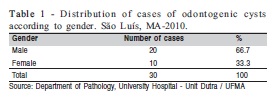
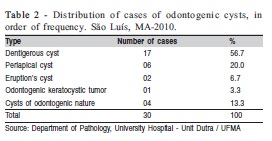
RESULTS: Thirty cases of odontogenic cysts were identified, and dentigerous cysts were the most frequent (n=17). Most occurrences were in males (66.7%) and the most frequent site was the posterior mandible (73.3%).
CONCLUSIONS: Odontogenic cysts in children and adolescents are mostly developmental cysts, especially dentigerous cysts, occurring predominantly in males, with a predilection for the posterior mandible.
Keywords: odontogenic cysts, child, adolescent.
Introduction
Odontogenic cysts are pathological entities that correspond to cavities lined with epithelial tissue, containing in its interior semi solid or liquid material, whose formation is associated with proliferation of epithelial remnants of the enamel organ or even the development of embryonic processes maxillomandibular1.
These cysts constitute an important group of lesions in the maxillofacial complex, whose frequency varies from 7 to 13% of lesions diagnosed in this anatomic region2-3, affecting mainly the adult population2,4. These lesions are characterized by having slow growth and tendency to expand, despite the biological behavior of benign entities. However, odontogenic cysts can reach a considerable size if not diagnosed early and properly treated5. Because they are generally asymptomatic and some behave aggressively, this group of lesions has required special attention from dentists6.
Odontogenic cysts can be classified as to their etiology in inflammation and development, and are differentiated by histopathology, mainly because the clinical and radiographic characteristics are very similar. The differential diagnosis of these lesions includes cystic ameloblastoma, adenomatoid odontogenic tumor, odontogenic calcification cyst7.
It draws attention to the fact that several developmental processes occur in the maxillofacial area during childhood, including bone growth and odontogenesis,which could be associated with cyst formation8. Nevertheless, the literature on odontogenic cysts includes studies mainly in the adult population, with few studies in children and adolescents. Considering these aspects, the purpose of this study was to investigate the distribution of odontogenic cysts in children and adolescents aged 0 to 18, referred to Department of Pathology of the University Hospital of the Federal University of Maranhão-UFMA, in São Luis, MA, Brazil, to determine the most common types of lesions and their distribution according to gender and anatomic site involved, since the occurrence of these lesions is poorly known in the study population.
Material and methods
This study followed the fundamental scientific and ethical requirements of Resolution 196/96 (Standards for Research involving Human Subjects) of the Brazilian National Health Council, and was approved by the institutional Ethics Committee (protocol #33104-649/2007; approval #407/2007). This research consisted of a retrospective, cross-sectional, and descriptive study of medical records of children and adolescents referred to the Department of Pathology, University Hospital Presidente Dutra / UFMA, São Luis, MA, Brazil.
The sample included all medical records with a histopathologic diagnosis of odontogenic cyst, as defined by the Medical Subject Headings - Mesh, of patients aged 0 to 18 referred to the service above, diagnosed during the period from 1990 to 2010.
As a tool for collecting data, we used a database of lesions classified as odontogenic cysts that were indicated for surgical removal and histopathological analysis. Data regarding gender, patient age at the time of diagnosis, anatomical site involved and histopathologic diagnosis were investigated. Data were subjected to descriptive statistics and presented in tables.
Results
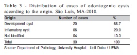
Thirty cases of odontogenic cysts were found in children and adolescents aged 2 to 16. The males were the most affected (66.7%), as shown in Table 1. The predominant histopathological type was the dentigerous cyst (56.7%), followed by periapical cyst (20%), eruption cyst (6.7%) and odontogenic keratocystic tumor (3.3%), as shown in Table 2. Regarding the origin, 66.7% were developmental cysts while 20% were inflammatory cysts. In addition, 13.3% of cysts could not be identified according to the methodology of this study (Table 3).
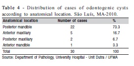
Concerning the anatomic site involved, the posterior mandible was the most frequently affected region (73.3%), followed by the anterior maxilla (16.7%), posterior maxilla (6.7%), and anterior mandible (3.3%) (Table 4).
Discussion
In the present study, the predominant histopathological type was the dentigerous cyst (56.7%), followed by periapical cyst, eruption cysts and odontogenic keratocystic tumor. The higher incidence of dentigerous cysts was also reported by some authors8-10. Prockt et al.11 reinforced these data showing a frequency of 42.46% of dentigerous cysts in patients aged 10 to 19. This frequency can be explained by the large number of impacted third molars and canines in this age group, which are teeth associated with the etiology of these cysts.
Regarding the origin, the fact that most cysts were developmental cysts (66.7%) is in accordance with the literature, which shows a lower incidence of inflammatory cysts in children and adolescents8-9, unlike the adult population, which is more affected by cystic lesions of inflammatory origin5,12-13.
The difference in the distribution of odontogenic cysts among adults, children and adolescents, in particular developmental cysts, is probably due to the fact that theorigin of these lesions occurs during infancy and is associated with continued growth and subsequent changes in teeth, going to the end of puberty12. The occurrence of inflammatory cysts in permanent teeth are also infrequent in children and adolescents, since their pathogenesis starts from the pulp necrosis14, and recently erupted permanent teeth are generally healthy, not showing the condition necessary for the development of cystic lesion. In primary teeth, the inflammatory stimulus would not have enough time to act as a chronic irritant15-16.
Males were more affected (66.7%) and this result corroborates with the literature9-10. This finding can be explained by the fact that boys are usually more prone to trauma and have poorer oral hygiene than girls17. In contrast, Bodner8 found no significant differences between genders in a sample of pediatric patients.
Regarding the anatomic site, the posterior mandible was the most frequently affected region, with 73.3% of cases, followed by the anterior maxilla (16.7%), as also observed by Godoy et al.9. Low incidence of lesions in the anterior mandible is probably due to the fact of the anterior teeth present less fissures and grooves than the posterior teeth, being less susceptible to caries and, consequently, to periapical inflammatory lesions9.
From the obtained results, it was possible to conclude that odontogenic cysts in children and adolescents are mostly developmental cysts, particularly dentigerous cysts, with a predominance of male patients and mostly located in the posterior mandible. The importance of the early diagnosis of these lesions by dentists is providing a better treatment for patients. Further research on this nature is needed, considering the scarcity of studies in this age group.
References
1. Nanami R, Sampaio C, Olivete J, Pizzatto E, Moresca R, Giovanini AF. Prevalence of cysts of the jaws diagnosed on a Brazilian center of reference. RSBO. 2009; 6: 143-6. [ Links ]
2. Jones AV, Craig GT, Franklin CD. Range and demographics of odontogenic cysts diagnosed in a UK population over a 30-year period. J Oral Pathol Med. 2006; 35: 500-7. [ Links ]
3. Ledesma-Montes C, Hernández-Guerrero JC, Garcés-Ortýz M. Clinicopathologic study of odontogenic cysts in a Mexican sample population. Arch Med Res. 2000; 31: 373-6. [ Links ]
4. Koseoglu BG, Atalay B, Erdem MA. Odontogenic cysts: a clinical study of 90 cases. J Oral Sci. 2004; 46: 253-7. [ Links ]
5. Ochsenius G, Escobar E, Godoy L, Peñafiel C. Odontogenic Cysts: Analysis of 2.944 cases in Chile. Med Oral Patol Oral Cir Bucal. 2007; 12: 85-91. [ Links ]
6. Pereira JVP, Figueiredo DU, Souza EA, Holmes TSV, Gomes DQC, Cavalcanti AL. Prevalence of odontogenic cysts and tumors in patients treated at the Paraíba Health Assistance Foundation: a retrospective study. Arq Odontol. 2010; 46: 75-81. [ Links ]
7. Pozzer L, Jaimes M, Chaves Neto HDM, Olate S, Barbosa JRA. The Odontogenic Cyst in Children: Analysis of the Surgical Decompression in 2 Cases. Rev. Cir Traumatol Buco-maxilo-fac. 2009; 9: 17-22. [ Links ]
8. Bodner, L. Cystic lesions of the jaws in children. Int J Pediatr Otorhinolaryngol. 2002; 62: 25-9. [ Links ]
9. Godoy PG, Silveira EJD, Gordón-Núñez MA, Queiroz LMG, Gomes DMD. Jaw cysts in children: a clinical analysis. Rev ADM. 2007; 64: 226-9. [ Links ]
10. Iatrou I, Theologie-Lygidakis N, Leventis M. Intraosseous cystic lesions of the jaws in children: A retrospective analysis of 47 consecutive cases. Oral Surg Oral Med Oral Pathol Oral Radiol Endod. 2009; 107: 485-92. [ Links ]
11. Prockt AP, Schebela CR, Maito FDM, Sant'Ana Filho M, Rados PV. Odontogenic Cysts: Analysis of 680 Cases in Brazil. Head and Neck Pathol. 2008; 2: 150-6. [ Links ]
12. Tay AB. A 5 year survey of oral biopsies in an oral surgical unit in Singapore: 1993 -97. Ann Acad Med Singapore. 1999; 28: 665-71. [ Links ]
13. 13 Mosqueda TA, Irigoyen-Camacho ME, Diaz-Franco MA, Torres-Tejero MA. Odontogenic cysts. Analysis of 856 cases. Med Oral. 2002; 7: 89-96. [ Links ]
14. Mehgji S, Qureshia W, Hendersona B, Harrisa M. The role of endotoxin and cytokines in the pathogenesis of odontogenic cysts. Archiv Oral Biol. 1996; 41: 523-31. [ Links ]
15. Mass E, Kaplan I, Hirshberg A. A clinical and histopathological study of radicular cysts associated with primary molars. J Oral Pathol Med. 1995; 24: 458-61. [ Links ]
16. Lustig JP, Schwartz-Arad D, Shapira A. Odontogenic cysts related to pulpotomized deciduos molars: Clinical features and treatment outcome. Oral Surg Oral Med Oral Pathol Oral Radiol Endod. 1999; 87: 499-503. [ Links ] 17. Meningaud JP, Oprean N, Pitak-Arnnop P, Bertrand J-C. Odontogenic cysts: a clinical study of 695 cases. J Oral Sci. 2006; 48: 59–62.
 Correspondence:
Maria Carmen Fontoura Nogueira da Cruz
Rua dos Rouxinóis, Condomínio Alphaville,
bloco I, apto 102 - Renascença II
CEP: 65075-630, São Luís - MA - Brazil
E-mail: ma.carmen@uol.com.br
Received for publication: September 22, 2011
Accepted: March 22, 2012
