


Serviços Personalizados
Artigo
 pdf em Inglês
 Artigo em XML
 Referências do artigo
 Enviar este artigo por email
Links relacionados
Compartilhar

 Permalink
Brazilian Journal of Oral Sciences
versão On-line ISSN 1677-3225
Braz. J. Oral Sci. vol.11 no.3 Piracicaba Jul./Set. 2012
ORIGINAL ARTICLE
Sealing ability of gutta-percha/Nano HA versus Resilon/Epiphany after 20 months using an electrochemical model – an in vitro study
Salma B. AbdoI; Aziza Al DarratII; Sam'an Malik MasudiIII; Norhayati LuddinIV; Adam HusienIV
IBDS, MSc, PhD Student, Department of Restorative Dentistry, School of Dental Sciences, University Sains Malaysia, Malaysia
IIBDS, MSc, PhD, Department of Restorative Dentistry, College of Dentistry, University of Sharjah, United Arab Emirates
IIIDDS, MS, School of Dental Sciences, Department of Restorative Dentistry, University Sains Malaysia, Malaysia
IVBDS , Department of Restorative Dentistry, School of Dental Sciences, Universiti Sains Malaysia, Malaysia
ABSTRACT
AIM: To evaluate the sealing ability of gutta-percha-nano-HA and Resilon-Epiphany by electrochemical method and micro-computed tomography (CT) scan at 48 h and 20 months using three different obturation techniques (cold lateral condensation technique, warm vertical condensation - System B, and warm vertical condensation with vibration - Down-Pak).
METHODS: 150 human mandibular single-rooted premolars were prepared and randomly allocated into 6 groups of 25 specimens each, and filled with either gutta-percha-nano HA or Resilon-Epiphany with the three different obturation techniques (cold lateral, warm vertical - System B, and warm vertical with vibration - Down-Pak). Electrochemical microleakage method was used to measure the microleakage after 48 h and after 20 months, and a micro-CT Scanner 1072 was used to evaluate the quality of obturation after 48 h.
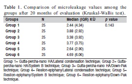
RESULTS: Group 6 (Resilon-epiphany/Down-Pak technique) had the highest microleakage value, followed by Group 2 (Gutta-percha-nano HA/System B technique), Group 4 (Resilon-Epiphany/Lateral condensation technique), Group 3 (Gutta-percha-nano HA/Down-Pak technique), Group 5 (Resilon-Epiphany/System B technique), and Group 1 (Gutta-percha-nano HA/Lateral condensation technique) with the values of 4.69 (6.06) KÙ, 3.88 (2.92) KÙ, 3.77 (3.75) KÙ, 3.38 (3.92) KÙ, 2.64 (2.90) KÙ, and 2.44 (4.34) KÙ, respectively. No significant difference in the quantity of leakage was observed for each root in each group between the two tested filling materials and their sealers (p=0.143). Micro CT scan investigations revealed more micro-voids in the Resilon-Epiphany Group obturated with Down-Pak technique.
CONCLUSIONS: Nano-hydroxyapatite sealer with gutta-percha filling material provided a reasonable seal compared with Epiphany sealer and Resilon filling material.
Keywords: Resilon-epiphany, gutta-percha-Nano HA sealer, root canal filling, electrochemical
test, micro-CT scan.
Introduction
Obturation of the root canal is the final step of the endodontic treatment. The success of this step depends mainly on the root filling materials and techniques, which should have the ability to eliminate leakage from the oral cavity and periodontal tissues after cleaning and shaping. A number of obturationtechniques have been used over the years1-7. It is believed that the excellence of obturation seal is primarily related to the cohesive and adhesive interaction of filling materials to the root canal walls8-10. Recently, a new sealer has been introduced by the School of Dental Sciences, Universiti Sains Malaysia, Malaysia, known as Nano-Hydroxyapatite (Nano-HA) sealer11. The nano HA crystals, which range from 40-60 nm in size, are synthesized by wet chemical method using calcium hydroxide [Ca(OH)2] and phosphoric acid (H3PO3) as Ca and P precursors, respectively. The sealer is composed of nano-hydroxyapatite as a filler, bismuth (III) oxide as a radiopacity component and hexamethylenetetramine as a polymerization activator, and the liquid is Bisphenol A diglycidyl ether12. Previous investigations have shown no significant difference between the sealing ability of nano HA compared with AH 26, using dye penetration technique11-12.
On the basis of the in-vitro and in-vivo data available, several leakage studies have been conducted comparing Resilon-Epiphany with gutta-percha and conventional sealers. Onay et al.13 (2009) using the fluid filtration method and Kaya et al.14(2007) using glucose penetration method have shown that the quality of apical seal achieved with guttapercha and AH Plus or AH 26 in combination is similar to that of Epiphany-Resilon. However, other investigators have shown, using the same techniques, that the better quality of apical seal is achieved with Epiphany-Resilon combination due to superiority of this bonding system14-16. As the bonding systems have improved, so has the resistance to bacterial and fluid penetration of the materials. However, no study has yet been conducted to evaluate the sealing ability of different materials and obturation techniques using electrochemical and micro-computed tomography (CT) scan. Therefore, the aims of this study were to evaluate the sealing ability of gutta-percha-nano-HA and Resilon-Epiphany by electrochemical method and micro-CT scan after 48 h and 20 months, using three different obturation techniques (cold lateral condensation technique, warm vertical condensation - System B, and warm vertical condensation with vibration - Down-Pak).
Material and methods
Selection of teeth
Extracted human single-rooted mandibular premolars were obtained and stored at room temperature in sealed vials containing saline immediately after extraction. The teeth were examined under digital stereomicroscope (Motic Digital Microscope, LTD, France) to discard those with any preexisting root fractures. A sample of 150 teeth were selected and sectioned at the cementoenamel junction using a diamond disc at a high-speed handpiece under continuous water spray coolant, according to the following criteria:
• The same root curvature; between 0º-5º (using Schneider technique)
• Diameter of apical foramen equals to K-file size 15
• Dentin thickness standardized between 2.5-3.5 mm from the apical foramen to the cervical orifice.
Preoperative periapical digital radiographs from buccolingual and mesiodistal directions were taken to ensure that root samples had normal canal shape and enough thickness of dentinal walls. Then the samples were randomly divided into 6 groups of 25 roots and kept in separate plastic containers. Each sample was given a unique number.
Sample preparation
Working length for each root canal was established using a size 15 K Flex file (Dentsply Tulsa Dental Specialties, Tulsa, OK, USA). File was placed and advanced into the root canal until its tip was visualized at the apical foramen. The working length was set at 1 mm shorter of the apical foramen and it was equal to 15 mm. All canals were instrumented with Pro file Ni-Ti (Dentsply-Maillefer, Ballaigues, Switzerland) rotary instruments up to a size 35 master apical file following manufacturer's instructions and using crown down pressureless technique.
During preparation and between each profile file, the canals were irrigated with 2 mL of 5.25% NaOCL (Farmácia Amazon, São Carlos, SP, Brazil). After instrumentation, the canals were rinsed initially with 5 mL of 17% EDTA to remove the smear layer and followed by 5 mL of distilled water to remove any residues of NaOCL.
Sample obturation
Group 1 (n=25): The prepared root canals were obturated with gutta-percha and Nano-HA endodontic sealer using cold lateral condensation technique. A master cone size 35 Profile was fitted to the working length and presence of tug-back was confirmed. Nano-HA sealer was mixed according to manufacturer's instructions to a creamy consistency and applied into the canals using lentulo spiral (Dentsply-Caulk, Milford, DE, USA) which was inserted within 2 mm short of the working length. After placement and condensation of the master cone at the appropriate working length, accessory cones were placed and condensed using a finger spreader (Miltex, Inc., York, PA, USA). The excess of gutta- percha was removed with a heated instrument and condensed vertically with plugger (Nordent, USA) to the level of the canal orifice.
Group 2 (n=25): Similar to group 1, the prepared root canals were obturated with gutta-percha and Nano-HA endodontic sealer using vertical condensation technique System B (Analytic, Sybron Dental Specialties, Orange, CA, USA).
Group 3 (n=25): Similar to group 1, the prepared root canals were obturated with gutta-percha and Nano-HA endodontic sealer using vertical and vibration condensation technique Down-Pak (EI-Endo, Hu-Friedy, Chicago, IL, USA).
Group 4 (n=25): The prepared root canals were obturated with Resilon/Epiphany self-etching primer and Epiphany sealer (Pentron Clinical Technologies LLC, Wallingford, CT, USA). First, the primer was inserted into the root canals and excess was removed with paper point (Dentsply Tulsa Dental Specialties). Subsequently, Epiphanysealer was mixed according to the manufacturer's instructions and inserted into the root canals with a lentulo spiral. Resilon master cone size 35 was placed into the root canal. Following the application of the sealer, root canal filling was completed by inserting accessory cones dipped in Epiphany sealer and laterally condensing with a finger spreader. Excess Resilon cones were removed with a heated instrument and condensed vertically with a plugger to the level of the canal orifice.
Group 5 (n=25): Similar to group 4, the prepared root canals were obturated with Resilon and Epiphany using vertical condensation technique System B (Analytic, Sybron Dental Specialties, CA, USA).
Group 6 (n=25): Similar to group 4, the prepared root canals were obturated with Resilon and Epiphany using vertical and vibration condensation technique Down-Pak (EIEndo, Hu-Friedy).
Postoperative radiographs were taken to ensure complete and void-free obturation. All the samples were wrapped in wet piece of gauze to assure 100% humidity and were stored individually in screw-capped glass vials in an incubator at 37°C for 48 h and 20 months.
III) Quantitative microleakage measurement
Apical leakage of the obturated root canals was assessed by ac-impedance technique at 48 h and 20 months. A PVCinsulated copper wire with a 5.0mm bared end was inserted coronally into the obturated canal of each root and sealed in position with sticky wax. Thereafter, the coronal 2/3 of the external roots surfaces as well as the root/wire junctions were sealed with three layers of nail varnish. Each root was immersed in an electrolyte solution (0.9% sodium chloride solution) at room temperature. Then the ac-current was applied between the electrodes.
The current flow, denoting onset of leakage, was measured by IR drop across a 10-Ohm resistor placed in series with the electrodes and power source. Then after 20 months, 2 samples from each group were securely placed and fixed into the sample holder of the SkyScan micro-CT scanner 1072 (Micro Photonics Inc., Allentown, PA, USA) at 100 kV and source current at 120 MA mps, beam hardening set to 10. Each sample was placed for roughly 2 h to produce 1000 projections in TIFF. These images were then converted to tomograms (cross sections) saved in BMP, using NRecon (version 1.4.3; SkyScan). Next, image were examined for microleakage using image analysis programs provided by SkyScan (T-view and CT-an) using ANT for the 3D reconstruction for creating the 3D model.
Results
The results from this study seem to suggest that, Group 6 had the highest microleakage value, followed by Group 2, Group 4, Group 3, Group 5, and Group 1 with the values of 4.69 (6.06) KÙ, 3.88 (2.92) KÙ, 3.77 (3.75) KÙ, 3.38 (3.92) KÙ, 2.64 (2.90) KÙ, and 2.44 (4.34) KÙ, respectively. No statistically significant difference in the quantity of leakage was observed for each root in each group between the two tested filling materials and their sealers (p=0.143), as shown in Table 1.
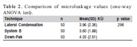
Down-Pak technique group exhibited the highest microleakage value (4.33 ± 2.61) KÙ, followed by the lateral condensation technique group (3.96 ± 2.36) KÙ, and System B group (3.60 ± 1.88) KÙ. In addition, no significant difference was observed among all experimental groups based on the obturation technique used (p=0.296). as shown in Table 2.
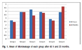
The results of comparison between microleakage measured at 48 h and 20 months was not statistically significant (p<0.05) as shown in Figure 1.
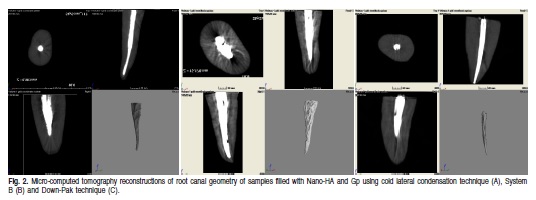
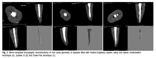
Figure 2 (A, B and C) depict Micro-computed tomography reconstructions of root canal geometry of samples obturated with Nano-HA and Gp using cold lateral condensation technique, System B and Down-Pak technique. While, Micro-computed tomography reconstructions of root canal geometry of samples obturated with Resilon-Epiphany system using the same three obturation techniques are shown in Figure 3 (A, B and C). Micro CT scan investigations showed micro-voids were observed more in the Resilon-Epiphany Group obturated with the Down-Pak technique.
Discussion
The results of the present study indicate that there was no significant difference in the sealing ability between the two tested filling materials and their sealers (p=0.143) using the three obturation techniques. This may be due to Nano-HA epoxy resin, which has been shown to have a reasonable sealing ability12,17-18. In addition, it was reported that Nano-HA sealer was not affected by heat application during continuous waves of condensation technique19-20 unlike the Resilon material. The present experimental work indicated that Resilon-Epiphany root canal fillings when compared with gutta-percha/Nano-HA have no significant difference in the sealing ability when the whole length of root canal was filled with lateral, vertical and vibration techniques measured after 20 months of obturation. However, Shemesh et al.21 (2008) found that, the apical 4 mm of Resilon-Epiphany root canal fillings allowed more glucose penetration than gutta-percha. Shipper et al.16 (2004) detected faster bacterial leakage in gutta-percha and AH 26 filling when compared with Resilon/Epiphany during a period of 31 days for the entire root length.
This study showed that there is no significant difference in the sealing ability of Resilon-Epiphany using 3 three different obturation techniques compared with gutta-perchananoHA within 48 h. However, after 20 months, sealer dissolution resulted in gap formation between root dentin and root filling material. According to De Munck et al.22 (2005), after 3 months, all dentin adhesives under investigation exhibited mechanical and morphological evidence of degradation. As the bond degrades, interfacial microleakage increases, which resembles the in-vivo ageing effect. In the present study, the ageing effect through storage of the specimens over 20 months possibly evokes the current limitations of dentin bonding in the root canal system. In addition, the polymerization shrinkage of the Epiphany sealer and water sorption and solubility play an important role concerning the increased microleakage in the long term23. The solubility values a reported by Versiani et al.24 (2006) were 3.41% for Epiphany and 0.21% for AH Plus. Bonding is further compromised in sclerotic dentin, which is found more often in the apical area of permanent teeth25.
Electrochemical leakage tests offer advantages in terms of speed, accuracy and efficiency, as well as, its ability to operform longitudinal studies. The results of the present study corroborate those of previous investigations, even though, those studies used different methods for measuring microleakage, such as dye penetration26, bacterial leakage27, fluid filtration method17and glucose penetration method21. All of these studies were short-term experiemnts of no more than 3 months, except for one study with duration of 16 months25. The present study had duration of 20 months. Micro-CT scan investigation of samples showed more micro-voids for Resilon groups obturated by Down-Pak technique than in gutta-percha groups. Sealing ability of both Nano-HA sealer and Epiphany sealer was similar by using three techniques: cold lateral, heat applied by System B or heat and vibration applied by Down Pak pressure. As it can be seen clearly in micro-CT reconstructions in Figures 1 and 2, none of the techniques was superior in creating a hermetic seal of root canals. Generally, the micro CT scan view of root obturated with Resilon-Epiphany Down-Pak System showed poor adaptation of Resilon-Epiphany with a massive amount of voids, which coincide with the result of microleakage using electrochemical test. The electrochemical test showed that this group has the highest amount of leakage, 4.69 KÙ. However, statistically it cannot be compared between the two tests because the sample size in the micro-CT scan analysis was too small, and the machine is very expensive and time consuming test.
Nano-hydroxyapatite sealer with gutta-percha filling material provides a reasonable seal as compared to epiphany sealer and Resilon filling material. As such, it could be used as an alternative to the commercial available sealer whereas techniques and duration have no effect on the results. None of the filling materials and techniques used in the present study provided a complete tight seal in a three-dimensional manner.
Further in vitro studies should be conducted to evaluate the effect of increasing temperature and vibration of Down-Pak obturation technique on adhesion, dimensional stability and setting time properties of the tested obturation materials (Nano-HA with gutta-percha or Epiphany with Resilon).
References
1. Gulsahi K, Cehreli ZC, Kuraner T, Dagli FT. Sealer area associated with cold lateral condensation of gutta-percha and warm coated carrier filling systems in canals prepared with various rotary NiTi systems. Int Endod J. 2007; 40: 275-81. [ Links ]
2. Schilder H. Filling root canals in three dimensions. J Endod. 2006; 32: 281-90. [ Links ]
3. Kurtzman GM, von Fraunhofer JA. Leakage resistance of a self-etch sealercone obturation system. Compend Contin Educ Dent. 2008; 29: 246-8. [ Links ]
4. Boussetta F, Bal S, Romeas A, Boivin G, Magloire H, Farge P. In vitro evaluation of apical microleakage following canal filling with a coated carrier system compared with lateral and thermo-mechanical Gutta-Percha condensation techniques. Int Endod J. 2003; 36: 367-71. [ Links ]
5. Buchanan LS. The continuous wave of obturation technique: 'centered' condensation of warm gutta-percha in 12 seconds. Dent Today. 1996; 15: 60-2, 64-7. [ Links ]
6. Gencoglu N, Garip Y, Bas M, Samani S. Comparison of different guttapercha root filling techniques: Thermafil, Quick-fill, System B, and lateral condensation. Oral Surg Oral Med Oral Pathol Oral Radiol Endod. 2002; 93: 333-6. [ Links ]
7. Marciano MA, Bramante CM, Duarte MAH, Delgado RJR, Ordinola-Zapata R, Garcia RB. Evaluation of single root canals filled using the lateral compaction, Tagger's Hybrid, Microseal and Guttaflow techniques. Braz Dent J. 2010; 21: 411-5. [ Links ]
8. Nunes VH, Silva RG, Alfredo E, Sousa-Neto MD, Silva-Sousa YTC. Adhesion of Epiphany and AH Plus Sealers to Human Root Dentin Treated with Different solutions Braz Dent J. 2008; 19: 46-50. [ Links ]
9. Williams C, Loushine RJ, Weller RN, Pashley DH, Tay FR. A comparison of cohesive strength and stiffness of Resilon and gutta-percha. J Endod. 2006; 32: 553-5. [ Links ]
10. Carvalho-Junior JR, Guimaraes LF, Correr-Sobrinho L, Pecora JD, Sousa-Neto MD. Evaluation of solubility, disintegration, and dimensional alterations of a glass ionomer root canal sealer. Braz Dent J. 2003; 14: 114-8. [ Links ]
11. Alshakhshir J. The apical sealing ability evaluation of a new experimental nano hydroxyapatite-filled epoxy resin based endodontic sealer- in vitro study. Malays J Med Sci. 2010; 17: 110-5. [ Links ]
12. Farea M, Masudi S, Wan Bakar WZ. Apical microleakage evaluation of system B compared with cold lateral technique: In vitro study. Aust Endod J. 2010; 36: 48-53. [ Links ]
13. Onay EO, Ungor M, Unver S, Ari H, Belli S. An in vitro evaluation of the apical sealing ability of new polymeric endodontic filling systems. Oral Surg Oral Med Oral Pathol Oral Radiol Endod. 2009; 108: e49-54. [ Links ]
14. Kaya BU, Kececi AD, Belli S. Evaluation of the sealing ability of guttapercha and thermoplastic synthetic polymer-based systems along the root canals through the glucose penetration model. Oral Surg Oral Med Oral Pathol Oral Radiol Endod. 2007; 104: 66-73. [ Links ]
15. Kim YK, Grandini S, Ames JM, Gu LS, Kim SK, Pashley DH, et al. Critical review on methacrylate resin-based root canal sealers. J Endod. 2010; 36: 383-99. [ Links ]
16. Shipper G, Orstavik D, Teixeira FB, Trope M. An evaluation of microbial leakage in roots filled with a thermoplastic synthetic polymer-based root canal filling material (Resilon). J Endod. 2004; 30: 342-7. [ Links ]
17. 17-Wedding JR, Brown CE, Legan JJ, Moore BK, Vail M. M. An in vitro comparison of microleakage between Resilon and gutta-percha with a fluid filtration model. J Endod. 2007; 33: 1447-9. [ Links ]
18. Sousa-Neto MD, Passarinho-Neto JG, Carvalho-Junior JR, Cruz-Filho AM, Pecora JD, Saquy PC. Evaluation of the effect of EDTA, EGTA and CDTA on dentin adhesiveness and microleakage with different root canal sealers. Braz Dent J. 2002; 13: 123-8. [ Links ]
19. Tagger M, Tagger E, Tjan AH, Bakland L. K. Measurement of adhesion of endodontic sealers to dentin. J Endod. 2002; 28: 351-4. [ Links ]
20. Wu MK, Van Der Sluis LW, Wesselink PR. Fluid transport along guttapercha backfills with and without sealer. Oral Surg Oral Med Oral Pathol Oral Radiol Endod. 2004; 97: 257-62. [ Links ]
21. Shemesh H, Souza EM, Wu MK, Wesselink PR. Glucose reactivity with filling materials as a limitation for using the glucose leakage model. Int Endod J. 2008; 41: 869-872. [ Links ]
22. De Munck J, Van Landuyt K, Peumans M, Poitevin A, Lambrechts P, Braem M et al. A critical review of the durability of adhesion to tooth tissue: methods and results. J Dent Res. 2005; 84: 118-32. [ Links ]
23. Tay FR, Loushine RJ, Lambrechts P, Weller RN, Pashley DH. Geometric factors affecting dentin bonding in root canals: a theoretical modeling approach. J Endod. 2005; 31: 584-9. [ Links ]
24. Versiani MA, Carvalho-Junior JR, Padilha MI, Lacey S, Pascon EA, Sousa-Neto MD. A comparative study of physicochemical properties of AH Plus and Epiphany root canal sealants. Int Endod J. 2006; 39: 464-71. [ Links ]
25. Paque F, Sirtes G. Apical sealing ability of Resilon/Epiphany versus gutta-percha/AH Plus: immediate and 16-months leakage. Int Endod J. 2007; 40: 722-9. [ Links ]
26. Kamalini R, Mithra NH, Priyadarshini H. Apical sealing ability of newer resinbased pulp spacesealers - An in vitro study. Endodontology. 2008; 16-21. [ Links ]
27. Baumgartner G, Zehnder M, Paque F. Enterococcus faecalis type strain leakage through root canals filled with gutta-percha/AH Plus or Resilon/Epiphany. J Endod. 2007; 33: 45-7. [ Links ]
 Correspondence:
Salma B Abdo
Tawam Dental Centre - Tawam Hospital Office
P.O. Box: 15258 Abu Dhabi, United Arab Emirates
E-mail: salma114@hotmail.com
Received for publication: May 18, 2012
Accepted: September 13, 2012
