


Serviços Personalizados
Artigo
 pdf em Inglês
 Artigo em XML
 Referências do artigo
 Enviar este artigo por email
Links relacionados
Compartilhar

 Permalink
Brazilian Journal of Oral Sciences
versão On-line ISSN 1677-3225
Braz. J. Oral Sci. vol.11 no.3 Piracicaba Jul./Set. 2012
ORIGINAL ARTICLE
Analysis of canine dimorphism in the estimation of sex
Yuri Trigueiro Faustino da CostaI; Laíse Nascimento Correia LimaII; Patrícia Moreira RabelloIII
IDDS, Federal University of Paraíba, João Pessoa, PB, Brazil
IISpecialist in Forensic Dentistry, MSc in Forensic Dentistry and Deontology, Piracicaba Dental School, University of Campinas, Piracicaba, SP, Brazil
IIIAdjunct Professor, Forensic and Ethical Dentistry and Dental Legislation, Federal University of Paraíba, João Pessoa, PB, Brazil
ABSTRACT
AIM: To evaluate the sexual dimorphism of mandibular and maxillary canines among dental students of the Federal University of Paraiba, Brazil.
METHODS: This was an observational, blind and cross-sectional study with comparative and statistical-descriptive procedure. Fifty-one pairs of plaster models belonging to undergraduate dental students aged 18-29 years were analyzed. Quantitative data were organized and processed by means of the Statistical Package for the Social Sciences (SPSS) software, version 15.0. This research has followed the guidelines of 196/96 Brazilian Resolution of the National Health Council, Ministry of Health.
RESULTS: All measures were found to show statistically significant differences between sexes (p<0.001) according to Student's t-test. Regarding the difference between the four canines for each sex separately, it was found difference only between mandibular and maxillary canines (p<0.001) according to the F test (ANOVA), but with no significant difference between the right and left sides.
CONCLUSIONS: Odontometric techniques allowed concluding that canine teeth present statistically significant sexual dimorphism, and that they may be useful in the estimation of sex in complementary methods during body identification. The data obtained in this study were compared with those of other studies to provide information about sexual dimorphism be specific for each population.
Keywords: canine tooth, forensic anthropology, forensic dentistry, odontometry, sex dimorphism.
Introduction
The estimation of sex is an important step to build the biological profile of unidentified human remains, especially when an accurate outcome is achieved, considering that about half of the population would be automatically excluded during search operations. Although DNA analysis provides irrefutable evidences on sex identification in human remains, such a technique is relatively prolonged and quite exhaustive if compared to assessment of skeletal parameters1.
It is known that morphological and metric aspects of the skeleton allow a more reliable diagnosis of sex. The more measurements and data obtained, the more reliable will be the result. For instance, sex may be estimated by means of bones that constitute the pelvic girdle since it is the skeleton segment which most presents sexual dimorphism. It also may be analyzed the morphology and dimension of long bones of the body. Additionally, estimation of sex may still be performed through observation of qualitative or morphological and quantitative or metric aspects present in the skull2.
Accordingly, estimation of sex does not represent a problem when a complete skeleton is found. Nevertheless, if only the mandibular bone along with the teethis found or mandible fragments or even the teeth by themselves are available in the site, so estimation of sex may be performed with the help of teeth dimensions3.
With this approach, numerous authors have studied intensively sexual dimorphism present in teeth by means of odontometric analyses, and most studies have showed statistically significant differences in the permanent dentition4-6.Thus, this work aimed to analyze the degree of sexual dimorphism of mandible and maxillary canines among undergraduate dental students of the Federal University of Paraiba, using odontometric techniques described in literature, as well as to provide arguments for the debate about dimorphism being specific for each population.
Material and methods
This research project was conducted in accordance with the 196/96 Brazilian Resolution of the National Health Council, Ministry of Health, which regulates research involving human beings. It was submitted to and approved by the Research Ethics Committee of Lauro Wanderley University Hospital (protocol #360/10).
An observational, blind and cross-sectional study was performed, applying comparative and statistical-descriptive procedure. The procedure method employed was an intensive direct observation by means of examination of plaster models of the upper and lower arches.
The sample was composed by plaster models of the upper and lower arches belonging to undergraduate dental students of the Federal University of Paraiba. Fifty-one students, aged 18-29 years, being 26 female, took part of the research, which encompassed a total of 102 plaster models.
A digital caliper with thin beaks (Digimess®, 150 mm/6" 100.250, 0.01 mm/.0005 resolution", São Paulo, SP, Brazil) was used to measure the mesiodistal (MD) and buccolingual (BL) distances of crowns of the maxillary and mandible canines, as well as their diagonal, mesiobuccal-distolingual (MB-DL) and distobuccal-mesiolingual (DB-ML) dimensions.
The measurements in the MD and BL directions were obtained following the Acharya and Mainali's method7 applied only for the canine teeth, proceeding as follows:
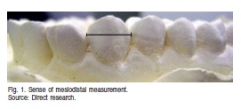
MD measurements
The greatest distance between the proximal surfaces of the tooth crown was registered by means of the caliper beaks incisally positioned along the long axis of the tooth. In cases of bad position or giroversions, measurements should be taken between the points of the proximal surfaces of the crown, where contact with the adjacent tooth would normally occur (Figure 1).
BL measurements
The greatest distance between the buccal and lingual surfaces of the tooth crown was obtained with the caliper located incisally and forming a right angle in relation to the MD sense (Figure 2).
For the measurements in the MB-DL and DB-ML senses,the methodology described by Karaman4 was employed, being performed in the canine teeth, proceeding as follows:

MB-DL measurements
The greatest distance between the MB and DL corners of the tooth crown was obtained with the caliper positioned parallel to the incisal edge (Figure 3).
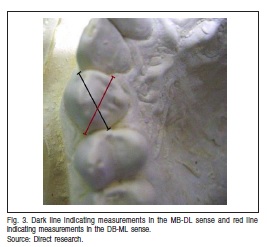
DB-ML measurements
The greatest distance between the DB and ML corners of the tooth crown was achieved with the caliper positionedparallel to the incisal edge (Figure 3).
Were excluded from the sample: all models lacking one of the teeth of interest to the research (upper and lower canines, right and left sides) and models exhibiting fillings in the proximal and free surfaces, bad tooth positioning (impossible to obtain the measurement), and damaged or broken teeth. Were also excluded the students who: wore orthodontic appliances, felt nausea during impression procedures, and those who expressed the feeling of being withdrawn from the study, after theoretical approach about the subject in question.
Quantitative data attained in the study were organized and processed by means of the Statistical Package for the Social Sciences (SPSS), version 15.0 (SPSS Inc., Chicago, IL, USA). It was conducted a descriptive and inferential analysis through specific statistical methods, in which were obtained: mean and standard deviation, Student's t with equal or unequal variances and F (ANOVA) tests for repetitive measures with Bonferroni's comparisons. Verification of the equal variances hypothesis was performed using the Levene F test. The error margin used in the decision of the statistical tests was 5%. From the results, it was made the distribution of frequencies of all variables addressed in the study, presented in tables, charts or graphs to characterize the sample and data description.
Results
Table 1 presents the means and standard deviations for the MD, BL, MB-DL and DB-ML measurements, according to tooth and sex. In this table, it was verified whether existed or not statistical difference between means of the measurements for each tooth in relation to sex. Statistically significant differences indicated by Student's t test were observed for all teeth and in all measurements (p(1) < 0.001 or p(1)=0.001). It was also investigated in that table whether there was significant difference or not among the 4 canines, for each sex separately, in relation to each one of the measurements analyzed. To this end, F test (ANOVA) was employed for repetitive measures with Bonferroni's comparisons, considering a 5% error margin. No statistically significant difference was found between the right and left sides, either in the upper or lower arches, in none of the measures analyzed. Nevertheless, were noted differences between the upper and lower measurements (p(2)<0.001).
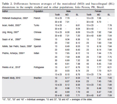
Table 2 expresses the comparison of dimensions of the MD and BL measurements in the considered sample in relation to other studies. It is verified that teeth dimensions in the MD and BL senses resembles the Portuguese population's measures.

Discussion
Numerous odontometric studies performed in human dental arches attest the fact that there are differences in teeth size patterns between sexes, and that those differences vary among populations, reaching significance or not. Canines have demonstrated higher degree of sex dimorphism among the populations10-11 and are considered as one the most resistant teeth in the dentition, remaining intact in several post-mortem scenarios12.
MD and BL distances
According to Table 1, the MD and BL distances were found to show statistically significant differences for all canines, being higher for males than females, which is a finding that corroborates the results verified in other populations. Comparing with measurements obtained in other studies (Table 2), it was noted that, for both sexes and measures, the present study assimilates with the upper canine teeth of the Portuguese6 population sample, but are higher than the Indian13 population also for both sexes and measures. Nonetheless, it is observed that for other populations under comparison, the means of measurements for both sexes vary a lot, being higher for a given measure and sex and lower for another measure and sex, e.g., when the present investigation is compared with the study by Suazo et al.14, it is verified that Brazilian's canine dimensions are higher for male and lower for the female gender. Carefully, of course, we can assign these apparent differences in measurements due to such things as differences in techniques of tooth measurement and degree of wear.
Garn et al.15 report that genetic influence tends to keep low/high magnitude dimorphism in interrelated racial groups originating from the same geographical region. That may explain the differences between tooth size in the diverse populations. However, those differences should also be in part assigned to the degree of miscegenation that some populations present.
Thus, it makes sense that the sample studied resembles with the sample surveyed in the Portuguese population, despite the fact that only the upper teeth were assessed in the present study.
Regarding the difference between right and left sides (asymmetry), it was found no significant difference for both sexes, evidenced by the minimum difference between the averages of right and left sides (Table 1). It is suggested that there is no tendency for the dimensions of the crown being larger on one side than on the other side, so confirming some literature findings16-17. However, Pereira et al.6 showed that, for females, in the MD measurement, there was statistically significant difference between right and left sides of the upper canine (p <0.005), using the paired t test.
MB-DL and DB-ML distances
According to the results obtained in the analysis of diagonal measures undertaken in the course of this study, males' teeth were considered higher than females', and such a difference was statistically significant between sexes (p<0.001). These findings are consistent with other studies described in literature4,18, which affirm canines to be the most dimorphic elements in these diagonal lengths. No statistically significant difference was observed between the right and left sides, in upper and lower arches, similarly to the MD and BL measurements.
This way, although the completion of odontometric measurements in this field is relatively new, these results corroborate with those found in literature, confirming that these dimensions suggest a high level of reliability, and may be used in conjunction with other measures.
The present investigation showed significant differences between dimensions of canines in both sexes, suggesting a high reliability rate for using these teeth in the estimation of sex.
References
1. Acharya AB, Prabhu S, Muddapur MV. Odontometric sex assessment from logistic regression analysis. Int J Legal Med. 2011 Mar.; 125(2): 199-204. [ Links ]
2. Vanrell J. Odontologia legal e antropologia forense. 2º Ed, Rio de Janeiro: Guanabara Koogan; 2009. 420 p. [ Links ]
3. Kavitha B. Sex determination in teeth. [Dissertation]. Chennai: The Tamil Nadu Dr. M.G.R. Medical University; 2005. 23p. Master of Dental Surgery. [ Links ]
4. Karaman, F. Use of Diagonal Teeth Measurements in Predicting Gender in a Turkish Population. J Forensic Sci. 2006 May.; 51(3): 630-635. [ Links ]
5. Astete JC, San Pedro VJ, Suazo GI. Sexual Dimorphism in the Tooth Dimensions of Spanish and Chilean peoples. Int J Odontostomatol. 2009 Jan-Jun.; 3(1): 47-50. [ Links ]
6. Pereira C, Bernardo M, Pestana D, Santos JC, Mendonça MC. Contribution of teeth in human forensic identiûcation – Discriminant function sexing odontometrical techniques in Portuguese population. J Forensic Leg Med. 2010 Feb.; 17(2): 105-110.
7. Acharya AB, Mainali S. Univariate sex dimorphism in the Nepalese dentition and the use of discriminant functions in gender assessment. Forensic Sci Int. 2007 Nov.; 173(1): 47-56. [ Links ]
8. Pettenati-Soubayroux I, Signoli M, Dutour O. Sexual dimorphism in teeth: discriminatory effectiveness of permanent lower canine size observed in a XVIIIth century osteological series. Forensic Sci Int 2002 May.; 126: 227–232.
9. Ling JYK, Wong RWK. Tooth dimensions of Southern Chinese. Homo 2007 Mar.; 58(1): 67-73. [ Links ]
10. Iscan MY, Kedici PS. Sexual variation in bucco-lingual dimensions in Turkish dentition. Forensic Sci Int. 2003 Nov.; 137(2-3): 160-164. [ Links ]
11. Lund H, Mörnstad H. Gender determination by odontometrics in a Swedish population. J Forensic Odontostomatol. 1999 Dec.; 17(2): 30-34. [ Links ]
12. Acharya AB, Aangali PV, Prabhu S, Nagnu S. Validity of the Mandibular canine index (MCI) in sex prediction: Reassessment in an Indian sample. Forensic Sci Int. 2011 Jan.; 204(1-3): 207.e1-207.e4. [ Links ]
13. Prabhu S, Acharya AB. Odontometric sex assessment in Indians. Forensic Sci Int. 2009 Nov.; 192(1-3): 129.e1-129.e5. [ Links ]
14. Suazo GI, Cantín LM, Lopez FB, Sandoval MC, Torres MS, Gajardo RP, Gajardo RM. Sexual dimorphism in mesiodistal and bucolingual tooth dimensions in Chilean people. Int J Morphol. 2008 Sep.; 26(3): 609-614. [ Links ]
15. Garn SM, Lewis AB, Swindler DR, Kerewsky RS. Genetic Control of Sexual Dimorphism in Tooth Size. J Dent Res. 1967 Sep.; 46(5): 963-972. [ Links ]
16. Sherfudhin H, Abdullah MA, Khan N. A cross-sectional study of canine dimorphism in establishing sex identity. Comparison of two statistical methods. J Oral Rehabil. 1996 Sep.; 23(9): 627-631. [ Links ]
17. Kalia S. A Study of Permanent Maxillary and Mandibular Canines and Inter-Canine Arch Widths among Males and Females. [Dissertation]. Karnataka: Rajiv Gandhi University of Health Sciences; 2006. 175 p. Master of Dental Surgery. [ Links ]
18. Rai B, Anand SC. Gender determination by diagonal distances of teeth. The Internet Journal of Biological Anthropology [serial on the Internet] 2007; 1(1): [about 7 pages] [ Links ].
 Correspondence:
Yuri Trigueiro Faustino da Costa
Rua Francisco Timóteo de Souza, 577
CEP: 58052-130
Bancários, João Pessoa, PB - Brasil
E-mail: yuri.trigueiro@yahoo.com.br
Received for publication: May 16, 2012
Accepted: September 18, 2012
