


Serviços Personalizados
Artigo
 pdf em Inglês
 Artigo em XML
 Referências do artigo
 Enviar este artigo por email
Links relacionados
Compartilhar

 Permalink
Odontologia Clínico-Científica (Online)
versão On-line ISSN 1677-3888
Odontol. Clín.-Cient. (Online) vol.9 no.3 Recife Set. 2010
ORIGINAL ARTICE ARTIGO ORIGINAL
Pattern and distribution of salivary gland tumours in a Brazilian hospital
Padrão e distribuição dos tumores de glândulas salivares em um hospital Brasileiro
Francisco Carlos Sancio-GonçalvesI; Rogério Queiroz PachecoI; Marcelo Drummond NavesIII; Alvimar Afonso BarbosaII; Luiz César Fonseca AlvesIII; Lisette Lobato MendonçaI; Evandro Neves AbdoIII
IAlunos do curso de Especialização em Cirurgia e Traumatologia Bucomaxilofacial da Universidade Federal de Minas Gerais
IIProfessor da Faculdade de Medicina da Universidade Federal de Minas Gerais
IIIProfessores da Faculdade de Odontologia da Universidade Federal de Minas Gerais
ABSTRACT
OBJETIVE:document the distribution of salivary gland tumours (SGT) diagnosed in a period of 5 years.
PATIENTS AND METHODS: A descriptive and retrospective study was conducted of SGT from a Brazilian hospital between January 1999 and December 2003. Data concerning gender, age, disease evolution, gland, histological tumour diagnosis and type of Health Care Service (private or public) was retrieved.
RESULTS: The sample consisted of 121 cases of SG. Parotid (62%) and minor salivary glands (21.5%) were the most common glands involved. The benign tumours comprised 69.5%. Pleomorphic Adenoma (61.5%) and the Mucoepidermoid Carcinoma (11.5%) were the most prevalent tumours. The tumours aff ected mostly females (71.4%). Malignant tumours aff ected older patients (55.6 years). The prevalence of malignant tumours was significantly higher in the minor salivary glands. Individuals from National Public Health Service had a longer waiting period for treatment (18.5 months ± 12.5) in comparison to patients from Private Health Service (10.7 months ± 9.4) (p= 0.015). The prevalence of SGT was 7.1%.
CONCLUSION: Malignant tumour was more prevalent in older patient. It would make a diff erence for patients if the National Health Service, both public and private, changed their management policies in order to reverse the treatment delay.
Keywords: salivary glands; tumours; neoplasia
RESUMO
OBJETIVO: documentar a distribuição de tumores de glândulas salivares (TGS) diagnosticados em um período de 5 anos.
PACIENTES E MÉTODO: estudo descritivo retrospectivo dos casos de TGS de um hospital brasileiro, entre janeiro de 1999 e dezembro de 2003. Dados obtidos: gênero, idade, evolução da doença, glândula envolvida ou sitio anatômico, diagnóstico histológico e tipo de atendimento (público ou privado).
RESULTADOS: a amostra consistiu 121 TGS. Parótida (62%) e glândulas salivares menores (21,5%) foram as mais envolvidas. Os tumores benignos compreenderam 69,5%. Adenoma Pleomórfico (61,5%) e Carcinoma Mucoepidermóide (11,5%) foram os mais prevalentes. Os tumores foram mais frequentes em mulheres (71,4%). Tumores malignos afetaram os pacientes mais idosos (55,6 anos). A prevalência de tumores malignos foi mais alta em glândulas salivares menores. Indivíduos do Sistema Público apresentaram um tempo de evolução da doença mais longo (18,5 meses ± 12,5) em comparação com o Sistema Privado (10,7 meses ± 9,4) (p= 0.015). A prevalência de TGS foi de 7,1%.
CONCLUSÃO: tumores malignos de glândulas salivares são mais prevalentes em pacientes idosos. Faria diferença para o paciente se os Serviços de Saúde, público e privado, mudassem suas políticas para reverter a demora no tratamento.
Descritores: glândulas salivares; tumores; neoplasia
INTRODUCTION
The salivary gland tumours (SGT) are rare and they correspond to 2% of tumours in neck and head region1. An incidence of 1.22 cases per 100,000 inhabitants per year has been estimated in epidemiological studies2. The prevalence studies of salivary tumours in Brazil are scarce3,4.
Usually, the first symptom observed in these tumours is an increase of intra-oral volume5, reason why the dentist may be the first one to observe it6. Therefore, dentist should be able to do a proper and early diagnose of benign and malignant tumours, increasing the chances of a successful treatment.
This study aims to do a survey on this condition in a hospital of Belo Horizonte, a large city in Brazil.
PATIENTS AND METHODS
Luxemburgo Hospital is a reference, located in Belo Horizonte city, Brazil, for diagnose and treatment of benign and malignant tumours.
This descriptive study was conducted through a retrospective data collection from all cases of head and neck's tumour from Luxemburgo Hospital between January 1999 and December 2003.
Data concerning gender, age, disease evolution, gland or anatomic site in case of minor glands, histological tumour diagnosis and type of Health Care Service (private or public) was retrieved.
To verify the role of variables such as gender and aff ected gland on malignancy or benignancy the Chi Square test was used. The t Test of Student for independent sample was utilized for comparing groups with tumours in relation to age, patient delay and Health Care Service. The significance level was established at 0.05. The local ethical committee approved this research.
RESULTS
Out of 1664 patients treated with head and neck's tumours in Department of Head and Neck Surgery, in the period studied, 119 individuals were diagnosed with SGT (7.1%). More than one tumour was found in two individuals increasing the number of SGT in this sample to 121, which 69.5% were benign and 30.5% malignant.
Tumour incidence in male population (28.6%) was smaller than in female one (71.4%) and malignant tumours amongst males (27.8%) were also slightly inferior to those in females (29.4%), however this difference was not significant.
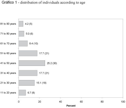
Tumours were more prevalent in age between 31 to 60 years (Gráfico 1). Benign tumours were more prevalent at a mean age of 41.2 (± 15.3) years and malignant tumours occurred at a mean age of 55.6 (± 18.4) years. This difference was statistically significant (p 0,001- t Test of Student).
According to the variable "National Health Service" 33.9% of patients were from National Public Health Service and 66.1% were from Private Health Service.
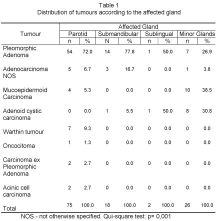
Histological distribution of tumours can be found in (Gráfico 2). The parotid gland was the most aff ected (62%) followed by minor glands (21.5%) and submandibular gland (14.9%) as shown in Table 1.

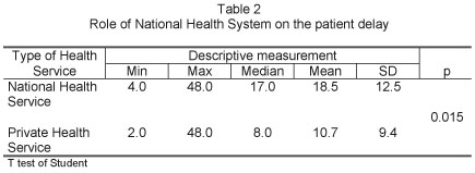
The time period between first symptom and diagnose could only be retrieved from 59 cases and indicated a big gap ranging from 2 to 96 months (15.3 months - SD 15.5). The patient delay from individuals coming from National Public Health Service (18.5 months) was longer than those from Private Health Service (10.7 months) as presented in table2.
DISCUSSION
The prevalence of SGT found in our study (7.1%) is higher than that reported in the literature1,2 . However, it next to the prevalence of 6.3% found by Ladeinde et al. 7. The diff erence is probably due to the methodology used since the sample reflects the characteristics of the hospital and not the population prevalence.
According to the variable "gender", most of the studies reviewed show that women are more affected by SGT3,8,9,10. In this study, the prevalence of SGT related to gender varies greatly: 71.4% were females and male-female ratio of 2.4:1, being in agreement with the literature.
The distribution of malignant tumours in females was 29.4% and in males 27.8% (p= 1.0), similar to other studies5,8. However, other studies have reported a higher prevalence of malignant tumors in male population2,3,10,11,12. This may be due to regional differences or the size of the samples used in each study.
The SGT distribution according to age in this sample shows that the tumours were more prevalent among 31 to 60 years but the higher occurrence of tumours (25.2%) was amongst those with 41 and 50 years of age. These results are similar to other ones reported in Brazil3,4.
The mean age of individuals with malignant tumours (55.6± 18.4 years) was bigger than those ones with benign tumours (41.2 ± 15.3 years), (p= 0.001). Other studies also show similar pattern12,13. However, studies in Brazil did not find any statistical difference in relation to age and tumour malignancy9,14. Loyola et al.9 studied only tumors of minor salivary glands and Vargas et al.4 analyzed a sample which had a small number of tumors of minor salivary glands.
The present data show a higher prevalence of benign tumours (69.5%) compared to malignant tumours (30.5%), as also reported in other studies3,4,10,11,14,15. The percentage of benign compared to malignant tumors varies among diff erent studies, showing regional differences and methodological.
The frequency of malignant tumors in minor salivary glands ranges from 38% to 76% as reported in the literature9,10,12. This study reveals a higher prevalence of malignant tumours in the minor salivary glands (73.1%) in comparison to parotid (17.4%) and submandibular (22.2%) (P < 0.001) as showed in table 1. Although the percentage found in our study is within the range reported in the literature, the fact that this hospital be a reference for the treatment of cancer may have contributed to it.
In the present study, pleomorphic adenoma was the most common condition (62,8%) and it corresponds to 90.4% of all benign tumours. Other findings confirm these results3,4,11,14,16. However, among the minor salivary glands, the most common condition was the mucoepidermoid carcinoma (38.5%), also reported by Jansisyanont et al.17. Other findings suggest that pleomorphic adenoma is the most common occurrence in the minor salivary glands9,10,12,17. This difference can be explained by our study was conducted in a referral hospital for treatment of cancer.
Considering only the malignant tumours, the mucoepidermoid carcinoma was the most common condition in this sample (11.5%), followed by adenoid cystic carcinoma (8.2%). Apparently there is no doubt in the literature that pleomorphic adenoma is the most common benign tumour. However, when malignant tumors are considered, some studies suggest that the mucoepidermoid carcinoma is the most common occurrence, as well as in our study3,9,10,11,14,17. Other researches show the adenoid cystic carcinoma as the most frequent malignant tumour5,8,12,19. Perhaps we can look at these diff erent findings as a result of socio-demographic influences and specificities playing an important role in the determination of SGT.
The Brazilian Health System comprises two categories; one is a National Public Health Service, free to all citizens, and the other one is a Private Health Service, administrated mainly by health insurance companies.
The treatment delay in the Public Health Service is high (18.5 ± 12.5 months) in comparison to the Private Health Service (10.7 ± 9.4 months), (p < 0.015). According to Rocha and Simões20, there are fewer beds in private hospitals destined to patients coming from the National Public Health Service and clearly the private hospitals favor patients coming from the Private Health Service. That fact generates a repressed demand and long waiting for the treatment procedures.
CONCLUSION
The prevalence of SGT at the Luxemburgo Hospital in Belo Horizonte, Brazil, was 7.1%. The mean age of individuals suff ering from malignant tumours is higher than those with benign tumours, contrary to other results reported in Brazil.
Individuals coming from the National Public Health Service wait much longer to be seen by doctors than those individuals coming from the Private Health Service. That suggests a great need to review management policies of the National Health Service, in public and in private too, in order to reverse the situation depicted.
REFERENCES
1. Leegaard T, Lindeman H. Salivary gland tumours: clinical picture and treatment. Acta Laryngol. 1969; 263(suppl): 155-9. [ Links ]
2. Frade Gonzalez C, Lozano Ramirez A, Garcia Caballero T, Labella Caballero T. Epidemiological study of salivary gland tumours. Rev Laryngol Otol Rhinol. 1999; 120(5): 331-6. [ Links ]
3. Ito FA, Ito K, Vargas PA, de Almeida OP, Lopes MA. Salivary gland tumours in a Brazilian population: a retrospective study of 496 cases. Int J Oral Maxillofac Surg. 2005 Jul; 34(5): 533-6. [ Links ]
4. Vargas PA, Gerhard R, Araújo Filho VJF, de Castro IV. Salivary gland tumours in a Brazilian population: a retrospective study of 124 cases. Rev Hosp Clin Fac Med Univ São Paulo. 2002 Nov-Dec; 57(6): 271-6. [ Links ]
5. Satko I, Stanko P, Longauerová I . Salivary gland tumours treated in the stomatological clinics in Bratislava. J Craniomaxillofac Surg. 2000 Feb; 28(1): 56-61. [ Links ]
6. Lotufo MA, Júnior CA, Mattos JP, França CM. Pleomorphic adenoma of the upper lip in a child. J Oral Science. 2008 Jun; 50(2): 225-8. [ Links ]
7. Ladeinde AL, Adeyemo WL, Ogunlewe MO, Ajayi OF, Omitola OG. Salivary gland tumours: a 15-year review at the Dental Centre Lagos University Teaching Hospital. Afr J Med Med Sci. 2007 Dec; 36(4):299-304. [ Links ]
8. Lima SS, Soares AF, de Amorim RF, Freitas Rde A. Epidemiologic profile of salivary gland neoplasms: analysis of 245 cases. Braz J Otorhinolarygol. 2005 May-Jun; 71(3): 335-40. [ Links ]
9. Loyola AM, de Araújo VC, de Sousa SO, de Araújo NS. Minor salivary gland tumours: a retrospective study of 164 cases in a Brazilian population. Eur J Cancer B Oral Oncol. 1995 May; 31B(3): 197-201. [ Links ]
10. Rivera-Bastidas H, Ocanto RA, Acevedo AM. Intraoral minor salivary gland tumours: a retrospective study of 62 cases in a Venezuelan population. J Oral Pathol Med. 1996 Jan; 25(1): 1-4. [ Links ]
11. Li LJ, Li Y, Wen YM, Liu H, Zhao HW. Clinical analysis of salivary gland tumour cases in West China in past 50 years. Oral Oncol. 2008 Feb; 44(2): 187-92. [ Links ]
12. Toida M, Shimokawa K, Makita H, Kato K, Kobayashi A, Kusunoki Y, Hatakeyama D, Fujitsuka H, Yamashita T, Shibata T. Intraoral minor salivary gland tumours: a clinicopathological study. Int J Oral Maxillofac Surg. 2005 Jul; 34(5): 528-32. [ Links ]
13. Wang D, Li Y, He H, Liu L, Wu L, He Z. Intraoral minor salivary gland tumours in a Chinese population: a retrospective study of 737 cases. Oral Surg Oral Med Oral Pathol Oral Radiol Oral Endod. 2007 Jul; 104(1): 94-100. [ Links ]
14. Otoh EC, Johnson NW, Olasoji H, Danfillo IS, Adeleke OA. Salivary gland neoplasms in Maiduguri, north-eastern Nigeria. Oral Dis. 2005 Nov; 11(6): 386-91. [ Links ]
15. Stramandinoli RT, Sassi LM, Pedruzzi PA, Ramos GH, Oliveira BV, Ogata DC, Ioshii SO. Accuracy, sensitivity and speciticity of fine needle aspiration biopsy in salivary gland tumours: a restrospective study. Med Oral Patol Oral Cir Bucal. 2010 Jan; 15(1):E32- 7. [ Links ]
16. Fonseca I, Martins AG, Soares J. Epithelial salivary gland tumours of children and adolescents in southern Portugal: a clinicopathologic study of twenty-four cases. Oral Surg Oral Med Oral Pathol. 1991 Dec; 72(6): 696-701. [ Links ]
17. Jansisyanont P, Blanchaert RH Jr, Ord RA . Intraoral minor salivary gland neoplasm: a single institution experience of 80 cases. Int J Oral Maxillofac Surg. 2002 Jun; 31(3): 257-61. 1 [ Links ]
8. Pons Vicente O, Almendros Marqués N, Berini Aytés L, Gay Escoda C.. Minor salivary gland tumours: A clinicopathological study of 18 cases. Med Oral Patol Oral Cir Bucal. 2008 Sep; 13(9):E582-8. [ Links ]
19. Yih WY, Kratochvill FJ, Stewart JC. Intraoral minor salivary gland neoplasms: review of 213 cases. J Oral Maxillofac Surg. 2005 Jun; 63(6): 805-10. [ Links ]
20. Rocha JS, Simões BJ. Study of public and private hospital care on a population basis, 1986-1996. Rev Saude Publica. 1999 Feb; 33(1): 44-54. [ Links ]
 Endereço para correspondência
Evandro Neves Abdo
Universidade Federal de Minas Gerais - Faculdade de Odontologia
Av. Antônio Carlos, 6627 - Belo Horizonte – Minas Gerais - Brasil - CEP: 31270-901
E-mail address: evandro.abdo@gmail.com
Recebido para publicação: 21/05/10
Aceito para publicação: 09/07/10
