


Serviços Personalizados
Artigo
 pdf em Inglês
 Artigo em XML
 Referências do artigo
 Enviar este artigo por email
Links relacionados
Compartilhar

 Permalink
Odontologia Clínico-Científica (Online)
versão On-line ISSN 1677-3888
Odontol. Clín.-Cient. (Online) vol.12 no.4 Recife Out./Dez. 2013
Artigo Original / Original Article
Comparison of distance and symmetry in the mental foramen obtained in situ and in radiographic images of Brazilians
Comparação da distância e simetria dos forames mentonianos obtidos em loco e em imagens radiográficas na população brasileira
Yuri Nejaim I; Amaro Ilídio Vespasiano Silva I; Anna Gabriella Camacho Pressoto II; Francisco Haiter Neto III; Paulo Henrique Ferreira Caria IV
I Ph.D student in Oral Radiology, Dental School, Dental School, Department of Radiology, University of Campinas (UNICAMP), Piracicaba, SP, Brazil
II Graduated student, Dental School, University of Campinas (UNICAMP), Piracicaba, SP, Brazil
III Associate professor, Department of Radiology, University of Campinas (UNICAMP), Piracicaba, São Paulo, SP, Brazil
IV Associate professor, Department of Morphology, University of Campinas (UNICAMP), Piracicaba, São Paulo, SP, Brazil
ABSTRACT
The precise location of the mental foramina is of paramount importance when using implantology to carry out oral rehabilitation procedures not affecting crucial anatomic structures. Panoramic radiography has become the imaging method of choice in many of these cases because of its lower cost and the fact that with it a general assessment of the maxillofacial complex can be made. This study set out to evaluate the distance between the mental foramina and the relationship between them and the mandibular base, and the posterior border of the mandible ramus, as well as the relationship between the bilateral mandibular foramina. The morphometric procedures were carried out by three examiners experienced in Dental Radiology, firstly on 58 human mandibles, with the aid of digital calipers and then on the respective panoramic radiographs of the mandibles, with Image J software, with and without the aid of artificial markers for delimitating the areas of interest. Using Friedman's statistical analysis, there were statistically significant differences between various measurements analyzed and compared to the gold standard, which points to the need to use more accurate imaging tests.
Keyword: Panoramic radiography; Mental foramen; Mandible.
RESUMO
A localização precisa do forame mentoniano tornou-se uma necessidade premente para a execução de procedimentos de reabilitações orais, empregando a implantodontia, que não afetem estruturas anatômicas importantes, sendo a radiografia panorâmica o método de imagem de escolha em muitos desses casos por ter baixo custo e pela possibilidade de avaliação geral do complexo maxilo facial. Este trabalho teve como objetivo avaliar a distância entre os forames mentonianos e a sua relação com a base mandibular, borda posterior do ramo da mandíbula e a relação entre os forames mandibulares bilateralmente. As morfometrias foram realizadas por três experientes avaliadores em Radiologia Odontológica, primeiramente em 58 mandíbulas humanas, com o auxílio de paquímetro digital e depois nas respectivas radiografias panorâmicas das mandíbulas, com o software Image J, com e sem o auxílio de marcadores artificiais para delimitação das áreas de interesse. Por meio da utilização da análise estatística de Friedman, observaram-se diferenças estatísticas significativas em diversas medidas analisadas, quando comparadas ao padrão ouro, fato que demonstra a necessidade da utilização de exames por imagem mais precisos.
PALAVRAS-CHAVE: Radiografia panorâmica; Forame Mentoniano; Mandíbula.
Introduction
The mental foramen is located in the lower premolar region through which nerve endings and blood vessels pass. The mental nerve is a branch of the inferior alveolar nerve which emerges through the mental foramen, and supplies sensory innervation for the lower lip, buccal surface and gingival tissue as far as the mesial region of the 1st lower molar.1
The precise identification of the mental foramen's location is important for the correct application of nerve block anesthesia, a differential diagnosis of bone pathologies, forensic dentistry, preoperative planning for implant placement and the surgical treatment of endodontic failure.1,5 It is valid to say that any procedure involving this region demands indepth anatomical knowledge to prevent problems involving accurate diagnosis and provide positive prognosis in clinical interventions.2
From the literature it can be seen that panoramic radiography continues to be the most widely used in studies on the mental foramen because of its easy access and low radiation dose, despite the fact that horizontal distortions have already been proven, which makes it unviable for linear measurements.3 Different classifications of the mental foramen on panoramic radiographs have been described, but the most commonly used refer to four types: in the first, the mental foramen marks the ending of the mandibular canal; in the second, the foramen is distinctly separated from the mandibular canal; in the third type, the border of the foramen is diffused, while the fourth is called the unidentified type, when the foramina cannot be seen on the panoramic radiograph.4,6
Another important aspect analyzed in studies is the symmetry of the mental foramina. Some authors have found an average of 70 to 80% symmetry between the positions of the foramina on both left and right sides which means it is a useful aid in different interventions carried out in the area where they are located.7,8 With this as a background, this study set out to compare the position of the mental foramina in panoramic radiographs with dry mandibles in situ and to analyze the existence of bilateral symmetry between the structures being measured.
Materials and methods
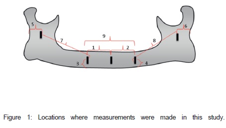
This study was begun after approval by the Research Ethics Committee at the School of Dentistry in Piracicaba - UNICAMP, protocol 081/2012. A total of 58 macerated mandibles were selected at the Department of Morphology. Those presenting any type of shape anomaly and/or bone fractures were excluded. The mandibles were analyzed morphometrically in four different phases with and without the aid of indelible markers. In the first phase, the mandibles were measured in situ (gold standard) using a Digital Electronic Caliper 0-150 mm, Stainless Hardened ® with markers in the following positions (Figure 1): 1 (from the center of the mandibular symphysis to the medial aspect of the mental foramen on the right side), 2 (from the center of the mandibular symphysis to the medial aspect of the mental foramen on the left), 3 (from the base of the right mental foramen to the base of the mandible), 4 (from the base of the left mental foramen to the base of the mandible) 5 (from the posterior aspect of the mandibular foramen on the right side to the posterior aspect of the ascending ramus of the mandible on the same side), 6 (from the posterior aspect of the mandibular foramen on the left side to the posterior aspect of the ascending ramus of the mandible on the same side), 7 (from the medial aspect of the mental foramen on the right side to the posterior aspect of the mandibular foramen on the same side), 8 (from the medial aspect of the mental foramen on the left side to the posterior aspect of the mandibular foramen on the same side), and 9 (from the mesial of the mental foramen on the right side to the mesial of the mental foramen on the left).
For the next phases, digital panoramic radiographs were taken with the Instrumentarium Orthopantomograph 100 D ® (General Electric, Tuusula, Finland) with and without gutta percha markers. After the panoramic radiographs had been obtained, they were then measured in the same positions mentioned above, after calibrating the pixel size of each image with the Image J 1.45s ® software (NIH, USA) in the second and third phases. The fourth phase of evaluation was the identification of the location of the mental foramen in relation to the teeth; this relation was classified in six possible positions described as follows: 1 (anterior to 1st premolar), 2 (in line with the longitudinal axis of the 1st premolar ), 3 (between the 1st and 2nd premolars), 4 (in line with the longitudinal axis of the 2nd premolar), 5 (between the 2nd premolar and 1st molar) and 6 (in line with the longitudinal axis of the 1st molar). All steps were carried out by three examiners with expertise in Dental Radiology, at two different times in order to reduce statistical errors. After taking the measurements, all the data were tabulated in Microsoft Excel ® and analyzed using the Friedman statistical test, with a significance level of 5%, on the BioStat ® 5.0 software.
Results
According to the Kappa values for intra-observer agreement, it can be said that there was excellent reproducibility in the study, as the examiners obtained values of 0.808, 0.812 and 0.833, respectively. There was also inter-observer agreement; and these data validate this study.
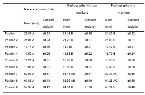
In the morphometric analysis, when the standard deviation is considered, there was symmetry between the measurements carried out on both right and left sides, with the exception of measurement 9, as it was the only measurement in the mandibular symphysis region (Tabela 1).
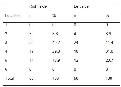
In evaluating the position of the mental foramen in relation to the lower teeth, there was a predominance of position 3 (between the 1st and 2nd premolars), followed by position 4 (in line with the longitudinal axis of the 2nd premolar), position 5 (between the 2nd premolar and 1st molar) and position 2 (in line with the longitudinal axis of the 1st premolar). Positions 1 and 6 were not found in any of the 58 mandibles (Tabela 2).
According to the Friedman statistical test, there was a statistically significant difference (p <0.05) between the gold standard (in situ measurements) and the radiographs with and without markers in measurements 1, 2, 3, 4, 5, 6 and 9, but there was no statistical difference when the radiographs were compared with one another.
For measurements 7 and 8, there was a statistically significant difference (p <0.05) when the radiographs with and without markers were compared with one another, but there were no statistical differences when compared to the gold standard.
Discussion
According to IBGE 9, Brazil is one of the countries with the highest rate of miscegenation, which would indicate the possibility of asymmetry and divergence in the condition being studied. However, according to the data analyzed in this study, when the means and standard deviation are considered, both in situ and in the radiographs with and without markers, there was symmetry between both sides in the majority of the measurements performed when the radiographs were compared with one another.
In assessing the position of the mental foramen in relation to the lower teeth, this study presented a greater prevalence of position 3 (43.2% on the right side and 41.4% on the left), followed by positions 4 (29. 3% on the right and 31.0% on the left), 5 (18.9% on the right and 20.7% on the left) and 2 (8.6% on the right and 6.9% on the left) while positions 1 and 6 (0%) were not visualized. In studies 4,10, positions 3 and 4 were also the most prevalent, but, according to these authors, after positions 3 and 4, the next most prevalent location of the mental foramen would be position 2 (in line with the longitudinal axis of the 1st premolar), followed by positions 5 (between the 2nd premolar and 1st molar) and position 1 (anterior to the 1st premolar), a fact which is at odds with the findings in this study.
Also with regard to the location of the mental foramen in relation to the lower teeth, was reported differences between its position on the right and left sides 2. The mental foramen on the right side is commonly located in positions 3 and 4 and on the left side in position 3. This difference in position between the two sides was not seen in this study, as both the right and left sides presented the same prevalence sequence.
This studies also used panoramic radiographs as they are preferred by many professionals for planning in implant dentistry, especially in the region of the lower premolars 4,11,12,13. These authors claim that panoramic radiography is a flawed exam, as there are distortions inherent to the technique, and this increases the risk of failure and transoperative complications. This can be proved in the analysis of measurements 1, 2, 3, 4, 5, 6 and 9 which presented statistically significant differences, which were overestimated in the panoramic radiographs with and without markers, when compared to the gold standard (measured in situ).
In 2004, among other parameters, Smajilagi assessed the distance from the mental foramen on both sides to the median sagittal plane and found values of 24.05 mm on the right and 24.15 mm on the left side for the Bosnian and Herzegovinan population 5. These same measurements were carried out in this study with the Brazilian population (measurements 1 and 2) where similar values of 23.83 mm on the right and 24.01 mm on the left were found in macerated mandibles.
In 2009, was described the relationship between the mental foramen and mandibular border and alveolar ridge and found both foramina located approximately 12.96 mm from the mandibular base and 12.82 mm from the alveolar ridge 2. In this study, the relationship between the mental foramen and the mandibular base on both sides (measurements 3 and 4) was measured where values of 11.14 mm for the right side and 11.52 mm for the left were found for the macerated mandibles - the gold standard.
In contrast to other study, who found statistically significant differences for measurements from the mandibular foramen to the posterior border of the mandible ramus on both left and right sides, this study showed that there was no such statistical difference between the sides, thereby proving they were symmetrical. However, there was a statistically significant difference when these same measurements (5 and 6), made on radiographs with and without markers were compared to the gold standard 14. These data are consonant with a study which also evaluated the relationship of the mandibular foramen to the posterior ascending branch of the mandible on both sides 15.
In contrast to the study which found that the values of the relationship of the mandibular canal with the posterior border of the ascending branch of the mandible (measurements 5 and 6) were lower in the radiographs with markers than in the gold standard 15, in this study, in the same comparison using radiographs both with and without markers, the averages were higher than those of the gold standard and were statistically significant in this analysis.
According a study 16, the mandibular canal is normally a unique structure with bilateral symmetry. The symmetry they reported was also found in this study, when the standard deviation of measurements 7 and 8 are taken into consideration.
This study also undertook to measure the inter-foramina (measurement 9) in the three assessment modules, where there was a statistically significant difference with a reduction in the averages measured in the radiographic exams when compared with radiographs with and without markers and measurements in situ. This fact is explained by the horizontal distortion factor in the panoramic radiographic image 17.
According to the Friedman test which was performed, there were no statistical differences in the measurements, with the exception of 7 and 8, when the radiographs with and without markers were compared to one another. This data where the use of markers did not influence the location of the anatomical structure being assessed 15.
Conclusion
From the data analyzed, it can be concluded that the anatomical structures in the anterior mandible and posterior ascending branches are easily identified on panoramic radiographs, but do not correspond to the actual location of these structures in situ.
With regard to the location of the mental foramina when compared to the lower teeth, it was concluded that they present a greater prevalence in the region between the lower premolars and the apex of the 2nd pre molar, which demands greater care when using image tests and carrying out possible clinical procedures.
Thus, this study highlights a real need for the use of image tests which provide a more accurate and adequate location of the anatomical structures being studied.
Fontes de Financiamento
Os autores declaram que a pesquisa não recebeu financiamento para a sua realização.
Conflito de interesses
Os autores informam que não houve qualquer potencial conflito de interesse, incluindo interesses políticos e ou financeiros, associados a patentes ou à propriedade, provisão de materiais e ou insumos e equipamentos utilizados no estudo pelos fabricantes.
References
1. Guedes OA, Rabelo LEG, Porto OCL, Alencar AHG, Estrela C. Avaliação radiográfica da posição e forma do forame mentual em uma subpopulação Brasileira. Rev Odontol Bras Central 2011;20(53). [ Links ]
2. Oliveira Júnior EM, Araújo ALD, Da Silva CMF, Sousa-Rodrigues CF, Lima FJC. Morphological and Morphometric Study of the Mental Foramen on the M-CP-18 Jiachenjiang Point. Int J Morphol 2009;27(1):231-238.
3. Moraes MEL, Manhães Júnior LRC, Moraes LC, Medici Filho E, Castilho JCM, Varoli FP, Xavier J. Localização vertical e horizontal do forame mentual em relação ao segundo pré-molar inferior pelo método radiográfico. RGO 2008; 56:47-52.
4. Gungor K, Ozturk M, Semiz M, Brooks SL.A Radiographic Study of Location of Mental Foramen in a Selected Turkish Population On Panoramic Radiograph. Coll Antropol 2006;30(4):801–805.
5. Smajilagi A, Dilberovi F. Clinical and anatomy study of the human mental foramen. Bosnian journal of basic medical sciences 2004; 4 (3): 15- 23.
6. Yosue T, Brooks SL. Study of Location of Mental Foramen. Oral Surg Oral Med Oral Pathol 1989;68:360.
7. Al Jasser NM, Nwoku AL. Radiographic study of the mentalforamen in selected Saudi population. DentomaxillofacRadiol 1998;27(6):341-3.
8. Ngeow WC, Yuzawati Y. The location of the mentalforamen in a selected, Malay population.J Oral Sci. 2003;45(3): 171-5.
9 . I B G E , B r a z i l . h t t p : / / www. i b g e . g ov. b r / h o m e / estatistica/populacao/tendencia_demografica/analise_ populacao/1940_2000/default.shtm
10. Amorim MM, Borini CB, Lopes SLPC, Haiter-Neto F, Caria PHF. Morphological Description of Mandibular Canal in Panoramic Radiographs of Brazilian Subjects: Association Between Anatomic Characteristic and Clinical Procedures. Int J Morphol 2009;27(4):1243-1248.
11. Al-Khateeb T, Al-Hadi Hamasha A, Ababneh KT. Position of the mental foramen in northern regional Jordain population. Surg Radiol Anat 2007; 29(3): 231-7.
12. Sakakura CE, Loffredo LC, Scaf G. Diagnostic agreement of conventional and dinverted scanned panoramic radiographs in the detection of the mandibular canal and mental foramen. J Oral Implantol. 2004; 30(1): 2-6.
13. Moraes MEL, Manhães Junior LRC, Moraes LC, Medici Filho E, Castilho JCM, Varoli, FP, Xavier J. Vertical and horizontal location of the mental foramen in relation to themandibular second premolar by the radiograph method. RGO 2008; 56(1):47-52.
14. Mendoza CC, Vasconcelos BCE, Sampaio G, Caiuás M, Batista JEM. Localização topográfica do forame mandibular: estudo estudo comparativo em mandíbulas humanas secas. Revista de Cirurgia e Traumatologia Buco-Maxilo-Facial 2004; 4(2):137-42.
15. Iwaki LCV, Iwaki Filho L, Silva MC, Candido GC, Kiyochi Júnior HJ, Aita T. Estudo morfométrico da distorção do forame mandibular em radiografias panorâmicas como auxiliar na osteotomia vertical intrabucal do ramo mandibular. RFO, Passo Fundo, v. 16, n. 1, p. 36-42, jan.-abr. 20.
16. Nortj CJ, Farman AG, Grotepass FW. Variations in the normal anatomy of the inferior dental (mandibular) canal: a retrospective study of panoramic Radiographs from 3612 routine dental patients. British Journal of Oral Surgery 1977; 78:55-63.
17. Atieh MA. Diagnostic accuracy of panoramic radiography in determining relationship between inferior alveolar nerve and mandibular third molar. J Oral Maxillofac Surg 2010; 68:74-82.
 Endereço para correspondência:
Yuri Nejaim
Oral Radiology, School of Dentistry of UNICAMP
Av. Limeira, 901 - Areião. 13414-903
Piracicaba, São Paulo/Brazil
e-mail: ynejaim@hotmail.com
Recebido para publicação: 20/05/2013
Aceito para publicação: 25/06/2014
