


Serviços Personalizados
Artigo
 pdf em Inglês
 Artigo em XML
 Referências do artigo
 Enviar este artigo por email
Links relacionados
Compartilhar

 Permalink
IJD. International Journal of Dentistry
versão On-line ISSN 1806-146X
IJD, Int. j. dent. vol.9 no.1 Recife Jan./Mar. 2010
ORIGINAL ARTICLE ARTIGO ORIGINAL
Histological analysis of the actinic cheilitis: an interobserver approach
Análise histologica da queilite actínica: uma abordagem interexaminadores
Fernanda BertiniI; Flávia Celina SgarbiII; Tábata de Mello TeraIII; Adriana Aigotti Habberbeck BrandãoIV; Ana Sueli Rodrigues CavalcanteV
IDDS, PhD Student, Postgraduate Program in Biopathology and Department of Biosciences and Oral Diagnosis, São José dos Campos Dental School, São José dos Campos, São Paulo, Brazil
IIMS, Oral Biopathology - Department of Biosciesce and Oral Diagnosis, School of Dentistry of São José dos Campos, UNESP - São Paulo State University, São José dos Campos -SP - Brazil
IIIMS, Student, Postgraduate Program in Biopathology and Department of Biosciences and Oral Diagnosis, São José dos Campos Dental School, São José dos Campos, São Paulo, Brazil
IVPhD, Professor, Department of Biosciences and Oral Diagnosis -School of Dentistry of São José dos Campos, São Paulo State University)
VPhD, Associate Professor, Oral Medicine, Department of Biosciesce and Oral Diagnosis, School of Dentistry of São José dos Campos, UNESP - São Paulo State University, São José dos Campos -SP - Brazil
ABSTRACT
Actinic Cheilitis (AC) is a clinical entity caused by excessive solar radiation exposure. This disease affects almost exclusively fair-skinned people with outdoor ocupations or those who have activities under prolonged exposure to sunlight. Due to its slow progression, the patient attributes his condition to the ageing process, ignoring its evolutive and malignant nature. The scope of this study is to establish a criteria standardization for being used as a reference in the histological evaluation of the epithelial changes, levels of atypia and connective tissue behavior that compromise the lip epithelium with AC. 48 slides were analyzed by two independent observers with different levels of experience and academic backgrounds. In the connective tissue, solar elastosis was found in 100% of the patients with varied degrees of inflammatory infiltrate and vessel dilatation. The presence of epithelial dysplasia was also seen in the whole sample, but one. The inter-examiners agreemnet degree by Kappa varied from substantial to perfect. We concluded that the inter-examiners previous calibration as well as the criteria standardization helped in the inter-examiner agreement degree, and that the academic background as well as the slide preparation influenced the study.
Key words: Actinic; cheilitis; inter-examiners; epithelial; dysplasia; lips.
RESUMO
A queilite actínica (QA) é uma entidade clínica causada pela excessiva exposição à radiação solar. Esta doença afeta quase exclusivamente pessoas de pele clara que possuem ocupações externas ou aquelas que que fazem atividades expostas por longos períodos à luz solar. Devido à sua lenta progressão, o paciente atribui à sua condição ao processo de envelhecimento, ignorando sua natureza evolutiva e de malignidade. O objetivo deste estudo foi estabelecer uma padronização de critérios para serem usados como referência na avaliação histológica das mudanças epiteliais, níveis de atipia e comportamento do tecido conjuntivo que comprometem o epitélio labial com QA. 48 lâminas foram analisadas por dois examinadores independentes com diferentes níveis de experiência e formação acadêmica. No tecido conjuntivo foi encontrada elastose solar e 100% dos pacientes com graus variados de infiltrado inflamatório e dilatação vascular. A presença de displasia epitelial foi também vista em toda a amostra. O grau concordância inter-examinadores determinado pelo valor de Kappa variou de substancial a perfeito. Concluiu-se que a calibração prévia dos examinadores, assim como a padronização dos critérios auxiliou no grau de concordância inter-examinadores, e que tanto a formação acadêmica quanto a preparação da lâmina influenciaram o estudo.
Palavras-chave: Queilite actínica; inter-examinadores; epitelial; displasia; lábios.
INTRODUCTION
Actinic Cheilitis (AC) was first described in 1923 as a chronic inflammatory disorder of the lips apparently due to actinic or solar rays1.
Manganaro et al.2, defined AC as a premalignant condition that can be focal or diffuse, shows epithelial atrophy or/and erythema and occasionally can present ulcers and vesicles. It affects almost exclusively fair-skinned men with outdoor occupations or those who have activities under prolonged exposure to sunlight3,4, being more frequent on the protrusive and unprotected lower lip5. It is rarely recognized in its early stage and due to its slow progression; the patient attributes his condition to the ageing process that is why it is neglected until it reaches more advanced stages enabling the occurrence of neoplasia6.
Histological, the epithelium of the lip vermillion must be acanthotic, hyperplastic and/or atrophic with several degrees of keratinization and shows disorder in the maturation evidenced by cellular atypias and alteration of mitotic activity7. There is also the proposal for the unification of the "premalignant lesions" classification, taking in consideration the histological pattern and the clinical correlation adopted to the gynecological model histological characterized as intraepithelial neoplásica8.
The medical literature presents countless works that show a constant search in the evaluation agreement or/and interpretation of the epithelial dysplasia degrees, but there is no ideal standard in the study of the epithelial dysplasia degree agreement9-13. The Kappa index is one of the methods used to evaluate inter and intra observers agreement degree11.
AC, for being a premalignant disease strongly involved in the etiology of the lip cancer, with multifocal clinical and histopathologic findings, demands better defined criteria to evaluate the alterations of the epidermis and the subjacent dermis. In an attempt to search for criteria to be used as reference in the histological evaluation of the epithelial alteration, we have carried out an inter-observer preliminary study of the levels of atypia that compromise the lip epithelium with AC.
MATERIALS AND METHODS
48 "Research Subjects" with AC were studied. They were informed that the condition was a premalignant lesion and that the biopsy was indicated to the verification of the tissue histomorphological behavior and, in order to do that, their permission was needed to carry out the procedure of the histopathologic diagnosis, afterward helping in the prevention and following up AC. The place of the biopsy was chosen mainly a white area and an erythematous atrophic area, it means, with mottled aspect, although in most cases the white lesion was predominant.
All the histological cuts were read by two independent observers. We will identify observer 1 as E1 and observer 2 as E2. The gradation of atypia and dysplasia was standardized using the interpretation criteria of the cellular atypia gradation established by the WHO described by Pindborg et al.7 and of the epithelial dysplasia preconized by Banócz, Csiba14.
A record card was used to the register of the histomorphological readings and the inter-observer agreement was studied among the variables through the Kappa test.
The following characteristics of the epithelium were analyzed:
a) keratinization: Absent; Parakeratin: keratin layer in which the epithelial nuclei are retained; Hyperparakeratin: increase of the parakeratin layer thickness; Orthokeratin: keratin layer in which there is loss of epithelial nuclei; Hyperorthokeratin: increase of the orthokeratin layer thickness.
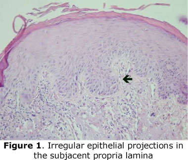
b) covering epithelium changes related to the cellular proliferation. Atrophy: decrease of cell layers and consequently of the epithelium thickness, usually at the cost of the prickle layer; Hyperplasia: noticeable, deep, irregular epithelial projections in the subjacent propria lamina (Fig. 1), increased number of epithelium layers, this change can extend and cover various epithelium layers; Acanthosis: thickening of the prickle layer due to the increased number of cells; Normal: epithelium with normal thickness.
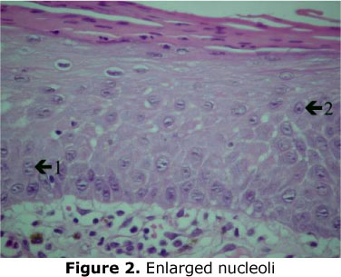
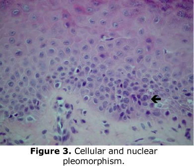
c) histological criteria of epithelial atypia -the presence of cellular changes related to changes in the proliferation and differentiation of the epithelial cells: Loss of basal cell polarity; Duplication of the basal layer; Drop-shaped rete pegs; Increased nucleus/cytoplasm ratio; Nuclear hypercromatism; Enlarged nucleoli (Fig. 2); Increased number of mitotic figures; Atypical mitosis; Mitotic figures in the middle portion of the epithelium; Cellular and nuclear pleomorphism (Fig. 3); Loss of epithelial stratification; Loss of cellular cohesion; Individual keratinization or keratinization in the group of cells in the prickle layer or in deeper layers.
d) connective tissue: presence of solar elastosis: characterized by the basophilic degeneration of collagen fibers.
e) inflammatory infiltrate: it is considered as a result of the immunologic reaction against the lesion: characterized by mild, moderate and severe infiltrate.
RESULTS
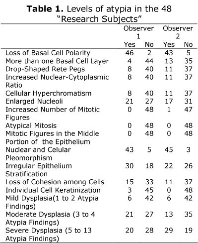
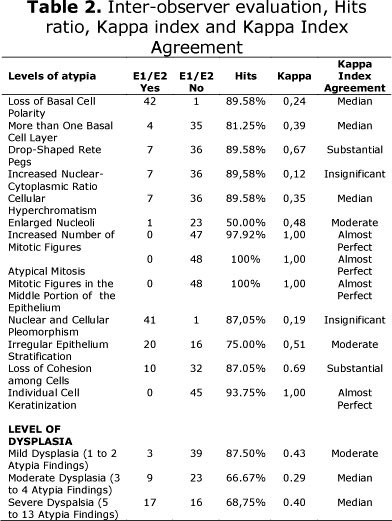
The results of the microscopic readings carried out by observers E1 and E2 are shown on Table 01.
DISCUSSION
The medical literature has shown in the various works involving histopathologic interpretation of the epithelial dysplasia that there is subjectivity in the precognition of the correct diagnosis. In the AC case, because of its multifocal aspect, if we don't improve the calibration of the clinical and histological criteria it will be impracticable to have an appropriate orientation of the patient. The interpretation in the meaning of epithelial dysplasia is consequence of the lack of objectivity in the established criteria evaluation; arbitrary division of the graduations; lack of criteria calibration and graduation and lack of enough knowledge about which criteria is important to the measurement of the malignant potential15.
Individual criteria to the measurement of the malignant transformation in studies of adjacent epithelial dysplasia and oral carcinomas are based on the presumption of the dysplasic findings in this region are truly premalignant16, and the presence of mild to moderate epithelial dysplasia on the borders of the excional oral squamous carcinoma carries a significant risk of local reoccurrence17. If we consider that in the last years we have had as reference of premalignant lesion the presence of dysplasia, these statements confirm a thesis still accepted in the medical literature. However, conditions like reactive hyperplasia (hyperplasia inducted by prosthesis), chronic inflammatory lesion (lichen planus) and benignant neoplasia (papilloma of squamous cells) can show mild degrees of dysplasic findings18 and the presence of inflammation can modify the reliability in the diagnosis of oral lesions because the inflammatory process seems to induct reactive atypia and still be associated with dysplastic alterations and/or reduce the ability of the pathologist in these observations12.
In inter-observer studies, the histopathologic diagnosis of the epithelial dysplasia is variable because some works that evaluate the agreement among pathologists have shown that they themselves don't agree with previous opinions19. In many studies, the Kappa test has been used to evaluate the reliability intra and inter observer when you are studying quantitative and categorical variables20. We have chosen the Kappa test to measure the agreement among the observers because the subjective answers can many times coincide by chance, taking in consideration the premise that the observers have already a preconceived opinion in reason that previously there is a probability to agree or disagree21.
For highly misbalanced situations, the Kappa test will have low results independently of the observers. It occurs because when the marginal totals are asymmetrically disbalanced22, similar result in our research (Table 2) referring to the loss of basal cells polarity in which the inter-observer agreement was of 89,58% and the Kappa index was 0,24, considering the median Kappa index agreement, almost insignificant.
Karabulut et al.13 in 1995, investigated the gradation agreement of the epithelial dysplasia among pathologists with same academic background, and pathologists with different academic backgrounds. They examined one hundred slides of oral leukoplakia, varying from dysplastic absence to Ca in situ. The inter-observer agreement varied from 49% to 69%, and the measured Kappa value increased from 27% to 45%, showing that the agreement level among the pathologists varied from poor to moderate. The authors concluded that the divergences occurred were due to the differences in academic background although there are divergences in the epithelial dysplasia diagnosis even among experienced professionals with the same academic background. Because of that, the previous calibration of the histological aspects in our research was of great importance, specially because the observers involved had different academic backgrounds and work experience. Similar data was observed by Brothwell et al.11.
Odell and Morgan23, declared that several studies of calibration revealed lack of reproducibility in the definition of oral epithelial dysplasia among pathologists in different occasions. In our study, the inter-observer agreement varied from substantial to perfect depending on the histological aspect evaluated. In our study, the inter-observer agreement varied from one end to the other, it means, it varied from insignificant to almost perfect depending of the level of atypia evaluated. It led us to accept the considerations of Warnakulasutriya15 when he says that the interpretation problem in the meaning of the epithelial dysplasia is consequence of the lack of objectivity in the evaluation of the established criteria; arbitrary division of the graduations; lack of criteria calibration and graduation and lack of enough knowledge about which criteria is important to the measurement of the malignant potential.
Epstein et al.24, declared that the histological interpretation is subjective and varies among the pathologists, so this variation can also induct to the improper diagnosis and treatment.
Pinto25, still emphasized the importance of the experience that the observer must have in the identification of the parameters to be studied, once that histotechnical factors can interfere in their evaluation.
Based on the results obtained and based on the medical literature we conclude that the inter-observer previous calibration as well as the criteria standardization help in the inter-observer agreement degree, diminishing the possible lack of reproducibility in the histological definitions among pathologists and also that the same inter-observer academic background as well as the slide preparation can influence the studies involving agreement analysis.
REFERENCES
1. Ayres S. Chronic actinic cheilitis. J Am Med Assoc 1923; 81(14):1183- 6. [ Links ]
2. Manganaro AM, Will MJ, Poulos E. Actinic cheilitis: A premalignant condition. Gen Dent 1997;45(5):492- 94. [ Links ]
3. Stender IM, Wulf HC. Photodynamic therapy with 5-aminolevulinic acid in the treatment of actinic cheilitis. Br J Dermatol 1996;135(3):454-6. [ Links ]
4. Gooris PJJ; Maat B, Vermey A, Roukema JA, Roodenburg JL. Radiotherapy for cancer of the lip. A long-term evaluation of 85 treated cases. Oral Surg Oral Med Oral Pathol Oral Radiol End 1998; 86(3):325-30. [ Links ]
5. Girard KR, Hoffman BL. Actinic cheilitis: report of a case. Oral Surg Oral Med Oral Pathol 1980; 50(1):21-24. [ Links ]
6. Main JHP, Pavone M. Actinic cheilitis and carcinoma of the lip. J Can Dent Assoc 1994; 60(2):113- 6. [ Links ]
7. Pindborg JJ, Reichart PA, Smith CJ, van der Waal I. Histological typing of cancer and precancer of the oral mucosa. Berlin: Springer. 1997; p. 21-31. [ Links ]
8. Küffer R, Lombardi T. Premalignant lesions of the oral mucosa. A discussion about the place of oral intraepithelial neoplasia (OIN). Oral Oncology 2002; 38:125-130. [ Links ]
9. Abbey LM, Kaugars GE, Gunsolley JC; Burns JC, Page DG, Svirsky JA, Eisenberg E, Krutchkoff DJ, Cushing M. Intraexaminer and interexaminer reliability in the diagnosis of oral epithelial dysplasia. Oral Surg Oral Med Oral Pathol Oral Radiol Endod 1995 80:188-191. [ Links ]
10. Abbey LM, Kaugars GE, Gunsolley JC, Burns JC, Page DG, Svirsky JA, Eisenberg E, Krutchkoff DJ. The effect of clinical information on the histopathologic diagnosis of oral epithelial dysplasia. Oral Surg Oral Med Oral Pathol Oral Radiol Endod 1998; 85:74-77. [ Links ]
11. Brothwell DJ, Lewis DW, Bradley GLewis DW, Bradley G, Leong I, Jordan RC, Mock D, Leake JL. Observer agreement in the grading of oral epithelial dysplasia. Community Dent Oral Epidemiol 2003; 31:300-5. [ Links ]
12. Fischer DJ, Epstein JB, Morton HJ, Schwartz SM. Interobserver reliability in the histopathologic diagnosis of oral pre-malignant and malignant lesions. J Oral Pathol Med 2004; 33:65-70. [ Links ]
13. Karabulut A. Reibel J, Therkildsen MH, Praetorius F, Nielsen HW, Dabelsteen E. Observer variability in the histologic assessment of oral premalignant lesions. J Oral Pathol Med 1995; 24:198-200. [ Links ]
14. Bánóczy J, Csiba A. Ocurrence of epithelial dysplasia in oral leukoplakia: analysis and follow-up study of 12 cases. Oral Surg Oral Med Oral Pathol 1976; 42(6):766-74. [ Links ]
15. Warnakulasuriya S. Histological grading of oral epithelial dysplasia: revisited. J Pathol 2001;194:294-7. [ Links ]
16. Wright JM, Shear M. Epithelial dysplasia immediately adjacent to oral squamous cell carcinoma. J Oral Pathol 1985; 14(7):559-64. [ Links ]
17. Weijers M; Snow GB, Bezemer PD, van der Wal JE, van der Waal I. The clinical relevance of epithelial dysplasia in the surgical margins of tongue and floor of mouth squamous cell carcinoma: an analysis of 37 patients. J Oral Pathol Med 2002; 31:11-5. [ Links ]
18. Macdonald DG, Rennie JS. Oral epithelial atypia in denture induced hyperplasia, lichen planus and squamous cell papilloma. Int J Oral Surg 1975; 4:4045. [ Links ]
19. Scully C, Sudbo J, Speight PM. Progress in determining the malignant potential of oral lesions. J Oral Pathol Med 2003; 32:251-256. [ Links ]
20. Cohen J. A coefficient of agreement for nominal scales. Educational and Psychological Meassurement 1960;20:37-46. [ Links ]
21. Abraira V. El índice kappa. Semergen 2000; 27:247-249. [ Links ]
22. Feinstein AR, Cicchetti DV. Hight agreement but low kappa: I. the problems of two paradoxes. J Clin Epidemiol 1990;43(6):543-9. [ Links ]
23. Odell EW, Morgan PR. Oral squamous carcinoma and premalignancy in: _. Biopsy pathology of the oral tissues. London: Chapman & Hall Medical. 1998; p.181-244. [ Links ]
24. Epstein JB, Zhang L, Rosin M. Advances in the diagnosis of oral premalignant and malignant lesions. J Can Dent Assoc 2002; 68:617-21. [ Links ]
25. Pinto DS. Critérios de avaliação do prognóstico -marcadores imuno-histoquimicos - em biópsias de carcinoma epidermoide de boca., Tese (Doutorado com área de Concentração em Patologia), Faculdade de odontologia da Universidade de São Paulo, São Paulo, 1991. [ Links ]
 Correspondence:
Fernanda Bertini
Rua Duque de Caxias, 188 sala 22, Centro
São Sebastião - SP - Brazil, 11600-000
Phones: 55-12-38931882 /FAX: 55-1238931921
Recebido em 22/02/2010
Aprovado em 24/03/2010
