


Serviços Personalizados
Artigo
 pdf em Inglês
 Artigo em XML
 Referências do artigo
 Enviar este artigo por email
Links relacionados
Compartilhar

 Permalink
Revista Odonto Ciência (Online)
versão On-line ISSN 1980-6523
Rev. odonto ciênc. (Online) vol.25 no.4 Porto Alegre Out./Dez. 2010
ORIGINAL ARTICLE
Incidence of second primary oral cancer tumors: a retrospective study
Incidência de segundos tumores primários de câncer de boca: um estudo retrospectivo
Laurindo Moacir SassiI; Onivaldo CervantesII; Juliana Lucena SchusselI; Roberta Targa StramandinoliI; Maria Isabela GueburI; Gyl Henrique Albrecht RamosII
IOral and Maxillofacial Surgery Department, Erasto Gaertner Hospital, Curitiba, PR, Brazil
IIHead and Neck Surgery Department, Erasto Gaertner Hospital, Curitiba, PR, Brazil
ABSTRACT
PURPOSE: Most head and neck malignant neoplasias are diagnosed in advanced stages. Another threatening element to the patients' survival chances and quality of life is the high risk of developing a second primary tumor (SPT). SPT significantly worsens prognosis, and for that reason patients must be monitored for early diagnosis. The main objective of this study was to analyze the occurrence of SPTs in patients with oral squamous cell carcinoma (OSCC) treated in Erasto Gaertner Hospital (EGH), Curitiba, PR, Brazil, in a period of 16 years.
METHODS: Design: retrospective study. The sample comprised patients with OSCC who developed SPT between January 1990 and December 2005. Demographic and clinical data were recorded form the patients' medical charts and analyzed with descriptive statistics.
RESULTS: During this period, 34,637 patients were admitted at EGH. A total of 1,637 (4.4%) patients were diagnosed with OSCC, and 37 (2.7%) developed a SPT. Patients who developed SPT were 29 (78.4%) male and 8 (21.6%) female, with a median age of 58 years old. The most frequent anatomical SPT site was the mouth, oropharynx and oesophagus, with an overall survival of 27%.
CONCLUSION: Patients treated from OSCC must be carefully monitored because of the increased risk of SPT, especially when there is a continuous history of tobacco and alcohol consumption.
Key words: Second primary tumor; squamous cell carcinoma; oral cancer; head and neck neoplasia
RESUMO
OBJETIVO: A maioria das neoplasias malignas de cabeça e pescoço é diagnosticada em estágios avançados. O alto risco de desenvolver um segundo tumor primário (STP) diminui a taxa de sobrevida dos pacientes. STP piora significativamente o prognóstico e estudos sobre ele devem ser realizados para se descobrir seus fatores de risco e melhor forma de tratamento. O principal objetivo deste estudo foi analisar a ocorrência de STP em pacientes com carcinoma epidermóide bucal (CEB) tratados no Hospital Erasto Gaertner (HEG), Curitiba, PRP, Brasil, num período de 16 anos.
METODOLOGIA: Um estudo retrospectivo foi realizado a fim de revisar os pacientes com CEB que desenvolveram STP entre Janeiro de 1990 e Dezembro de 2005. Os dados demográficos e clínicos foram coletados através dos prontuários médicos dos pacientes e foram analisados por estatística descritiva.
RESULTADOS: Neste período, 34.637 pacientes foram admitidos no HEG. Um total de 1637 (4,4%) pacientes recebeu diagnóstico de CEB, dos quais 37 (2,7%) desenvolveram STP. Os pacientes que desenvolveram STP eram 29 (78,4%) homens e 8 (21,6%) mulheres, com média de idade de 58 anos. Os sítios anatômicos mais frequentes para o STP foram a boca, orofaringe e esôfago; a taxa de sobrevivência foi de 27%.
CONCLUSÃO: Pacientes tratados de CEB devem ser examinados cuidadosamente e monitorados regularmente por causa do alto risco de desenvolver um STP, especialmente aqueles que mantêm os hábitos de tabagismo e etilismo.
Palavras-chave: Segundo tumor primário; carcinoma epidermóide; câncer bucal; neoplasia de cabeça e pescoço
Introduction
Second primary tumors (SPTs) have a significantly worse prognosis on patients with head and neck squamous cell carcinoma (HNSCC) (1). Usually they have a bad prognosis, because SPTs occur in important sites, such as the lungs or esophagus or in previously irradiated or operated areas (2). Nowadays, the leading cause of death amongst head and neck cancer patients is the development of a SPT (3,4). The interval between the initial disease and the SPT usually ranges from 2 to 4 years. Frequently, patients who develop SPTs are relatively young, presented a first tumor diagnosed in early stages at sites favourable to treatment, factors which, at the same time, increase the chance both of survival and SPT development (2).
The "field cancerization" hypothesis is considered the key model to explain the biological mechanism leading to second tumors (5,6). In a classical study, Slaughter et al. (6) described features of what they called "field cancerization" suggesting that oral and oropharyngeal cancer can develop precancerous changes in multifocal areas, presenting abnormal tissue surrounding the lesion. They also suggested that both cancers consist of multiple, independent lesions that sometimes coalesce, and the presence of abnormal tissue explains SPT and local recurrence.
The main objective of the present study was to analyze the incidence of SPTs in patients with oral cancer admitted to the EGH, in the city of Curitiba, PR, in the South of Brazil, between January 1990 and December 2005.
Methods
The study protocol of this retrospective study was approved by the institutional review board according to Brazilian regulations and international recommendations. The sample comprised patients diagnosed with oral squamou cell carcinoma (OSCC) that developed SPT, between January 1990 and December 2005, admitted at EGH, in Curitiba.
The eligibility criteria for patients to be included in the present study were: confirmed histopathologic diagnosis of OSCC; development of a SPT; and primary tumor(s) treated through curative protocols – including patients who received palliative radiotherapy with radiation doses equivalent to those of curative radiotherapy. Tumors were classified according to staging system proposed by the UICC (International Union Against Cancer) and the AJCC (American Joint Committee on Cancer). Subjects were excluded if they did not conclude or receive any treatment, those treated in other institutions or patients who underwent only chemotherapy.
Demographic data was collected from patients' medical records, such as: age, gender, risk factors (tobacco and alcohol), staging, topography, treatment and SPT survival. SPTs diagnose was performed following Warren and Gates classification (5), i.e., each neoplasia should present well-defined characteristics of malignancy, be distinct, and the probability of one being a metastasis of the other must be excluded. Multiple neoplasia were classified as simultaneous (tumors diagnosed at the same time), synchronous (tumors discovered within 6 months of their first detection), and metachronous (tumors detected more than 6 months apart). Recurring tumors that appeared either in adjacent areas to surgical incision, or in areas that received radiation, or in lymph nodes located in the drainage area of the first tumor were not included in this study.
Active lesions that appeared from 6 months after treatment at the same site or adjacent area to the index tumor were considered local recurrence. In contrast, residual lesions were considered those that persisted after treatment or, in case of surgical treatment, whereby tissue was compromised, those that reappeared within the surgical margins. Finally, patients' medical records for screened for presence of regional recurrence (i.e., metastases in regional lymph nodes) and distant metastases. Data were analyzed by using descriptive statistics.
Results
From the patients admitted at EGH, during the period from January of 1990 and December 2005, thirty-seven patients developed SPTs, 29 (78.4%) were male and 8 (21.6%) female. The mean age was 58 years-old, ranging from 43 to 86 years-old. As for the risk factors, 31 patients (83.8%) were smokers and 4 (10.8%) non-smokers – for the other two (5.4%) this piece of information was not available. Furthermore, 23 patients (62.2%) reported alcohol consumption.
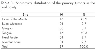
Primary tumors sites are described in Table 1. Most of the patients were diagnosed in advanced stage (13.5% CSIII and 35.1% CS IV), 8 patients (21.7%) were diagnosed in CS I and 11 (29.7%) in CS II.
Concerning the treatment of the index tumor, 7 patients (18.9%) underwent exclusive surgery, 23 patients (62.2%) were treated combining radiotherapy and surgery, 6 (16.2%) received only radiotherapy, and one patient (2.7%) was treated through a combination of surgery, chemotherapy and radiotherapy.
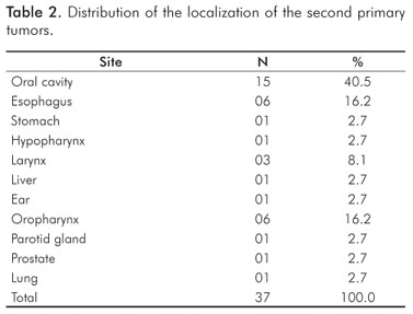
The most frequent site for SPT was the oral cavity in 15 cases (40.5%) (Table 2). The most common oral site for SPT was certainly the tongue, in 55.5% of the cases, followed by the floor of the mouth (22.2%), dental alveolus (11.1%), hard palate (5.6%), retromolar (2.8%) and buccal mucosa (2.8%).
Patients follow-up confirmed that 10 (27%) were alive and did not present any signs of disease; 5 (13.5%) were alive and still suffered from SPT; 16 (43.2%) had died because of the disease; 2 (5.4%) had died but the cause was not mentioned; and another 2 (5.4%) failed to attend follow-up or could not be located by the Department of Statistics and Medical Records of the EGH. The average interval between index tumor diagnosis and SPT was 65 months. Four cases had to be excluded because the patients failed to attend follow-up.
Discussion
SPTs are a life threatening risk for patients diagnosed with HNSCC. They are often fatal, and develop in 10 to 40% of patients with HNSCC (7,8). Tumor topography, particularly larynx, hypopharynx, oropharynx, major salivary glands and nasopharynx, and age can be considered risk factors for SPTs development (9).
The present study found a SPT incidence rate of 2.2% with low survival rates after diagnosis (43.2%). According to Rapport et al. (10), the risk of developing multiple tumors of the upper airways and digestive tract is ten fold high for those patients suffering from HNSCC in comparison to general population. Despite the advances made in the treatment of HNSCC, the incidence of SPT in past decades has not decreased (5). Similarly, little or no improve has been seen in survival rates, which remained at 50% for 5-year survival (2). Rennemo et al. (2) observed the impact of a SPT in 2,000 patients suffering from HNSCC over a 10-year period of time, and concluded that primary tumors located in oral cavity and larynx diagnosed at earlier stages constitute a paramount risk factor for the development of SPT.
The present study showed an average interval between diagnosis of first primary tumor and SPT of 65 months, demonstrating the necessity of long follow-ups with careful screening for new lesions. Indeed, authors suggest that patients may develop SPTs up to 48 months after the diagnosis of the first tumor (2).
Patients with HNSCC history have a greater chance of developing other malignancies due carcinogenic agents exposure (11), particularly tobacco and alcohol (12). SPTs account for about one third of head and neck cancer-related deaths, and 25% of all patients followed in a 10-year period will develop a SPT (13,14). Farshadpour et al. (15) have found greater positivity levels of the protein p53 in the adjacent tumor mucosa in patients suffering from HNSCC associated with tobacco and alcohol consumption when compared to those who were not exposed to risk factors.
The term "field cancerization" was first suggested by Slaughter et al. (6), who defend that certain characteristics in areas adjacent to tumors, may explain or indicate the development of both SPTs and local recurrences. Indeed, when index HNSCC is compared to SPT of the respiratory tract, they usually present identical patterns of genetic alteration (5). Particularly in the head and neck region, the epithelium may present countless genetic alterations, with no histopathological evidences of dysplasia whatsoever. For instance, one may find certain genetic alterations both in resected tumors of HNSCC and in the resection margins even though these very same margins are histologically cancer free (16). The fact that certain genetic alterations remain in histologically benign areas even after a complete tumor resection can be a predictive sign of local flaw or incorrect resection.
Vaamonde et al. (17) found that the oral cavity as most frequent SPT site, similarly to the present study. The most frequent SPT sites were the mouth, oropharynx and esophagus, with an average survival rate of 27%. The oral cavity is an easily accessed site for direct visualization, which could help early diagnosis of oral cancer. A comprehensive understanding of HNSCC SPT and metastasis occurrence must be made to improve patients' prognosis and survival rates. Furthermore, the incidence and fatal course of SPT must be taken into account for patient surveillance.
Acknowledgments
We thank Dr. Sérgio O. Ioshii, Dr. Paola A.G. Pedruzzi and Dr. José Luis Dissenha from the Erasto Gaertner Hospital for technical assistance and intellectual support, and Dr. Onivaldo Cervantes for helpful discussions and mentorship.
References
1. Braakhuis BJ, Tabor MP, Leemans CR, van der Waal I, Snow GB, Brakenhoff RH. Second primary tumors and field cancerization in oral and oropharyngeal cancer: molecular techniques provide new insights and definitions. Head Neck 2002;24:198-206. [ Links ]
2. Rennemo E, Zatterstrom U, Boysen M. Impact of second primary tumors on survival in head and neck cancer: an analysis of 2,063 cases. Laryngoscope 2008;118:1350-6. [ Links ]
3. Vokes EE, Weichselbaum RR, Lippman SM, Hong WK. Head and neck cancer. N Engl J Med 1993;328:184-94. [ Links ]
4. Tsou YA, Lin MH, Hua CH, Tseng HC, Chen SW, Yang SN et al. Survival outcome by early chemoradiation therapy salvage or early surgical salvage for the treatment of hypopharyngeal cancer. Otolaryngol Head Neck Surg 2007;137:711-6. [ Links ]
5. Ha PK, Califano JA. The molecular biology of mucosal field cancerization of the head and neck. Crit Rev Oral Biol Med 2003;14:363-9. [ Links ]
6. Slaughter DP, Southwick HW, Smejkal W. Field cancerization in oral stratified squamous epithelium; clinical implications of multicentric origin. Cancer 1953;6:963-8. [ Links ]
7. Sturgis EM, Miller RH. Second primary malignancies in the head and neck cancer patient. Ann Otol Rhinol Laryngol 1995;104:946-54. [ Links ]
8. Jones AS, Morar P, Phillips DE, Field JK, Husband D, Helliwell TR. Second primary tumors in patients with head and neck squamous cell carcinoma. Cancer 1995;75:1343-53. [ Links ]
9. Bhattacharyya N. An assessment of risk factors for the development of a second primary malignancy in the head and neck. Ear Nose Throat J 2006;85:121-5. [ Links ]
10. Rapoport A, Kowalski LP, Herter NT, Brandão LG, Walder F. Rastreamento, diagnóstico e tratamento do câncer de boca. Assoc Med Bras e Cons Fes Med 2001;1:3-5. [ Links ]
11. Sood S, Bradley PJ, Quraishi MS. Second primary tumors in squamous cell carcinoma of the head and neck: incidence, site, location and prevention. Curr Opin Otolaryngol Head Neck Surg 2000;8:87-90. [ Links ]
12. Tabor MP, Brakenhoff RH, van Houten VM, Kummer JA, Snel MH, Snijders PJ et al. Persistence of genetically altered fields in head and neck cancer patients: biological and clinical implications. Clin Cancer Res 2001;7:1523-32. [ Links ]
13. Leon X, Quer M, Diez S, Orus C, Lopez-Pousa A, Burgues J. Second neoplasm in patients with head and neck cancer. Head Neck 1999;21:204-10. [ Links ]
14. Lin K, Patel SG, Chu PY, Matsuo JM, Singh B, Wong RJ et al. Second primary malignancy of the aerodigestive tract in patients treated for cancer of the oral cavity and larynx. Head Neck 2005;27:1042-8. [ Links ]
15. Farshadpour F, Hordijk GJ, Koole R, Slootweg PJ. Head and neck squamous cell carcinoma in non-smoking and non-drinking patients with multiple tumors: etiologic significance of p53 and Ki-67 in non-tumorous epithelium. J Oral Pathol Med 2008;37:549-54. [ Links ]
16. Mao L, Lee JS, Fan YH, Ro JY, Batsakis JG, Lippman S et al. Frequent microsatellite alterations at chromosomes 9p21 and 3p14 in oral premalignant lesions and their value in cancer risk assessment. Nat Med 1996;2:682-5. [ Links ]
17. Vaamonde P, Martin C, del Rio M, LaBella T. Second primary malignancies in patients with cancer of the head and neck. Otolaryngol Head Neck Surg 2003;129:65-70. [ Links ]
18. Spector JG, Sessions DG, Haughey BH, Chao KS, Simpson J, El Mofty S et al. Delayed regional metastases, distant metastases, and second primary malignancies in squamous cell carcinomas of the larynx and hypopharynx. Laryngoscope 2001;111:1079-87. [ Links ]
 Correspondence:
Juliana L. Schussel
Hospital Erasto Gaertner
Rua Dr. Ovande do Amaral, 201
Curitiba, PR – Brazil
81.520-060
E-mail: julianals24@hotmail.com
Received: June 26, 2010
Accepted: September 30, 2010
Conflict of Interest Statement: The authors state that there are no financial and personal conflicts of interest that could have inappropriately influenced their work.
