


Serviços Personalizados
Artigo
 pdf em Inglês
 Artigo em XML
 Referências do artigo
 Enviar este artigo por email
Links relacionados
Compartilhar

 Permalink
Revista Odonto Ciência (Online)
versão On-line ISSN 1980-6523
Rev. odonto ciênc. (Online) vol.25 no.4 Porto Alegre Out./Dez. 2010
ORIGINAL ARTICLE
Effect of two models of stress associated with ligature-induced periodontitis on hematological parameters in rats
Efeito de dois modelos de estresse associados à periodontite induzida por ligadura sobre parâmetros hematológicos em ratos
Leonardo Stephan CaporossiI; Aurélio Rosa da SilvaI; Tereza Aparecida Delle V. SemenoffII; Fábio Miranda PedroIII; Álvaro Henrique BorgesII,III; Alex Segundo Semenoff-SegundoII
IFaculty of Dentistry, University of Cuiabá, Cuiabá, MT, Brazil
IIDiscipline of Hospital Dentistry, School of Dentistry of University of Cuiabá, Cuiabá, MT, Brazil
IIIDiscipline of Endodontics, School of Dentistry of University of Cuiabá, Cuiabá, MT, Brazil
ABSTRACT
PURPOSE: To evaluate the effect of two models of chronic stress in rats and their association with induced periodontitis on hematological parameters: mean corpuscular volume (MCV), mean corpuscular hemoglobin (MCH), mean corpuscular hemoglobin concentration (MCHC), hematocrit (Ht), erythrocytes (Hm), hemoglobin (Hg) and leukocytes (Lk).
METHODS: Forty-eight adult Wistar rats were randomly distributed into four groups (n=12): physical stress (PSG), variable stress (VSG), ligature (LG) and control (CG) and then started the test of physical stress (restraint and exposure to cold) and variable stress (exposure to flashing light, isolation, examination of the oral cavity, congested environment, the smell of blood and noise). After 10 days of the stress test, the animals in Groups PS, VS and L were anesthetized, and a silk thread was adapted around the upper right second molar; subsequently, the stress test continued for 50 days. The animals were anesthetized and held up the incision and visualization of the posterior vena cava for blood puncture vacuum in tubes containing EDTA. Data were collected by blinded and trained examiners and were statistically analyzed by means of ANOVA and Bonferroni's test at the significance level of 0.05.
RESULTS: The two models of stress changed all of the hematological parameters tested, with the exception of VCM.
CONCLUSION: The stress associated with periodontitis is able to modify blood parameters in rats.
Key words: Stress; rats; periodontitis
RESUMO
OBJETIVO: Avaliar o efeito de dois modelos de estresse crônico - físico e variável - em ratos, associados à periodontite induzida, sobre parâmetros hematológicos: volume corpuscular médio (VCM), hemoglobina corpuscular média (HCM) e concentração hemoglobínica corpuscular média (CHCM) hematócrito (Ht), hemácias (Hm), hemoglobina (Hg) e leucó- citos (Lc).
METODOLOGIA: Selecionaram-se 48 ratas adultas da linhagem Wistar divididas aleatoriamente em 4 grupos (n=12): estresse físico (GEF), estresse variável (GEV), ligadura (GL) e controle (GC). Iniciou-se o ensaio de estresse físico (contenção e exposição ao frio) e estresse variável (exposição à luz piscante, isolamento, exame da cavidade bucal, ambiente congestionado, odor de sangue e barulho). Decorridos 10 dias do início do ensaio de estresse, os animais dos Grupos EF, EV e L foram anestesiados e um fio de seda foi adaptado em volta do segundo molar superior direito, sendo o ensaio de estresse mantido por mais 50 dias. Os animais foram anestesiados e procedeu-se a incisão e visualização da veia cava posterior. Realizou-se a punção sanguínea a vácuo, em tubos com EDTA. Os dados foram coletados por examinadores cegos e treinados e analisados por ANOVA e teste de Bonferroni, ao nível de significância de 0,05.
RESULTADOS: Os dois modelos de estresse alteraram todos os parâmetros hematológicos do estudo com exceção do VCM.
CONCLUSÃO: O estresse associado à periodontite é capaz de modificar parâmetros sanguíneos em ratas.
Palavras-chave: Estresse; ratos; periodontite
Introduction
It is known that there is a link between infectious inflammatory processes in the periodontium and the systemic body. Some studies in humans have shown that the inflammatory process is capable of modifying CBC and markers, such as interleukin 6, C-reactive protein, fibrinogen and other more specific markers (1-3). It is estimated that periodontitis in an adult results in approximately 15 to 20 cm2 of injury in periodontal tissues.
The association between stress and periodontitis in rats has been shown to cause greater destruction of the periodontal structures (4). These findings are consistent with epidemiological studies and current clinical trials (5,6); studies that identify systemic markers are increasingly important, and they can assist in the treatment of both situation, quite common in the population.
From Hans Selye's classical works on stress (7) to current studies (8-11), investigators have noted the ability of stress to change the indicators of the CBC and inflammatory markers. The association between stress and the periodontitis-induced can arise leading to findings that, in the future, may assist in the diagnosis of these two prevalent diseases.
This study aimed to evaluate the effects of physical and variable models of chronic stress in rats and how they are associated with induced periodontitis on hematological parameters.
Methodology
Forty-eight female rats of the species Rattus Novergicus from the Wistar line (average initial weight of 232.81 ± 24.45 g) were selected and underwent a one-week adjustment period to the new environment. The animals were housed in six boxes (polyethylene 16x40x30), with standard chow and water ad libitum under a 12-hour light/dark cycle, controlled temperature of 24 ± 2ºC and relative humidity of 40 ± 4%. The experiment was approved and registered by the university ethics committee for animal experimentation (protocol number 74/05).
Study design
Initially, the animals were randomly distributed into the following groups (n=12 per group): physical stress group (PSG), variable stress group (VSG), ligature group (LG) and control group (CG). The animals of PSG and VSG were subjected to physical stress and variable stress, respectively. After 10 days of stress, the PSG, VSG and LG groups underwent the induction of experimental periodontitis. The CG group received no intervention but was kept in the same environment as the others.
Experimental periodontal disease
For the induction of periodontal disease, the PSG, VSG and LG groups received general anesthesia by intramuscular administration of 0.1 mL of ketamine hydrochloride (Dopalen, Agribrands. Saúde Animal, Paulínia, SP, Brazil) associated with 0.05 mL of ketamine hydrochloride (Rompun, Bayer Saúde Animal, São Paulo, SP, Brazil) per 100 g of body weight. After anesthesia was administered, a sterile suture, thread number 4 (Ethicon, Johnson e Johnson, São Paulo, Brazil), was placed around the right maxillary second molar.
Models of stress
The models chosen for the PSG were immobilization and immobilization with exposure to cold, used alternately six times a week at varying times of day, for two months.
Immobilization: Animals were exposed to an average temperature of 26ºC, placed in PVC (polyvinyl chloride) tubes, consistent with their size and then stopped to pass on both sides with wire. This procedure allowed the respiration of animals, keeping them immobilized, and lasted 4 hours.
Immobilization and cold exposure: The animals were exposed to an average temperature of 7ºC for a period of 4 hours. The immobilization was made in the same manner as described before. The models chosen for the variable stress were exposure to flashing light, isolation for 24 hours, examination of the oral cavity, congested environment, noise and smell of blood (10). One type of stress variable was taken each day.
Exposure to flashing light: The animals were placed in their box-dwelling within a framework that precluded the entry of light. A 60-watt bulb, previously installed with an adapter caused the light to flash intermittently. The animals were subjected to this type of stress for 4 hours.
Isolation: The animals were separated into new, individual boxes-dwelling for a period of 24 hours and maintained with food and water ad libitum.
Ligature examination
The animals were caught one by one and gently restrained by the researcher. After a few moments, the animals calmed down and allowed their oral cavity to be opened. The molar teeth of the rats were inspected with the aid of a spatula number 7.
Congested environment: The animals were grouped (n=12) in a new box for a period of 24 hours and maintained with food and water ad libitum.
Odor of blood: Two plastic test tubes measuring 5 mL with small holes at the top were used. Rat blood with the anticoagulant ethylenediaminetetracetic acid (EDTA) was placed inside the tubes so that it exuded the smell inside the box. This vessel was positioned to prevent the rats from having contact with the product. This procedure lasted 4 hours.
Noise: The animals were placed in their boxes-dwelling within a framework that prevented the entry of light. They were exposed to noise generated by a portable music device at an intensity of 90 decibels for four hours.
Blood sampling
After 60 days of the experiment, the animals were anesthetized using intramuscular injection of 0.1 mL of ketamine hydrochloride (Dopalen, Agribrands. Saúde Animal, Paulínia, SP, Brazil) associated with 0.05 mL of ketamine hydrochloride (Rompun, Bayer. Saúde Animal, São Paulo, SP. Brazil) per 100 g of body weight. After the administration of the anesthesia, the skin and the abdominal wall were incised at the base of the abdomen on the diagonal forming a "V". After the displacement of the flap, the access to the abdominal cavity was obtained. The internal organs were displaced to enable the visualization of the posterior vena cava. Blood was collected by a vein puncture needle 25x7 (Vacutainer – Becton Dickinson, Plymouth, UK) in a 5-mL tube with the anticoagulant EDTA. The sample was thoroughly homogenized.
Hematological processing
The hematological parameters that were analyzed were the following: total count of erythrocytes and leukocytes; hemoglobin; hematocrit determination and the hematological indices, including mean corpuscular volume (MCV), mean corpuscular hemoglobin (MCH) and mean corpuscular hemoglobin concentration (MCHC).
Data were collected by two trained examiners who were blinded with respect to the experimental groups. For hemoglobin measurements, the photocolorimetric method was used by using a spectrophotometer (Femto 700S, São Paulo, SP, Brazil). The sample was diluted in liquid Drakin (Newprov, Pinhais, PR, Brazil), and the calibration was done with a standard solution of hemoglobin (Bioclin-Quibasa, Belo Horizonte, MG, Brazil). To determine the hematocrit, the microhematocrit technique with reading table was used (Coleman, Santo André, SP, Brazil). The hematological indices were calculated from the total count of erythrocytes, hemoglobin and hematocrit determination.
Data analysis
Descriptive statistics, one-way ANOVA and post-hoc Bonferroni's test were used for data statistical analysis. The alpha error was set at 5% for all analyses.
Results
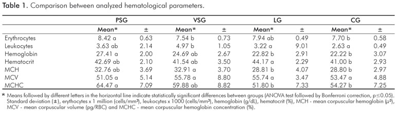
Regarding the erythrocyte count, LG showed a similar count to PSG, VSG and CG, with no statistical difference (p>0.05). For PSG, there were significantly greater differences (p<0.05) when compared with VSG and CG. When comparing VSG and LG, no statistical differences (p>0.05) were observed, as shown in Table 1.
Regarding the total leukocyte count, PSG showed statistical similarity (p<0.05) to VSG, LG and CG. Statistical differences were observed between PSG and LG and CG, with higher counts when compared with LG and CG. There was no statistical difference between LG and CG (Table 1).
In determining the concentration of hemoglobin and MCHC, no differences were observed (p>0.05) between PSG and the others groups. Statistical differences (p<0.05) were demonstrated between PSG, with greater value compared to LG and CG. No statistical difference (p>0.05) was found between LG and CG (Table 1).
There were significant differences (p<0.05) in the hematocrit counts between LG and CG (Table 1). In relation to the erythrocyte index, no statistical difference (p>0.05) was found between PSG and VSG, LG or CG. VSG presented the greater result with statistical difference (p<0.05), compared to LG and CG. LG and CG were not different from each other (p>0.05) (Table 1). The results obtained from the analysis of the hematological parameters showed no statistical differences, with the exception of the MCV index (Table 1).
Discussion
There is strong evidence for the alteration of inflammatory markers and CBC in the presence of generalized perio- dontitis (1-3), and similar condition occurs in patients suffering from stress. In a randomized clinical trial using stressed people, there were greater difficulties in responding to the healing of wounds in the arm (12). This study also highlighted systemic inflammatory markers similar to those presented in periodontitis (8).
The present study results showed that the stress associated with induced periodontitis can modify the normal pattern of hematological parameters. These results are similar to the findings of a recent study in humans (9) that showed psychosocial factors can affect the endothelial inflammatory response by altering inflammatory markers of heart disease (2,9,10). Other data also confirm the link between stress and periodontitis, confirming some similarities between their etiopathogeneses (11).
By the stress models selected in this study it was expected to observe changes in the standardized tests used. The results showed that the two models of stress acted in different ways in the body. In general, it is observed that physical stress causes serious damage to the periodontium (12,13). In the variable stress model, there seems to be a "protective" mechanism in the periodontium in relation to the destruction of structures (4,14). One hypothesis is that in the anatomy of the periodontium, the relationship between the external and internal medium can somehow respond to stress differently than in other parts of the body. These data can be confirmed by the effect of stress on the periodontium and evaluating the immune-inflammatory response around the junctional epithelium. In this analysis, there is evidence of no change in the inflammatory infiltrate during stress associated with ligature-induced periodontitis, which is in agreement with the literature (4,12,13,15).
Stress has three physiological stages. The first stage is reaction to alarm, when the body will quickly return to homeostasis. The second stage is adaptation phase. The aim of the stress model was achieved this second stage, avoiding being closer to the stage of exhaustion, when the mammal's body collapses (7,15). The body weight parameter showed that there were no differences among groups, which indicates that the animals remained healthy during the experiment. The organs that were stressed (thymus, spleen and adrenal glands) were weighed, showing the effectiveness of the stress (data not shown).
Especifically in relation to the hematological variables, only the hematocrit showed modifications in the animals with periodontitis induced by ligature compared with the control group. The ethiopathogenesis is not clear, and the lack of more specific hematological exams is a limitation to interpret these findings (16,17). The hematocrit increase can be related to chronic respiratory disease, which may be linked to the sawdust into the box-dwelling of the rats (18). However, in this experiment, the animal boxes were lined with shredded and sterilized paper, and no clinical symptoms of chronic respiratory disease were observed.
Both models of stress associated with induced periodontitis showed changes in blood variables when compared with the control group. In the experimental groups the hematological parameters were often larger than in those in the negative control group, but with small differences to the positive control group (only periodontitis induction). There is no doubt that changes occurred in the blood organization of the animals during the tests. Possibly, the hemoconcentration elevated some figures in relation to CBC (19,20), but the literature is controversial on this theme (21). Peri- odontitis should be considered an infection with systemic effects (1,10,17), which may be related with the changes in the CBC found in the present study using a quite long stress model.
Female rats were used by convenience, and one may argue if the rat gender could alter the disease susceptibility. In another study using male and female rats to understand the susceptibility to periodontitis, no difference was observed as a function of gender (22). Furthermore, null association with gender was reported in humans (17) and other mammals (23) linking periodontitis and systemic inflammatory markers.
The absence of hormonal markers, such as cortisone and its derivatives, would improve the experimental model used. However, there is evidence that hormonal changes do occur with this amount of stress (4). Another methodological consideration is the non-inclusion of a group of rats subjected only to stress. This decision was based on a similar study in which changes in CBC were not observed (24), so unnecessary animals were not used for ethical reasons. The methodological steps used to minimize errors were: examiner training for blood collection and animal treatment during the tests, close control of the material collected and blinding of the examiner in relation to the experimental and control groups. It is known that stress is a complex issue involving many variables, which require further investigation of the inflammatory parameters to help to translate the animal research findings in useful information for our patients.
Conclusion
The stress associated with ligature-induced periodontitis can modify blood parameters in rats.
References
1. Marcaccini AM, Meschiari CA, Sorgi CA, Saraiva MC, de Souza AM, Faccioli LH et al. Circulating interleukin-6 and high-sensitivity C-reactive protein decrease after periodontal therapy in otherwise healthy subjects. J Periodontol 2009;80:594-602. [ Links ]
2. Loss BG. Systemic markers of inflammation in periodontites. J Periodontol 2005;76:2106-15. [ Links ]
3. D'Aiuto F; Nibali L; Mohamed-Ali V; Vallance P; Tonetti MS. Periodontal therapy: a novel non-drug-induced experimental model to study human inflammation J Periodont Res 2004;39:294-9. [ Links ]
4. Semenoff-Segundo A, Hennemman C, Fontanela VRC, Rösing CK. The role of psychoneuroimmune interactions in the pathogenesis of ligature-induced periodontal disease in Wistar rats. J Inter Acad Periodontol 2007;9:26-31. [ Links ]
5. Genco RJ, Ho AW, Grossi SG, Dunford RG, Todesco LA. Relationship of stress, distress, and inadequate coping behaviors to periodontal disease. J Periodontol 1999;70:711-23. [ Links ]
6. Peruzzo DC, Benatti BB, Antunes IB, Andersen ML, Sallum EA, Casati MZ et al. Chronic stress may modulate periodontal disease. A study in rats. J Periodontol 2008;79:697-704. [ Links ]
7. Selye H. The general adaptation syndrome and the diseases of adaptation. J Clin Endocrinology 1946;6:117-230. [ Links ]
8. Kiecolt-Glasser JK, Marucha PT, Malarkey WB, Mercado AM, Glasser R. Slowing of wound healing by psychological stress. Lancet 1995; 346:1194-6. [ Links ]
9. Fischer JC, Kudielka BM, Von Känel R, Siegrist J, Thayer JF.Joachim E. Fischer Bone-marrow derived progenitor cells are associated with psychosocial determinants of health after controlling for classical biological and behavioral cardiovascular risk factors. Brain, Behavior, and Immunity 2009;23:419-26. [ Links ]
10. Pasceri V, T. Willerson JT, Yeh ETH. Direct proinflammatory effect of C-reactive protein on human endothelial cells. Circulation 2000;102;2165-68. [ Links ]
11. Oikarinen K, Zubaid M, Thalib L, Soikkonen K, Rashed W, Lie T. Infectious dental diseases in patients with coronary artery disease: an orthopantomographic case-control study. J Can Dent Assoc 2009;75:35-35e. [ Links ]
12. Nakajima K, Hamada N, Takahashi Y, Sasaguri K, Tsukinoki K, Umemoto T. Restraint stress enhances alveolar bone loss in an experimental rat model. J Periodontol Res 2006;46:527-34. [ Links ]
13. Takada T, Yoshinari N, Sugiishi S, Kawase H, Yamane T, Noguchi T. Effect of restraint stress on the progression of experimental periodontitis in rats. J Periodontol 2004;75:306-15. [ Links ]
14. Susin C, Rosing CK. Effect of variable moderate chronic stress on ligature-induced periodontal disease in Wistar rats. Acta Odontologica Scandinavica 2003;61:273-7. [ Links ]
15. Selye H. Thymus and adrenals in the response of the organism to injuries and intoxication. Brit J Exper Pathol 1936;17:234-48. [ Links ]
16. Hutter JW, Van der Velden U, Varoufaki A, Huffels RAM, Hoek FJ, Loss BJ. Lower numbers of erythrocytes and lower levels of hemoglobin in periodontitis patients compared to control subjects. J Clin Periodontol 2001;28:930-6. [ Links ]
17. D'Aiuto F, Parkar M, Andreou G, Suvan J, Brett PM, Ready D et al. Periodontitis and systemic inflammation: control of the local infection is associated with a reduction in serum inflammatory markers. J Dent Res 2004;83:156-60. [ Links ]
18. Politi FAZ, Pietro RCLR, Salgado HRN. Caracterização de biotérios, legislação e padrões de biossegurança. Rev Ciênc Farm Básica Apl 2008;29:17-28. [ Links ]
19. Benoit D, Esa L, Ralph G. The driving license examination as a stress model Effects on blood picture, serum cortisol and the production of interleukins in man. Life Sciences 2001;68: 1641-7. [ Links ]
20. Mischler K, Fischer JE, Zgraggen L, Kudielka BM, Preckel D, von Känel R. The effect of repeated acute mental stress on habituation and recovery responses in hemoconcentration and blood cells in healthy men. Life Sciences 2005;77:1166-79. [ Links ]
21. Oishi K, Yokoi M, Maekawa S, Sodeyama C, Shiraishi T, Kondo R. et al. Oxidative stress and haematological changes in immobilized rats. Acta Physiol Scand 1999;165:65-9. [ Links ]
22. Breivik T, Opstad PK, Germo P, Thrane PS. Effects of hypothalamicpituitary-adrenal axis reactivity on periodontal tissue destruction in rats. Eur J Oral Sci 2000;108:115-22. [ Links ]
23. Arrouca ME, Miranda LB, Lopes RS, Takahira RK, Kohayagawa A, Ciarlini PC, et al. Valores hematológicos de capivaras (Hydrochoerus Hydrochaeris) criadas em cativeiro no município de Botucatu, SP. Cienc. Rural 2000;30:813-7. [ Links ]
24. Segundo AS, Hennemann K, Fontanella VR, Rösing CK. The role of psychoneuroimmune interactions in the pathogenesis of ligature-induced periodontal disease in Wistar rats. J Int Acad Periodontol 2007;9:26-31. [ Links ]
 Correspondence:
Leonardo Stephan Caporossi
Rua Brigadeiro Eduardo Gomes, 135
Edifício Rio Sena - apto 901
Cuiabá, MT – Brasil
78045-350
E-mail: leo_caporossi@hotmail.com
Received: January 11, 2010
Accepted: June 23, 2010
Conflict of Interest Statement: The authors state that there are no financial and personal conflicts of interest that could have inappropriately influenced their work.
