


Serviços Personalizados
Artigo
 pdf em Inglês
 Artigo em XML
 Referências do artigo
 Enviar este artigo por email
Links relacionados
Compartilhar

 Permalink
RGO.Revista Gaúcha de Odontologia (Online)
versão On-line ISSN 1981-8637
RGO, Rev. gaúch. odontol. (Online) vol.58 no.4 Porto Alegre Dez. 2010
ORIGINAL ORIGINAL
Influence of OrmocerTM component on the performance of dentin adhesive systems
Influência do componente Ormocer® no desempenho de sistemas adesivos dentinários
Fabrício Mezzomo CollaresI; Fabrício Aulo OgliariII; Vicente Castelo Branco LeituneI; Susana Maria Werner SamuelI,*
IFederal University of Rio Grande do Sul, School of Dentistry, Dental Materials Laboratory. Rua Ramiro Barcelos, 2492, 90035-003, Porto Alegre, RS, Brazil
IIFederal University of Pelotas, School of Dentistry, Department of Operative Dentistry. Pelotas, RS, Brazil
ABSTRACT
OBJECTIVE: Evaluate the influence of OrmocerTM component on the performance of dentin adhesive systems.
METHODS: Microtensile bond strength test in adhesion of adhesive and dentin and characterization of the adhesive interface was done. As control, an adhesive system based on a traditional monomeric system was used. The teeth were embedded in acrylic resin and had their vestibular enamel removed in order to expose the dentin surface closer to the enamel. The exposed dentin was polished with 600 SiC sandpaper for 30s in running water to produce a standardized smear layer. Lower bovine incisors were used in this study, divided in two groups, according the adhesive system used: OrmocerTM based adhesive system and Solobond M adhesive system. On the polymerized adhesive two increments of composite resin were placed to cover the dentin surface completely. After storage for 24 hours in distilled water at 37ºC, the samples were cut to produce two sticks for each tooth, with adhesive interface area of approximately 0.5mm2. Microtensile bond strength values are shown in MPa and fracture analysis with SEM was classified as adhesive, cohesive or mixed.
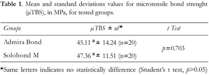
RESULTS: There were no significant differences between OrmocerTM group: 45.11(±14.24) and Solobond M group: 47.36(±11.51), Student's t test (p>0.05). The predominant failure pattern was mixed for the two groups.
CONCLUSION: The incorporation of OrmocerTM into dentin adhesives, in comparison with the conventional adhesive system tested did not influence the immediate bond strength.
Indexing terms: Dentin-bonding agents. Scanning electron microscopy. Tensile strength.
RESUMO
OBJETIVO: Avaliar a influência do Ormocer® no desempenho de adesivos dentinários.
MÉTODOS: Foi realizado o ensaio de microtração para se obter a resistência de união entre o adesivo e a dentina. A caracterização da interface também foi realizada. Como controle, utilizou-se um método de adesivo baseado em um sistema monomérico tradicional. Os dentes foram embutidos em resina acrílica e tiveram o esmalte vestibular removido para expor a dentina superficial que foi abrasionada com lixa d'água granulação 600 em água corrente, por 30 segundos para padronizar uma camada de lama dentinária. Neste estudo, foram utilizados incisivos inferiores bovinos divididos em dois grupos, de acordo com o sistema adesivo empregado: sistema adesivo com Ormocer® e o sistema adesivo convencional Solobond M. Sobre o adesivo polimerizado foram colocados dois incrementos de resina composta, cobrindo toda a superfície da dentina. Após a polimerização da resina, os dentes foram armazenados em água destilada a 37ºC por 24 horas e então seccionados, produzindo dois palitos por dente, com área adesiva de aproximadamente 0,5mm2. Os valores obtidos no ensaio de microtração são mostrados em megapascals e as fraturas analisadas em microscopia eletrônica de varredura são classificadas como adesivas, coesivas ou mistas.
RESULTADOS: Não foi encontrada diferença estatisticamente significativa entre os grupos Ormocer® e Solobond M (p>0,05), sendo os valores de 45,11(±14,24) e 47,36(±11,51), respectivamente. O padrão de fratura foi em sua maioria mista.
CONCLUSÃO: A adição do componente Ormocer® não influenciou na resistência de união imediata à dentina quando comparado a um sistema adesivo convencional.
Termos de indexação: Adesivos dentinários. Microscopia eletrônica de varredura. Resistência à tração.
INTRODUCTION
The achievement of restorative materials and tooth substrate that are resistant to oral cavity stress has been studied for the whole evolution of dentistry adhesive systems1. With the acid etching technique2, associated to the development of the first composite resins3, it became possible to perform a larger variety of procedures, as well as to determine a new less invasive restorative approach.
Acid conditioning has been effectively successful in bonding to enamel. When dentin tissue is involved a definite material has not yet been achieved, especially, when the restoration margins are at dentin4-6. Adhesion at dentin substrate is much more challenging, considering that dentin is a vital heterogenic tissue of low superficial energy, constantly moistened by dentin fluid and closely related to the pulp organ, than enamel7. The acid conditioning of dentin tissue determines the exposition of collagen fibrils of dentin through the removal of hydroxyapatite crystals. This allows the penetration of resin monomers in the collagen matrix and the sealing of dentinal tubules opened by demineralization, which constitutes the best and most effective way to bond a resinous material to the dentin8.
Attempting to improve the performance of adhesive systems at dentin, the OrmocerTM-based material technology was introduced into adhesive systems. This material presents a matrix formed by an inorganic skeleton of Si-O-Si ligations, which a part of polymerizable organic monomers and inorganic filler particles is added9. The aim was to reduce the polymerization shrinkage, and to improve the marginal adaptation10, because higher values of polymerization shrinkage results in stress on the bond between composite and tooth structure11. This stress can results in gaps formation and post-operative sensitivity12-13. The aim of this study was to evaluate the influence of OrmocerTM component on the behavior of dentin adhesives bond strength.
METHODS
Microtensile bond strength
Twenty bovine lower incisors stored at 4ºC in distilled water for less than three months were used. This study was approved by the Research Committee of School of Dentistry, Federal University of Rio Grande do Sul. The teeth were embedded in acrylic resin and had their vestibular enamel removed in order to expose the dentin surface closer to the enamel. The exposed dentin was polished with 600 SiC sandpaper for 30s in running water to produce a standardized smear layer14-15. The teeth were divided in two groups. In the OrmocerTM (OM) group, after the conditioning of the surface with phosphoric acid at 37% for 15s, OrmocerTM-based adhesive Admira Bond Single Dose (Voco, Cuxhaven, Germany) was applied for 15s. In the Solobond M (SO) group, it was used the adhesive system based on a conventional monomeric system Solobond M Single Dose (Voco, Cuxhaven, Germany), which was applied over the surface identically conditioned for 15s. In both groups, the adhesive was polymerized for 10s through a halogen-light photopolymerizing unit (3M Curing Light XL 2500) with light intensity of 550 mW/cm2 previously measured with a radiometer (Demetron, Model 100). On the polymerized adhesive two increments of composite resin (AB: Admira; SB: Top Arabesk) were placed to cover the dentin surface completely. The two increments were photoactivated for 40s each. After storage for 24 hours in distilled water at 37ºC, the samples were cut perpendicular to the adhesive interface, with a refrigerated disk in low rotation (Isomet, Buehler Ltd, Lake Bluff, IL, EUA), thus producing two sticks for each tooth, with adhesive interface area of approximately14-15 0.5 mm2. These sticks had their ends fixed to a device for microtensile tests with cyanoacrylate adhesive. The microtensile test were performed in a universal test machine Emic DL-2000 (Emic, São José dos Pinhais, Brazil) at a speed of 1 mm/min. Bond resistance values were calculated in MPa and differences among the groups were determined by Student's t test. The level of significance was 5%.
The half corresponding to dentin in each sample was removed from the device and put in a dissector for 24 hours at room temperature. The specimens were covered with gold and subjected to fracture mode in scanning electronic microscope. The failure pattern was classified as adhesive, cohesive in dentin and mixed.
RESULTS
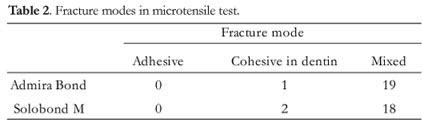
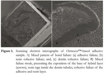
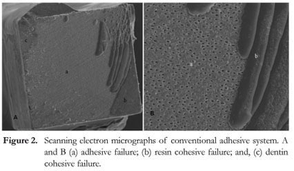
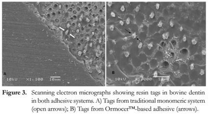
The values for microtensile bond strength, mean and standard deviation, are presented in Table 1. The statistical analysis using Students t test showed no significant differences between the OrmocerTM-based adhesive and the conventional monomeric system groups. The frequency of failure patterns is presented in Table 2. In the fracture mode of OM group, 19 samples presented the mixed pattern of bond failure (Figure 1A). The failure pattern was predominantly mixed and can be observed at higher magnification (Figure 1B), presenting the exposition of the base of hybrid layer, resin tags inside the dentin tubules, cohesive failure of the adhesive and resin layer. In SO group, the mixed failure pattern was also prominent (Figure 2A and 2B). Both the adhesive based on the traditional monomeric system and on the OrmocerTM, demonstrated their ability to form resin tags in bovine dentin (Figure 3A and 3B).
DISCUSSION
The results of this study show that the incorporation of OrmocerTM in a dentin adhesive has not led to higher bond strength, compared with the Solobond M adhesive system. The values obtained in both groups are consistent with other studies that employed adhesives using the acid conditioning of dentin16-19. Like these studies, this work used the microtensile bond strength methodology, which allows better distribution of stress in the bond interface, thus determining a predominance of failure in the resin/dentin interface and obtaining high bond strength values. The portions corresponding to the dentine examined by scanning electron microscopy showed a predominance of mixed failures (adhesive and cohesive in resin and cohesive in dentin). These results suggest that the failure started at the interface resin/dentin or resin/resin which is the weakest bond region20.
Clinical longitudinal studies showed that OrmocerTM based composite resin presents no difference in clinical longevity21-22 compared with traditional hybrid composites. One of the advantages claimed for the OrmocerTM based materials are its low shrinkage which could improve marginal adaptation restorative procedure. Laboratory results showed an increased gap formation of restorations with OrmocerTM based materials and suggested that these results could be due to the OrmocerTM based adhesive system used. Our results showed no significant differences between traditional bonding system and OrmocerTM based materials at immediate bond strength. This increased gap formation could also lead to fluid flow through the adhesive interface leading to pos operative sensitivity. Furthermore, if Admira Bond is unable to prevent the occurrence of the demineralized dentin region not infiltrated by resin it could promote the degradation of the collagen fibrils not encapsulated by the adhesive, and therefore the durability of the bond23. Despite this inability to penetrate this region or to prevent gap formation24, many dentinal tubules were obliterated by resin tags fractured during the microtensile test in both groups. This is important to the pulp protection, due to the prevention of bacteria products penetration. The scanning electron microscopy of beams used for microtensile bond strength showed the tubules in a longitudinal direction, thus confirming the fracture modes findings25-26. The bond quality and its consequent durability are closely related to monomers ability to penetrate the spaces previously occupied by hydroxyapatite, thus encapsulating and protecting the collagen fibrils, which, if exposed, will weaken the bond27.
Dentin organic matrix after demineralization not infiltrated by adhesive is susceptible to be degraded by metalloproteinase matrix (MMP). MMPs are a family of zinc-dependent proteolytic enzymes. These enzymes are present in dentin matrix and are activate by dentin etching and exposure of collagen fibrils23,28. This degradation should be avoided with a collagen layer completely filled by adhesive system or the use enzyme inhibitors, like chlorhexidine23,29. Longitudinal bond strength studies are needed to verify the degradation behavior of these interfaces, if this new chemical structure could avoid the hydrolytic degradation of interfaces. Hydrolytic degradation could lead to a plasticization of polymer matrix increasing the ester bonds degradation. With a more inorganic matrix, this OrmocerTM based materials could avoid this interface degradation.
In this study, no difference was observed at microtensile bond strength with the OrmocerTM component. Some longitudinal researches are needed to verify durability of this system and its resistance against hydrolytic degradation of the polymer and collagen.
CONCLUSION
OrmocerTM-based adhesive did not improve the bond strength to dentin compared to conventional monomeric adhesive system.
Collaborators
FM COLLARES and FA OGLIARI were responsible for conceiving the idea, conducting experimental tests, analyzing the data and writing the manuscript. VCB LEITUNE was responsible for conducting experimental tests, statistical analyses and writing the manuscript. SMW SAMUEL, conceiving the idea and statistical analyses, drafting and made a final review of the manuscript.
REFERENCES
1. Martins GC, Franco APGO, Godoy EP, Maluf DR, Gomens JC, Gomes OMM. Adesivos dentinários. RGO - Rev Gaúcha Odontol. 2008;56(4):429-36. [ Links ]
2. Buonocore MG. A simple method of increasing the adhesion of acrylic filling materials to enamel surfaces. J Dent Res. 1955;34(6):849-53. [ Links ]
3. Bowen RL. Properties of a silica-reinforced polymer for dental restorations. J Am Dent Assoc. 1963;66:57-64. [ Links ]
4. Sano H, Takatsu T, Ciucchi B, Horner JA, Matthews WG, Pashley DH. Nanoleakage: leakage within the hybrid layer. Oper Dent. 1995;20(1):18-25. [ Links ]
5. Paul SJ, Welter DA, Ghazi M, Pashley D. Nanoleakage at the dentin adhesive interface vs microtensile bond strength. Oper Dent. 1999;24(3):181-8. [ Links ]
6. Okuda M, Pereira PN, Nakajima M, Tagami J, Pashley DH. Long-term durability of resin dentin interface: nanoleakage vs. microtensile bond strength. Oper Dent. 2002;27(3):289-96. [ Links ]
7. Li H, Burrow MF, Tyas MJ. Nanoleakage patterns of four dentin bonding systems. Dent Mater. 2000;16(1):48-56. [ Links ]
8. Nakabayashi N, Kojima K, Masuhara E. The promotion of adhesion by the infiltration of monomers into tooth substrates. J Biomed Mater Res. 1982;16(3):265-73. [ Links ]
9. Gouvea CVD, Costa MF, Costa Neto CA, Weig KM, Magalhães Filho TR, Barros RN. Avaliação dos aparelhos fotoativadores utilizados em odontologia. RGO - Rev Gaúcha Odontol. 2008;56(4):399-403. [ Links ]
10. Moszner N, Salz U. New developments of polymeric dental composites. Prog Polym Sci. 2001;26(4):535-76. [ Links ]
11. Braga RR, Ferracane JL. Alternatives in polymerization contraction stress management. Crit Rev Oral Biol Med. 2004;15(3):176-84. [ Links ]
12. Brannstrom M. Communication between the oral cavity and the dental pulp associated with restorative treatment. Oper Dent. 1984;9(2):57-68. [ Links ]
13. Eick JD, Welch FH. Polymerization shrinkage of posterior composite resins and its possible influence on postoperative sensitivity. Quintessence Int. 1986;17(2):103-11. [ Links ]
14. Pashley DH, Ciucchi B, Sano H, Carvalho RM, Russell CM. Bond strength versus dentine structure: a modelling approach. Arch Oral Biol. 1995;40(12):1109-18. [ Links ]
15. Watanabe I, Nakabayashi N, Pashley DH. Bonding to ground dentin by a phenyl-P self-etching primer. J Dent Res. 1994;73(6):1212-20. [ Links ]
16. Sano H, Shono T, Sonoda H, Takatsu T, Ciucchi B, Carvalho R, et al. Relationship between surface area for adhesion and tensile bond strength-evaluation of a micro-tensile bond test. Dent Mater. 1994;10(4):236-40. [ Links ]
17. Phrukkanon S, Burrow MF, Tyas MJ. Effect of cross-sectional surface area on bond strengths between resin and dentin. Dent Mater. 1998;14(2):120-8. [ Links ]
18. Ceballos L, Camejo DG, Victoria Fuentes M, Osorio R, Toledano M, Carvalho RM, et al. Microtensile bond strength of total-etch and self-etching adhesives to caries-affected dentine. J Dent. 2003;31(7):469-77. [ Links ]
19. Hashimoto M, Ohno H, Sano H, Kaga M, Oguchi H. In vitro degradation of resin-dentin bonds analyzed by microtensile bond test, scanning and transmission electron microscopy. Biomaterials. 2003;24(21):3795-803. [ Links ]
20. Armstrong SR, Keller JC, Boyer DB. Mode of failure in the dentin-adhesive resin-resin composite bonded joint as determined by strength-based (muTBS) and fracture-based (CNSB) mechanical testing. Dent Mater. 2001;17(3):201-10. [ Links ]
21. Bottenberg P, Alaerts M, Keulemans F. A prospective randomised clinical trial of one bis-GMA-based and two ormocer-based composite restorative systems in class II cavities: three-year results. J Dent. 2007;35(2):163-71. [ Links ]
22. Bottenberg P, Jacquet W, Alaerts M, Keulemans F. A prospective randomized clinical trial of one bis-GMA-based and two ormocer-based composite restorative systems in class II cavities: Five-year results. J Dent. 2009;37(3):198-203. [ Links ]
23. Pashley DH, Tay FR, Yiu C, Hashimoto M, Breschi L, Carvalho RM, et al. Collagen degradation by host-derived enzymes during aging. J Dent Res. 2004;83(3):216-21. [ Links ]
24. Kijsamanmith K, Timpawat S, Harnirattisai C, Messer HH. Micro-tensile bond strengths of bonding agents to pulpal floor dentine. Int Endod J. 2002;35(10):833-9. [ Links ]
25. Kournetas N, Chakmakchi M, Kakaboura A, Rahiotis C, Geis-Gerstorfer J. Marginal and internal adaptation of Class II ormocer and hybrid resin composite restorations before and after load cycling. Clin Oral Investig. 2004;8(3):123-9. [ Links ]
26. Ogata M, Okuda M, Nakajima M, Pereira PN, Sano H, Tagami J. Influence of the direction of tubules on bond strength to dentin. Oper Dent. 2001;26(1):27-35. [ Links ]
27. Sano H, Yoshikawa T, Pereira PN, Kanemura N, Morigami M, Tagami J, et al. Long-term durability of dentin bonds made with a self-etching primer, in vivo. J Dent Res. 1999;78(4):906-11. [ Links ]
28. Tjaderhane L, Larjava H, Sorsa T, Uitto VJ, Larmas M, Salo T. The activation and function of host matrix metalloproteinases in dentin matrix breakdown in caries lesions. J Dent Res. 1998;77(8):1622-9. [ Links ]
29. Hebling J, Pashley DH, Tjaderhane L, Tay FR. Chlorhexidine arrests subclinical degradation of dentin hybrid layers in vivo. J Dent Res. 2005;84(8):741-6. [ Links ]
Received on: 13/5/2009
Final version resubmitted on: 14/10/2009
Approved on: 15/12/2009
* Correspondence to: SMW SAMUEL . E-mail: <samuelsp@adufrgs.ufrgs.br>
