Serviços Personalizados
Artigo
Links relacionados
Compartilhar
RGO.Revista Gaúcha de Odontologia (Online)
versão On-line ISSN 1981-8637
RGO, Rev. gaúch. odontol. (Online) vol.59 no.4 Porto Alegre Out./Dez. 2011
ARTIGO ORIGINAL / ORIGINAL ARTICLE
Influence of food supplementation with omega 3 on the remission of centrally mediated chronic myalgia
Influência da suplementação alimentar com ômega 3 na remissão da mialgia crônica centralmente mediada
Karine Aparecida Gois GUIMARÃESI; Josemar Parreira GUIMARÃESI ;Nádia Rezende Barbosa RAPOSOI
IUniversidade Federal de Juiz de Fora, Faculdade de Odontologia.
ABSTRACT
Objective
To evaluate the influence of food supplements with fatty acid omega 3 on the remission of a neurogenic inflammation denoted as centrally mediated chronic myalgia.
Methods
24 patients were selected between the ages of 19 and 42, divided into the Experimental Group (n = 12) and the Control Group (n = 12). The patients were evaluated for spontaneous and provoked pain, respectively, by means of a visual analog scale and palpation of the temporal and masseter muscles; they were also evaluated by means of pressure algometry in order to check muscular sensitivity. The data were recorded at T0 (start), T7 (seven days), T14 (fourteen days), T21 (21 days) and T28 (28 days) after the application of the methodology. The patients in the Experimental Group, simultaneously with the use of the neuromuscular myorelaxant splint, were supplied with 1 g of fish oil capsules, three times a day. The Control Group used only the neuromuscular myorelaxant. In order to monitor the omega 6 / omega 3 ratio, blood collections were performed in the Experimental Group, prior to therapy and again after using the supplement for 28 days. The analysis of the fatty acids was conducted using gas chromatography.
Results
The results demonstrated an improvement in pain in both groups, there being a significant statistical difference between them only for the evaluation via Pressure Algometry. The ingestion of omega 3 was effective for obtaining a better proportion of omega 6 and omega 3 and with the reduction in pro-inflammatory cytokines.
Conclusion
The anti-inflammatory potential of fatty acid omega 3 and also the effectiveness with pain remission was confirmed.
Indexing terms: Fatty acids. Pain. Supplementary feeding.
RESUMO
Objetivo
Avaliar a influência da suplementação alimentar com ácido graxo ômega 3 na remissão de uma inflamação neurogênica denominada de mialgia crônica centralmente mediada.
Métodos
Foram selecionadas 24 pacientes com idade entre 19 e 42 anos, divididas em Grupo Experimental (n = 12) e Controle (n = 12). Os pacientes foram avaliados quanto à dor espontânea e provocada, respectivamente, por meio de escala visual analógica e palpação dos músculos temporal e masseter; também foram avaliados por meio de algômetro de pressão para verificação da sensibilidade muscular. Os dados foram registrados em T0 (início), T7 (sete dias), T14 (14 dias), T21 (21 dias) e T28 (28 dias) da aplicação da metodologia. Foram fornecidas aos pacientes do Grupo Experimental, concomitantemente ao uso de férula neuromiorrelaxante, cápsulas de 1g de óleo de peixe, três vezes ao dia. O Grupo-Controle fez apenas uso da férula neuromiorrelaxante. Para monitorar a relação de ômega 6 e ômega 3, foram realizadas, no Grupo Experimental, coletas de sangue antes da terapêutica e após 28 dias do uso da suplementação. A análise dos ácidos graxos foi realizada por cromatografia gasosa.
Resultados
Os resultados demonstraram melhora da dor em ambos os grupos, havendo diferença estatisticamente significante entre os mesmos somente na avaliação por Algometria de Pressão. A ingestão do ômega 3 foi efetiva para se obter uma melhor proporção de ômega 6 e ômega 3 e na redução de citocinas pró-inflamatórias.
Conclusão
Foi comprovado o potencial anti-inflamatório do ácido graxo ômega 3 e também uma efetividade na remissão da dor.
Termos de indexação: Ácidos graxos. Dor. Suplementação alimentar.
INTRODUCTION
The sparse or unbalanced consumption of certain nutrients can exacerbate pain and inflammation. Therefore, research studies have suggested safer, more natural alternatives for the treatment of the signs and symptoms associated with these conditions, such as supplementing diet with essential fatty acids1.
Western diets favor the intake of a large quantity of omega 6 (n-6) as opposed to omega 3 (n-3), and this may be the primary cause of several chronic diseases that afflict the population2-3.
N-6 predominantly possesses linoleic acid which is metabolized into arachidonic acid (AA); with n-3 on the other hand, we get α-linolenic acid (ALA), metabolized into eicosapentaenoic acid (EPA) and docosahexaenoic acid (DHA)4.
The proper proportions of these acids, n-3 and n-6, in the diet, is essential for maintaining a good cellular metabolism and have an influence on various biological processes5; they can reduce the risk of diseases such as hypertension and cardiac problems4,6-9, and influence other chronic diseases such as rheumatoid arthritis; they improve mental health; in addition to the fact that this equilibrium in a 3:1 proportion is effective in controlling pain and inflammation10.
Centrally-mediated chronic myalgia is a chronic muscular disorder characterized by neurogenic inflammation and responds to treatment with antiinflammatory drugs11.
In this sense, the aim of the present study has been to evaluate the effects of omega-3 dietary supplements in the remission of pain in patients with centrally mediated chronic myalgia.
Painful facial diseases, of musculoskeletal origin, of the masticatory apparatus are grouped under the denomination Temporomandibular Disorders (TMD). Muscular abnormalities are pointed to as being one of the main causes of chronic pain and non-dental pain in the orofacial region12.
The treatment of TMD can be carried out by way of occlusion, drug, physical or psychological therapies. Parafunctional habits, stress and muscular hyperactivity appear to be involved in its cause. Almeida et al.13 conducted a study with the aim of comparing the effectiveness of occlusion therapy, using a stabilizing plate and pharmacology, by means of the use of antiinflammatory drugs, as well as the interaction of these
two behaviors in the remission of TMD-related cephalies. Patients were divided into three groups: Group "A": comprising patients who used a stabilizing plate; Group "B": composed of patients who used anti-inflammatory drugs for 20 days; Group "C": composed of patients who made use of both forms of therapy, under the same conditions as the other two groups. The patients made use of the plate for a period of 45 days and were evaluated 30 days after the conclusion of the therapy. Statistical analysis of the results made it possible to verify that the use of the stabilizing plate, whether or not in combination with antiinflammatory medication, showed a better behavior than the use of anti-inflammatory drugs by themselves.
Masticatory muscle dysfunctions are classified as either acute or chronic. Chronic pain may be linked to behavioral and psychosocial impairment14. Moreover, it can affect the Central Nervous System (CNS) and lead to certain muscular responses, such as a profile of myofascial pain. Another muscular disorder influenced by the Central Nervous System is referred to as centrally mediated chronic myalgia. This condition is dependent upon antidromically produced neurogenic inflammation (a condition in which the nervous stimulus moves in an opposite direction to normal) via central mechanisms11.
Centrally mediated chronic myalgia is a continuous chronic muscular disturbance predominantly originating from the effects of the Central Nervous System, which are felt peripherally in the muscle tissue. It is not characterized however by the classic signals associated with inflammation (hyperemia, edema). However, this clinical disturbance presents characteristics that are close to other inflammatory dysfunctions and responds to treatment with anti-inflammatory drugs11.
This pathology in a jaw-raising muscle (temporal, masseter and medial pterygoid) is accentuated during the movement of opening due to the stretching of the muscles and during chewing and occlusion of the teeth, due to the contraction of the painful muscle15.
The most common cause of centrally mediated chronic myalgia is prolonged muscular sensitivity. The longer the patient complains of myogenic pain, the greater the probability of developing muscular disturbance. The muscles are very sensitive to palpation and structural dysfunction is common. Another common clinical characteristic is the prolonged duration of the symptoms11.
The sensation of pain is not a single entity. As with other sensations (palate or temperature), pain can be described by the degree of intensity and unpleasantness, discomfort or anxiety16.
Rizzatti-Barbosa et al.17 concluded, in their study, that visual analog scales are effective in offering a practical contribution to the multidimensional evaluation of chronic pain and to the degree of unpleasantness that this may give to the patient, as they permit subjective aspects of chronic pain to be evidenced and to link them to the progress of the treatment.
Muscular palpation, however, is the most widely used clinical technique in evaluating muscular pain16,18, but the various Manual Palpation methods carry obvious difficulties: they are subjective and difficult to quantify or to establish a pattern16.
Much evidence has shown that the evaluation of sensitivity to muscle pain can be improved if, instead of using a finger or fingers, the examiner were to use an instrument that applies pressure to a specific area at a constant uniform rate (pressure algometer). Using this method, at the point at which pressure applied by the apparatus generates a painful sensation, the patient informs the professional. As well as obtaining the pressure pain threshold, it produces reliable, valid measurements of the pain threshold, both in patients with various musculoskeletal pain syndromes and in asymptomatic individuals16.
N-3 and n-6 are essential fatty acids because they are not synthesized by the organism and should be obtained through the diet. They are called α-linolenic acid and linoleic acid, respectively19,20.
The consumption of these fatty acids has been diminishing. So there is a need to return to a more physiological n-6/n-3 ratio of around 1-4/1 instead of 16- 20/1 obtained in Western diets6,21.
For a better n-3 and n-6 balance, it is necessary to reduce the use of vegetable oils rich in n-6, replacing them with oils rich in n-3, such as rapeseed oil, an increase in fish intake (two or three times a week) or n-3 supplementation by means of fish oil capsules21. This type of supplementation is a low-cost option and quite acceptable to patients, it being rare to experience any adverse side effects22.
Polyunsaturated fatty acids of the omega 3 family (PUFA n-3) act as antagonists to arachidonic acid23-24, and may affect both natural and acquired immunity components, including the production of cytokines, by modulating the amounts and types of eicosanoids produced6,21,24.
N-3 may be used as a therapy for acute or chronic inflammation3, and for disorders that involve the inappropriate activation of the immune response23,25-28.
Epidemiological studies have supported a connection between a diet rich in the consumption of fish and seafood and a fall in the prevalence of depression and other disturbances such as bipolar disorder, personality deviation and schizophrenia25-26.
Both the consumption of fish and high n-3 blood concentrations are linked to a reduction in the risk of cardiovascular diseases8-9,27.
However, Kew et al.28 conducted a study with the aim of determining the effects of diets enriched with ALA or EPA+DHA in the immunity responsible for the representation of the key functions of human neutrophils, lymphocites and monocites. They came to the conclusion that an ALA intake of less than or equal to 9.5 g per day or EPA + DHA less than or equal to 1.7 g per day, did not change the functional activity of neutrophils, monocites or lymphocites, but did change the composition of fatty acids in the mononuclear cells.
In a study by Wallace et al.29, on human beings, the immunological effects were investigated of ALA and EPA + DHA. Supplementation using ALA increased the EPA level, but not that of DHA.
Supplementation with fish oil reduced the proportions of linoleic acid and arachidonic acid and increased the proportions of EPA and DHA in the plasma phospholipids. They concluded that, with the exception of the production of IL-6, a modest increase in the intake of ALA or EPA + DHA did not influence the functional activity of mononuclear cells. Supplementation with 1.8 g per day of n-3 was reported as being beneficial in the treatment of inflammatory disease of the intestine, eczema, psoriasis and rheumatoid arthritis3,25.
Adverse effects cited include gastrointestinal discomfort with nausea, diarrhea and an aftertaste of fish in the mouth6,19-26.
The findings, in respect of n-3 supplementation, present notable limitations such as small samples, too short a period of monitoring to determine the effects of long-term use, as well as inconclusive research studies19.
Part of the inflammatory process is specifically modulated by prostaglandins and leukotrienes1. The type of prostaglandin or leukotriene produced during an inflammatory response is determined by the composition of lipids of the cellular membrane, which is directly influenced by the type of fats in the diet. By increasing the availability of DHA and EPA in the diet or as supplementation with capsules containing these fatty acids, it is possible that they competitively inhibit the release of arachidonic acid and consequently reduce the production of pro-inflammatory eicosanoids7.
The different activities of the eicosanoids derived from AA as opposed to those derived from EPA are one of the most important mechanisms explaining why PUFA n-3 exhibit anti-inflammatory properties in many diseases10.
In a study performed by Lacerda et al.10, patients of both sexes with a referral for the extraction of enclosed third molars were selected and divided into two groups: Control Group, with no change to diet; and the Experimental Group, in which diet therapy was proposed (rich in n-3). It was found that the balance between the fatty acids n-3 and n-6 obtained through the diet, over a period of 15 days, significantly reduced post-op pain and inflammation.
The evaluation of the intake of fatty acids may be conducted using questionnaires. However, a more accurate method for analyzing omega 3 is gas chromatography, used in various studies to analyze fatty acids in both biological and non-biological environments22.
METHODS
The sample comprised only female patients in order to obtain a homogeneous group and also by virtue of the fact that there is a greater demand for treatment by women, according to the literature11. They were attended in the emergency section of the Diagnostics Service and Orientation of Patients with Temporomandibular Disorder (TMD Service) at the Faculty Of Odontology at the Federal University of Juiz de Fora (in the Brazilian state of Minas Gerais). A total of 24 patients satisfying the criteria were selected for the sample composition, aged between 19 and 42, and who had begun TMD support treatment using neuromuscular myorelaxant splint. These patients were divided into the Experimental Group (n=12) and the Control Group (n=12).
As a specific criterion for the composition of the sample, the patient had to present a clinical profile of centrally mediated chronic myalgia, with constant myogenic pain, present at rest and exacerbated during function, and painful sensitivity to palpation in the masseter and/or temporal muscle11.
The following patients were excluded from the sample: those who used medication with anti-inflammatory and/or analgesic potential, patients with systemic illnesses, pregnant mothers or patients with cognitive deficits who did not correctly follow the proposed methodology were also excluded.
A clinical examination was conducted for diagnostic purposes and an evaluation of the pain level of the patients. The patients received information about the project and once they agreed to participate in this project, they signed the Free and Informed Consent Form to enable participation in the research project.
The patients were evaluated for spontaneous pain by means of visual analog scales and provoked pain by way of digital and instrumental palpation (pressure algometry) of the temporal and masseter muscles. The visual analog scale, which measured the subjective intensity of the pain felt by the patient, is graduated with whole numbers from 0 to 10, where "0" means no pain and "10" is maximum pain felt by the patient.
Pain from palpation was classified as 0 (no pain), 1 (mild pain), 2 (moderate pain) and 3 (severe pain). The palpation was carried out by one examiner using index and middle fingers, exerting pressure for 4 seconds18. A pressure algometer was also used for this evaluation of muscle sensitivity, by providing objective, quantitative documentation of the myalgia15-18.
The data were recorded at T0 (start), and T7 (7 days), T14 (14 days), T21 (21 days) and T28 (28 days) after the application of the methodology.
The patients in the Experimental Group were provided simultaneously with the use of the neuromuscular myorelaxant splint, capsules of 1g of fish oil (containing EPA, DHA and ALA), three times a day (after breakfast, lunch and dinner), for 28 consecutive days. The patients in the Control Group, on the other hand, were instructed not to take foods rich in n-3 and only used the neuromuscular myorelaxant splint.
In order to monitor the n-3/n-6 ratio, two collections of blood were carried out on the Experimental Group by a specialist, by means of an venous puncture using disposable needles and syringes, both at the start and after 28 days of supplement use. This collection took place in the morning, after fasting for 12 hours. The blood samples were stored at -80°C until the point of preparation. The fatty acid esters in the plasma were analyzed via gas chromatography in the Analytical Identification and Quantification Center of the Faculty of Pharmacy and Biochemistry at the Federal University of Juiz de Fora.
The preparation of the sample was carried out (separation of the plasma, lipid extraction and conversion of the fatty acids in methyl esters). The chromatographic analyses were carried out with a gas chromatograph (Agilent Technologies 6890N) equipped with a flame ionization detector and a split-splitless injection system. Helium gas was used as the carrier gas with a flow of 1ml/ min. To separate the compounds (EPA and DHA) the Supelco SP2380 chromatographic column was used (60mm long by 0.25mm internal diameter and 0.25μm porosity). The oven temperature gradient was programmed to go from 80°C for 3 minutes to 175°C (increasing by 10°C/minute) for 10 minutes and then up to 250°C (increasing 3.3°C/minute) for 4 minutes. The temperature of the detector was set at 280°C and the temperature of the injection door at 250°C. Analyses were carried out in split mode (50%) using a Supelco inlet liner with a 4mm internal diameter. Analyses were carried out in duplicate and results were expressed as mean ± standard deviation.
The following inflammatory markers were also analyzed: Interleukin 10 (IL-10), Interleukin 4 (IL-4), Interleukin 6 (IL-6), Interleukin 2 (IL-2) and Tumor Necrosis Factor (TNF). These were simultaneously quantified by way of flow cytometry using a Kit Multiplex (BD™ Cytometric Bead Array - CBA; Bioscience, San Jose, CA, USA). The reading was carried out at a wavelength of 650nm and the results were expressed in pg/ml. The tests were performed in duplicate.
Firstly, a normality test was applied to the data obtained. As a normal data distribution was not found, and also due to the fact that the sample was small (n<30), the data were statistically analyzed by means of nonparametric tests. To evaluate data within the same group, Friedman's test was employed. Comparison between the groups was made using the Mann-Whitney test at a significance level of 5% for all the statistical conclusions using SPSS software, version 13.0.
This study was approved by the Ethics In Research Committee at the Federal University of Juiz de For a, under no. 216/2007, on 27 March 2008.
RESULTS
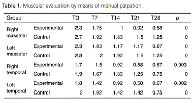
The descriptive values found in the muscular evaluation via Manual Palpation are shown in table 1, where the mean values can be found for the level of pain during palpation of the right-side masseter muscle, leftside masseter muscle, right-side temporal muscle and leftside temporal muscle.
The results showed a statistically significant improvement of the painful condition in the muscles evaluated, both in the Experimental Group and the Control Group, with the right masseter muscle in the Experimental Group presenting the highest rate of improvement.
When performing the statistical analysis to compare the degree of improvement, no statistically significant difference was found between the two groups, despite descriptively observing a slightly better result in the Experimental Group.
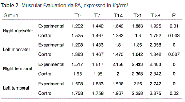
In the muscular evaluation using the Pressure Algometer (PA), the pressure pain threshold was found for the four muscle groups. Similarly to what took place during Manual Palpation, there was an improvement in results where the pressure pain threshold increased for both groups, taking into account the start and end arithmetic means. As for the weekly evaluation, there was a situation of worsening, as in the right masseter muscle in the Control Group, where the pain threshold only rose at the time of the third evaluation (21 days), and also cases where the clinical profile remained unaltered from one week to the next, such as with the right and left temporal muscle in the Control Group from T0 to T7, right temporal muscle in the Experimental Group from T21 to T28 (Table 2).
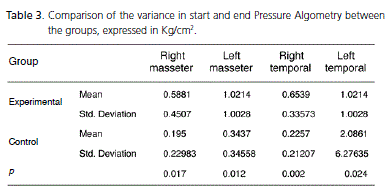
A comparison of the variance between the start and end Pressure Algometry between the groups was also established. When analyzing Tabela 3, it can be seen that the muscles on the left side showed an effective improvement when compared to those on the right, as higher arithmetic means could be found on the right side. The difference between the Experimental and Control groups was statistically significant (p<0.05).
A pain evaluation was also carried out using the visual analog scale, in which the subjective account of the patients' pain was assessed. A positive evolution was found in both the Experimental Group and the Control Group. The results of this descriptive analysis were statistically significant (p<0.05); in the Experimental Group better results were found for narrated pain remission when compared with the Control Group.
A comparison between the groups of the percentage variance of the start and ending visual analog scale was carried out by means of the Mann-Whitney statistical test, though no statistically significant difference was found between them (p=0.242). The mean of the Experimental Group was 0.7924 ± 0.31733 and in the Control Group 0.6965 ± 0.28843, demonstrating that the levels of improvement were very close in terms of the subjective accounts of pain of patients in both groups.
When the fatty acids present in the blood plasma were analyzed for the Experimental Group, it was possible to see that the mean EPA and DHA values had increased, in terms of statistical significance (p<0.005). The AA values on the other hand decreased, demonstrating that supplementation with the n-3 capsule was effective in establishing a more balanced proportion of n-6/n-3. The results were expressed based on the percentage of total fatty acids (Table 4).
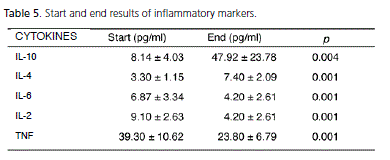
Table 5 shows the result of the mean values (pg/ ml) of the start and end inflammatory mediators (IL-10, IL-4, IL-6, IL-2 and TNF) and the standard deviations. A number of the standard deviations were high because the variance between patients was large. The pro-inflammatory mediators IL-6, IL-2 and TNF showed a reduction while the anti-inflammatory drugs showed a statistically significant increase (p<0.005), demonstrating the effectiveness of the n-3 in modulating the expression of pro- and antiinflammatory cytokines.


DISCUSSION
Centrally mediated chronic myalgia is not characterized by all of the cardinal signs of inflammation, principally the classic signs (hyperemia, edema), which could be observed during the period of the study. However, this clinical disturbance presents characteristics close to other inflammatory dysfunctions, and responds to treatment with anti-inflammatory drugs7. Because of this, it was proposed to carry out the study using n-3, quoted by various authors on account of its potential as an anti-inflammatory drug2,6,16,19,21,26, as therapy for cases of chronic inflammation as proposed by Calder23, Calder24, Simopoulos4, however, in this study, its performance was checked in parallel with the use of neuromuscular myorelaxant splints.
The improvement in muscle pain was achieved in the two groups tracked in this study, being greater in the Experimental Group. Although different muscular pain conditions were evaluated, the results of this study are in agreement with the statement by Almeida et al.13, when they reported that there was the same pattern of pain remission in the groups that made use of a stabilizing plate in isolation or in conjunction with the anti-inflammatory drug, however in the group that combined the two types of therapy, the plate and the anti-inflammatory drug, the treatment achieved a greater effect.
As far as adverse effects are concerned, Friedman & Moe19 believed that there was a risk of an increase in bleeding time in doses of fish oil greater than 3 g per day, but have no scientific proof of this. Other adverse effects quoted in the literature include gastrointestinal discomfort with nausea, diarrhea and an aftertaste of fish in the mouth15,22,27. In this study, however, there was no patient complaint of adverse reactions, perhaps because of the low dosage used. This corroborates what Covington6 believed when he argued that adverse effects are dose dependent.
There was no statistically significant difference between Control Group and Experimental Group, despite the observance, in the descriptive analysis, of a better result in the painful muscular condition in the Experimental Group. This difference could be statistically significant if the dosage used had been higher, in agreement with Walser & Stebbins27, who reported that high doses of n-3 (5g of EPA + DHA) would be required to improve the chances of obtaining measurable alterations. Calder24, on the other hand, stated that, with the ingestion of doses above 2.3g per day of EPA + DHA, there could be satisfactory anti-inflammatory effects. The comparison of doses of n-3 quoted by Walser & Stebbins27and Calder24, may not be applicable in the present study, as they deal with referrals for treatment for different purposes, in addition to the use of quite distinct methodologies.
According to Simopoulos21, there exists a need to return to a more physiological n-6/n-3 ratio of around 1-4/1 instead of 16-20/1 obtained at present with western diets. For Andrade et al.30 on the other hand, the PUFA n-3 in the organism, in a n-6/n-3 proportion of between 3:1 and 5:1, it would be necessary to show the beneficial effects. However, this proportion was not achieved in this study. Perhaps because of this, no statistically significant difference was achieved between the groups.
The study by Lacerda et al.10 revealed that the equilibrium between fatty acids n-3 and n-6 obtained through the diet, over a period of 15 days, significantly reduced post-op pain and inflammation in cases of the extraction of enclosed third molars. Patients of both sexes were selected and divided into two groups: Control Group, with no modification to diet and Experimental Group, where therapy with a diet rich in n-3 was proposed. The use of analgesics was reduced for 51% of the patients and 100% in the case of the use of antibiotics. In the group in which diet therapy was employed, there was no report of moderate or intense pain, just mild pain, which was measured by means of a visual analog scale. The result obtained in the study was quite significant, showing a large difference between the groups. The same was not true for this study, perhaps on account of a lower dosage of n-3 being used and due to the fact that Lacerda et al.10 had simultaneously linked the use of vitamin supplements.
Bemrah et al.22 proposed EPA + DHA doses between 3 and 4 g per day in order to have any nutritional benefit. Ross et al.26, on the other hand, by way of a systematic review, concluded that 1 to 2 g per day of n-3 would be required to obtain a positive response. The results of this study do not concur with the authors quoted above since, despite a lower dosage of these fatty acids having been used, beneficial results had been obtained in terms of the reduction of pain and the alteration in inflammatory mediators.
In this study, with its analysis of fatty acids present in the blood, an increase was found in the levels of EPA and DHA, a reduction in AA, an increase in the anti-inflammatory cytokine indices (IL-10 and IL-4) and a reduction in the expression of pro-inflammatory drugs (IL- 6, IL-2, TNF). This variance in the inflammatory mediators is probably the result of the action of the n-316-17,19. The reduction in available AA was responsible for the decrease in the pro-inflammatory mediators, which supports the views of Harris7 and Percival1, where they point to the fact that the AAs are direct precursors of pro-inflammatory mediators. These authors also stated that the type of prostaglandin or leukotriene produced is directly influenced by the type of fats in the diet. These considerations were also verified in this study, in which the increase in the availability of DHA and EPA by means of supplements using capsules containing these fatty acids, competitively inhibited the release of AA and consequently reduced the production of pro-inflammatory eicosanoids.
New anti-inflammatory substances would be those which have the property of selectively blocking the adhesion and migration of leukocytes, interfering with the expression of adhesion molecules, or of modulating the synthesis, release and effects of the main pro-inflammatory cytokines, such as TNF. In the case of the current study, n-3 fits this classification as an anti-inflammatory substance as the expression of cytokines was altered. Accordingly, corroborating the reports of Percival1, there was a tendency towards the predominance of anti-inflammatory mediators over pro-inflammatory ones, responsible for altering the maintenance of the inflammatory process.
The results obtained in this study also approximate those of Andrade et al.30, who demonstrated that the profile of plasma fatty acids altered in the group using the n-3 supplement, there was an increase in EPA and DHA and a reduction in AA. The methodologies applied, however, were different. Andrade et al.30 checked the effects of supplementation with 0.95 g per day of EPA and 0.5 g per day of DHA in men for a longer period of time (45 days).
Contrary to what was found in this study, where there was a reduction in the inflammatory mediators TNF, IL-6 and IL-2, Wallace et al.29, were not able to conclude in their research study any alteration in the production of TNF and interleukins, with the exception of IL-6, with dosages of 0.44, 0.94 or 1.09 g per day of EPA + DHA. The difference in the results may have been due to the researchers investigating the immunological effects of the EPA + DHA in male individuals between the ages of 18 and 39, monitored over 12 weeks.
In the present study, it was noted that all three types of evaluation quoted (visual analog scale, Manual Palpation, PA) were valid for verifying the degree of improvement in the painful condition, within each group. When comparing the Experimental and Control Groups, the difference between these groups was statistically significant in the evaluation with PA, however this did not occur in the Manual Palpation and the visual analog scale. This result concurs with the statements by Fricton & Dubner16, as the measurement was effective in symptomatic patients. The introduction of the use of PA was a way to minimize the subjective nature of Manual Palpation, confirming the reflections of Fricton & Dubner16 and Gomes et al.18.
CONCLUSION
Based on the results obtained in this study, it may be concluded that there was a statistically significant improvement (p < 0.05) in pain, in both the Experimental Group, which used n-3 and a neuromuscular myorelaxant splint, and the Control Group, which only used the latter device. Analyzing the results in a descriptive fashion, pain remission in the Experimental Group was more effective than in the Control Group. In addition, a statistically significant difference (p < 0.05) was obtained between the groups, only in the evaluation of pain via Pressure Algometry.
The use of the proposed n-3 dosage in the study was effective in reducing the n-6/n-3 proportion in the blood, with an increase in the plasma levels of EPA and DHA, and a reduction in arachidonic acid. The dosage ingested also provides a reduction in pro-inflammatory cytokines and an increase in the expression of anti-inflammatory cytokines, proving the anti-inflammatory potential of these polyunsaturated fatty acids.
Collaborators
KAG GUIMARÃES was responsible for the research, data collection, interpretation of results and the writing of the article. JP GUIMARÃES was responsible for the standardization of the article, guidance and editing of the article. NRB RAPOSO was responsible for the guidance during the interpretation of the results, for performing the gas chromatography examination and the editing of the article.
REFERENCES
1. Percival M. Understanding the natural management of pain and inflammation. Clin Nutr Insights. 1999;4(30):1-5. [ Links ]
2. SanGiovanni JP, Chew EY. The role of omega-3 long-chain polyunsaturated fatty acids in health and disease of the retina. Prog Retin Eye Res. 2008;24(1):87-138.
3. Wardhana, Surachmanto EE, Datau EA. The role of omega-3 fatty acids contained in olive oil on chronic inflammation. Acta Med Indones. 2011;43(2):138-43.
4. Simopoulos AP. Symposium: role of poultry products in enriching the human diet with n-3 PUFA: human requirement for n-3 polyunsatured fatty acids. Poult Sci. 2000;79(7):961-70.
5. Candela CG, Lopéz LMB, Kohen VL. Importance of a balanced omega 6/omega 3 ratio for the maintenance of health. Nutr Hosp. 2011;26(2):323-9. 6. Covington MB. Ômega-3 fatty acids. Am Fam Physician. 2004;70(1):133-40.
7. Harris WS. The omega-3 index as a risk factor for coronary heart disease. Am J Clin Nutr. 2008;87(6):1997S-2002S.
8. Christensen JH. Omega 3 polyunsaturated fatty acids and heart rate variability. Front Physiol. 2011;2(84):1-9.
9. Peairs AD, Rankin JW, Lee YW. Effects of acute ingestion of different fats on oxidative stress and inflammation in overweight and obese adults. Nutri J. 2011;10(1):1-22.
10. Lacerda ECD, Silva A, Pientznauer G, Pereira TF. Dietoterapia: o equilíbrio entre ômega 6 e ômega 3 no controle da inflamação e da dor bucofacial. Rev Bras Odontol. 2005;62(3/4):256-9.
11. Okeson JP. Dores bucofaciais de Bell: tratamento clínico da dor bucofacial. 6ª ed. São Paulo: Quintessence; 2006.
12. Siqueira JTT, Teixeira MJ. Dor muscle-esquelética do segmento cefálico. Rev Med. 2001;80(Pt 2):290-6.
13. Almeida FG, Mello EB, Irikura S. Terapêuticas farmacológica e oclusal em cefaléias relacionadas a desordens temporomandibulares. Rev Serviço ATM. 2003;3(1):22-9.
14. Maciel RN. ATM e dores craniofaciais: fisiopatologia básica. São Paulo: Editora Santos; 2003.
15. Lund JP, Sessle BJ. Mecanismos neurofisiológicos. In: Zarb GA, Carlsson GE, Sessle BJ, Mohl ND. Disfunções da articulação temporomandibular e dos muscles da mastigação. 2ª ed. São Paulo: Santos; 2000. p. 188-207.
16. Fricton, JR, Dubner R. Dor orofacial e desordens temporomandibulares. São Paulo: Ed. Santos; 2003.
17. Rizzati-Barbosa CM, Arana ARS, Cunha Jr AC, Morais ABA, Gil I A. Avaliação diária da dor na desordem temporomandibular. Rev Assoc Bras Odontol Nac. 2000;8(3):171-5.
18. Gomes MB, Guimarães JP, Guimarães FC, Neves ACC. Palpation and pressure pain threshold: reliability and validity in patients whith temporomandibular disorders. Cranio. 2008;26(3):1-9.
19. Friedman A, Moe S. Review of the Effects of omega-3 supplementation in dialysis patients. Clin J Am Soc Nephrol. 2006;1(1):182-92.
20. Kurzweil R, Grossman T. A medicina da imortalidade: as dietas, os programas e a inovações tecnológicas que prometem revolucionar nosso processo de envelhecimento. São Paulo: Aleph; 2006.
21. Simopoulos AP. Omega-3 fatty acids in inflammation and autoimmune diseases. J Am Coll Nutr. 2002;21(6):495-505.
22. Bemrah N. Fish and seafood consumption and omega 3 intake in French coastal populations: CALIPSO survey. Public Health Nutr. 2008;12(1):1-10.
23. Calder PC. Polyunsatured fatty acids and inflammation. Biochem Soc Trans. 2005;33(Pt 2):423-7.
24. Calder PC. Polyunsaturated fatty acids, inflammation and immunity. Lipids. 2002;56(3):14-9.
25. Logan AC. Omega-3 fatty-acids and major depression: a primer for the mental health professional. Lipids Health Dis. 2004;3(25):1-8.
26. Ross BM, Seguin J, Sieswerda LE. Omega-3 fatty acids as treatments for mental illness: which disorder and which fatty acid? Lipids Health Dis. 2007;6(21):1-19.
27. Walser B, Stebbins CL. Omega-3 fatty acid supplementation enhances stroke volume and cardiac output during dynamic exercise. Eur J Appl Physiol. 2008;144(3):455-61.
28. Kew S, Banerjee T, Minihane AM, Finnegan YE, Muggli R, Albers R. Lack of effect of foods enriched with plant-or marine-derived n-3 fatty acids on human immune function. Am J Clin Nutr. 2003;77(5):1287-95.
29. Wallace FA, Miles EA, Calder PC. Comparison of the effects of linseed oil and different doses of fish oil on mononuclear cell function in healthy human subjects. Br J Nutr. 2003;89(3):679-89.
30. Andrade PMM, Ribeiro G, Carmo MGT. Suplementação de ácidos graxos ômega 3 em atletas de competição: impacto nos mediadores bioquímicos relacionados com o metabolismo lipídico. Rev Bras Med Esporte. 2006;12(6):339-44.
 Endereço para correspondência:
KAG GUIMARÃES
Universidade Federal de Juiz de Fora,
Faculdade de Odontologia.
Rua Rei Alberto 108, sala 602, Centro,
36016-300, Juiz de Fora, MG, Brasil.
e-mail: kgois@hotmail.com
Recebido: 29/6/2010
Aceito: 16/3/2011


