


Serviços Personalizados
Artigo
 pdf em Inglês
 Artigo em XML
 Referências do artigo
 Enviar este artigo por email
Links relacionados
Compartilhar

 Permalink
RGO.Revista Gaúcha de Odontologia (Online)
versão On-line ISSN 1981-8637
RGO, Rev. gaúch. odontol. (Online) vol.59 no.4 Porto Alegre Out./Dez. 2011
ARTIGO ORIGINAL / ORIGINAL ARTICLE
Pull-out strength of endodontically treated teeth restored with glass fiber posts of different diameters
Resistência à tração de dentes tratados endodonticamente restaurados com glass fiber posts de diferentes diâmetros
Guilherme Pedrosa AMANAJÁS NETOI; Welyton Ramon Soares PINTOI ;Eliza Burlamaqui KLAUTAUI;Bruno Pereira ALVESI
IUniversidade Federal do Pará, Faculdade de Odontologia. Rua Augusto Corrêa
ABSTRACT
Objective
To evaluate the pull-out strength of endodontically treated teeth restored with glass fiber posts through different techniques in enlarged conduits.
Methods
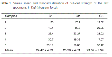
A total of 15 bovine teeth were endodontically treated, divided into three groups. Group 1- restored with number 1 glass fiber posts, Group 2- restored with number 3 glass fiber posts, Group 3- restored with number 3 glass fiber posts associated with accessory glass fiber posts. The pins in all groups were cemented with same self-adhesive resin cement. All test specimens were subjected to the pull-out strength test in the testing machine at a speed of 0.5mm per minute and the results obtained in kilogram-force.
Results
The pull-out strength values were as follows: group 1 - 24.47 kilograms-force; group 2 - 25.28 kilograms-force; group 3 - 23.59 kilogramsforce. The following standard deviations were observed: 4.33 kilograms-force for group 1, 4.03 kilograms-force for group 2 and 8.39 kilograms-force for group 3. No statistically significant differences between groups were found using the ANOVA test.
Conclusion
The decrease in the thickness of the cement film by using pins of larger diameter and/or accessories, does not interfere with the retention and stability prediction of the rehabilitation of the complex.
Indexing terms: Endodontics. Tensile strength. Post and core technique.
RESUMO
Objetivo
Avaliar a resistência à tração de dentes tratados endodonticamente restaurados com glass fiber posts através de diferentes técnicas com condutos alargados.
Métodos
Foram utilizados 15 dentes bovinos, tratados endodonticamente, sendo distribuídos em 3 grupos. Grupo 1- restaurados com pino de fibra de vidro número 1, Grupo 2- restaurados com pino de fibra de vidro número 3, Grupo 3- restaurados com pino de fibra de vidro número 3 associados com pinos acessórios. Os pinos de todos os grupos foram cimentados com o mesmo cimento resinoso auto-adesivo. Os espécimes foram submetidos ao teste de resistência a tração na máquina de ensaios a uma velocidade de 0,5 milímetros por minuto e os resultados obtidos em quilograma-força.
Resultados
Os valores de resistência a tração obtidos foram: Grupo 1 - 24,47 quilograma-força; Grupo 2 - 25,28 quilograma-força; Grupo 3 - 23,59 quilograma-força. Foram verificados os seguintes desvios-padrão: 4,33 quilograma-força para o Grupo 1, 4,03 quilograma-força para o Grupo 2 e 8,39 quilograma força para o Grupo 3. Sem diferença estatística significante entre os grupos pelo teste de ANOVA.
Conclusão
A diminuição da espessura da película de cimento com o uso de pinos de maior diâmetro e/ou acessórios não interfere de maneira efetiva no prognóstico da retenção e estabilidade do complexo reabilitador.
Termos de indexação: Endodontia. Resistência a tração. Técnica para retentor intra-radicular.
INTRODUCTION
The ever-growing pursuit of esthetics creates a need to improve materials and techniques in Odontology. As far as oral rehabilitation is concerned, this quest is significantly more accentuated. To this end, and as part of an evolutionary process, it is necessary for advances to be taking place in the field of esthetics that go hand in hand with the structural, functional piece. Caries, erosion, abrasion, prior restorations, endodontic traumas and accesses may all have a negative impact on all, or at least a large part of, the crown structure, so much so that reconstruction becomes a challenge for oral rehabilitation, resulting in the need for the use of intra-radicular posts, with the aim of increasing stability and resistance to the fracture of the remnants1-2. Factors such as structural quantity, clinical adaptation, health of the supporting tissue, esthetics and the prognosis for restoration are fundamental to the success and longevity of rehabilitation in endodontically treated teeth3. For a long time, cast metal cores were used as the technical solution for the reconstruction of endodontically treated teeth, however characteristics such as preparation which is far from being conservative, rigidity far superior to that of dentine, the need for the laboratory stage, probability of corrosion and retention impaired by lack of adhesion to the remnant tooth, has led to studies that have developed other retainers3-6. As an alternative to cast metal cores, non-metallic posts emerged, such as those made from zirconium, ceramics, carbon fiber and also glass fiber based posts1.
Glass fiber posts have been shown to be quite a viable alternative as they possess characteristics such as an elasticity module similar to that of dentine, they are biocompatible, they distribute masticatory force better, are highly durable, resistant to corrosion, do away with the laboratory stage and are esthetically superior as they have optical properties that provide greater translucence to the dental core4,7-9. Another notable aspect is the possibility of using resin cements that present dual polymerization, since they may be activated both physically and chemically as the retention may also be influenced by cementation10-11. In the sense of increasing pull-out strength and fracture resistance, glass fiber accessory posts have been used, however, as far as pull-out strength is concerned, further exploration is required if the use of posts with different diameters and glass fiber accessory posts result in considerable alterations12.
The aim of this study was to evaluate the pull-out strength of teeth treated endodontically with enlarged conduits restored with glass fiber posts of different diameters, in conjunction, or not, with accessory posts, and using the same dual polymerized cement.
METHODS
15 clean bovine central incisors were used with a periodontal curette (Trinity, São Paulo, Brazil), then sterilized in autoclave and stored in distilled water at a temperature of 37ºC, prior to and during the course of the experiment, thereby preventing dehydration. The specimens were marked 17mm from the root apex, corresponding to the mean length of the roots of the human upper central incisors and cut transversely with a double-faced diamond disc (KG Sorensen, São Paulo, Brazil), eliminating the crown and a part of the root. The cervical surfaces were filed using sandpaper under refrigeration, in order to obtain a surface perpendicular to the long axis of the tooth.
The teeth were treated endodontically using conventional techniques with the working length of 16mm, and cervical preparation being carried out with Gates-Glidden and Largo (Dentsply Maillefer, Ballaigues, Switzerland) drill bits, numbers 1 and 2, instrumentation up to K-file no. 80 (Dentsply Ind e Com., Petrópolis, Brazil), irrigation with 10ml of 0.5% sodium hypochlorite (Líquido de Dakin, Iodontosul, Porto Alegre, Brazil), and 10ml of 17% EDTA-T (Inodon, Porto Alegre, Brazil) as a chemical substance to assist with the final irrigation for 60 seconds. Subsequently, they were filled with no. 80 primary gutta-percha cones and accessories (Dentsply Ind e Com., Petrópolis, Brazil) and endodontically cemented (Sealer 26, Dentsply Ind e Com., Petrópolis, Brazil), using the lateral and vertical condensation technique, temporarily sealed (Coltosol - Vigodent, Bonsucesso, Brazil), and then stored for 24 hours in distilled water in an oven at 37ºC.
The removal of the gutta-percha from the canals was performed using Largo drill bits nos. 3 and 4 (Maillefer, Ballaigues, Switzerland) until 5mm of filling material was obtained in the apical region, followed by vertical condensation for the adaption of the filling to the canal. The root conduit was then expanded using drill bit 4137 (KG Sorensen, São Paulo, Brazil) as far as the active body. The 15 teeth were inserted into PVC tubes 40mm long and 12mm wide with self-polymerizing colorless acrylic resin(Vipi Flash, Pirassununga, Brazil), so that the long axis of the roots was perpendicular to the horizontal plane and centered in the block.
The present study was evaluated and approved by the Ethics Committee in Research with Animals (CEPAN) at the Evandro Chagas institute, approval opinion (Nº 008/2010/CEPAN/IEC/SVS/MS) under procedural record CEPAN - 002/2010.
Composition of the groups
The 15 test specimens, already with the filling removed to 5mm and enlarged, were divided at random depending on the restoration technique to be applied.
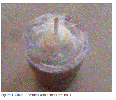
Group 1 - comprising five test specimens restored using a primary no. 1 glass fiber post (Reforpost - Ângelus, Londrina, Brazil). The specimens were irrigated with 17% EDTA for 1 minute and then washed copiously with distilled water, the excess being removed with absorbent paper cones. The posts were degreased using 96% alcohol, and subsequently applied with silane (Ângelus, Londrina, Brazil) over the entire surface with the help of a brush. After waiting for one minute, the silanized posts were gently air dried. For the cementation, the self-adhesive resinous cement RelyX U100 (3M, Sumaré, Brazil) was selected. Manipulation took place in equal proportions, in accordance with manufacturer's recommendations, and with the assistance of an insertion spatula (Duflex, Rio de Janeiro, Brazil). The post, covered in a layer of cement, was inserted into the conduit with the help of surgical pliers with cement being added to the conduit, so as to obtain the total filling of the conduit. After removing the excess cement, the test specimens were photopolymerized for 60 seconds.
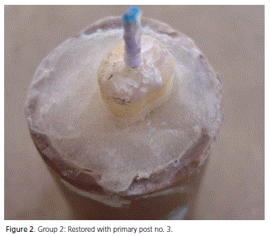
Group 2 - comprising five test specimens restored with no. 3 primary glass fiber post (Reforpost - Ângelus, Londrina, Brazil). The cementation protocol followed that used for group 1.
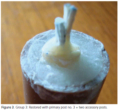
Group 3 - comprising five test specimens restored with no. 3 primary glass fiber post (Reforpost - Ângelus, Londrina, Paraná, Brazil) connected to two accessory glass fiber posts (Reforpin - Ângelus, Londrina, Brazil). The cementation protocol followed that used for the two previous groups, both for the primary post and for the accessory posts.
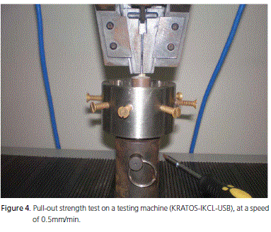
The test specimens for all 3 groups were stored in an oven containing distilled water, at 37ºC, for 24 hours. Once this period of time had elapsed, a pull-out strength test was performed on the 15 test specimens in a testing machine (KRATOS-IKCL-USB), at a speed of 0.5mm/min. The samples were placed in the machine on a device developed to eliminate lateral forces, so as to allow the axis of traction to remain equal all along the tooth axis. After the rupture, the Kgf values were noted and then sent for statistical analysis.
RESULTS
The presented values were ordered and tabulated, from which the groups' arithmetic means and standard deviations were obtained. The data obtained were subjected to the Lilliefors test, in which the normality of the sample was checked. They were then analyzed using the ANOVA variance by means of a criterion, in which no significant difference between the groups was found, with p=0.9034.
DISCUSSION
The success of the retention of the post to the root canal is measured by a series of factors. To eliminate any kind of influence from the cementation technique, the same cementing agent was chosen, namely selfadhesive RelyX U100, due to the fact that it presents the same adhesive resistance values for all of the thirds13, since the adhesive resistance between the dentine walls and the cementing agent is affected by the distribution of the cement along the cervical, medial and apical thirds during the cementation of glass fiber posts14. The weakest link of the adhesive bond is the region at the dentinecement interface, with defects being found in between 50% and 70% of those that commonly occur15. Despite the adhesion between the root dentine and the cementing agent being characterized as the most sensitive bond in the cementation of glass fiber posts, the post/cement and the post/dental core interfaces also require attention16.
The application of silane was used as the bonding agent since the coverage of the post with silane exhibits the highest retention values when compared to other types of surface treatment of the post12. Another important factor for retention is provided by the use of a double polymerization system of adhesion. The resin cement is perhaps not the most important factor for the retention of glass fiber posts in the root canal, as they have similar physical and mechanical properties, but more the use of a double polymerization system of adhesion, seeing as this system has the ability to penetrate into the dentinal tubules and to bond chemically and mechanically with the glass fiber post4.
Due to the need to evaluate if the increase in the post diameter and the use or non-use of accessories interferes with the retention of the glass fiber posts, the aim of the present study was to test the pull-out strength of teeth that are endodontically treated with enlarged conduits, restored using different techniques. In the absence of any significant difference between the values found in our study, it may be said that the use of glass fiber posts of different diameters and the use or nonuse of accessories in the reconstruction of teeth treated endodontically with enlarged conduits, does not interfere significantly with the pull-out strength of intra-radicular forces. This statement is corroborated by other studies that confirm that the increase in the diameter of the post does not represent a significant difference in the increase in retention and moreover makes the tooth remnant more fragile17-19, seeing that the resin cements present adequate retention and resistance even on thicker layers in enlarged conduits20.
The results obtained in this study can also be vouched for by the study in which the pull-out strength was evaluated of teeth restored with glass fiber posts enlarged with drill bits of different diameters and cemented with the same system of adhesion and cementing agent, with no significant statistical differences being found, which also indicated that the adaptation of the fiber posts in the root conduits is not essential for their retention due to the use of resin cements. Therefore, this study supports the idea of the applicability of glass fiber posts in enlarged conduits, in which a thicker layer of cement can be observed, possibly explained by an increase in the dentine surface in contact with the cement15. The same results were obtained when evaluating the effects of alterations in the film of resin cement with prefabricated posts and conduits of different diameters21 and also in the evaluation of the retention of teeth restored with different reinforcements, using the same cement, where there was no significant difference between the groups analyzed. Accordingly, the hypothesis that the use of glass fiber-based accessory posts would reduce the line of cementation and consequently promote better mechanical resistance of the dental retainer/ structure5,22-24 system, was not proven.
Studies can be found in the literature that contradict the results of this research, and despite other studies not obtaining statistically significant differences, the use of a primary post in conjunction with accessories is argued as they point to greater pull-out strength values than the restored teeth with just one primary glass fiber post or with cast metal5. This proposal is underpinned by the idea that this connection works like a single entity that provides an improvement in the adaptation to the prepared conduit and reduces the thickness of the cementing agent, to the detriment of the lack of adaptation of the post in relation to the conduit walls, resulting in a greater need for resin cement, bringing an increase in stress at the adhesive interface during the contraction of polymerization5,22. Prefabricated posts are not ideal as their adaptation is not perfect and the thick layer of the post produces an unfavorable prognosis25. However, prefabricated posts adapt well only in the apical third of the canal, exhibiting poor adaptation in the cervical third, depending on a large quantity of cement to hold the post in place26.
The production of anatomic posts and the use of accessory posts are recommended as they provide a thin cement layer between the post and the conduit wall, promoting retention and preventing adhesive defects, since it increases the amount of fibers23-24.
Despite clinical concerns being greater over the stability of the retainers in the root canals due to the higher failure rates, on account of the ineffective retention of the posts in the root conduit, it is important to underline the resistance to the fracture of endodontically treated teeth with enlarged conduits restored using glass fiber posts. When comparing cast metal cores, primary glass fiber posts, primary glass fiber posts wrapped in a matrix of glass fiber tapes, primary fiber posts allied to accessories and anatomic posts, higher fracture resistance values were found in the groups with cast metal core and those joined to accessory posts26, with the most favorable fracture patterns being found in teeth restored with glass fiber accessory posts25. These results may be explained by the combination of a primary glass fiber post with a variety of accessories in order to produce clinically a better biomechanical behavior in dental structures, by virtue of the reduction in the thickness of the cement line and the use of a larger quantity of material elastically similar to dentine, thereby reducing the rate of catastrophic fracture27.
CONCLUSION
Glass fiber posts are shown to be the best choice for the reconstruction of teeth treated endodontically with enlarged conduits. The reduced thickness of the cement film with the use of posts with larger diameters and/or accessories does not effectively interfere with the prognosis of retention and stability of the rehabilitative complex. Therefore, to attain effective adhesiveness, factors such as adhesive choice and the cementing agent require greater caution.
Collaborators
GP AMANAJÁS NETO took part in the bibliographical survey, the submission of the project to the Bioethics Committee, editing of the article and was responsible for all the laboratory stages, mainly the part involving the cementing of the posts. WRS PINTO took part in the bibliographical survey, the submission of the project to the Bioethics Committee, editing of the article and was responsible for all the laboratory stages, mainly the part involving the selection and endodontic treatment of the specimens. EB KLAUTAU was co-leader of the research, providing guidance and helping with the bibliographical survey and was responsible for the pull-out test, statistical analyses and results validation. She also took part in the editing of the article. BP ALVES provided guidance and coordinated the research, providing guidance and assistance with the bibliographical survey, methodology, editing of the article and in all the laboratory stages.
REFERENCES
1. Kaizer OB, Bonfante G, Pegoraro LF, Kaizer ROF, Reis KR. Resistência à fratura de dentes tratados endodonticamente, reconstruídos com pinos de fibra de polietileno e com pinos biológicos. RGO - Rev Gaúcha Odontol. 2009;22(1):19-25. [ Links ]
2. Christensen GJ. Posts and cores: state of the art. J Am Dent Assoc. 1998;129(1):96-7.
3. Scotti R, Ferrari M. Pinos de fibra considerações teóricas e aplicações clínicas. São Paulo: Artes Médicas; 2003.
4. Conceição AAB, Conceição EN, Silva RB. Resistência à remoção por tração de pinos de fibra de vidro utilizando-se diferentes tipos de agentes de cimentação. Rev Odonto Ciênc. 2002;17(38):409-14.
5. Silva PMB, Silva RVC, Andrade AM, Silva LM, Veronezi MC. Avaliação comparativa da resistência à tração entre pinos metálicos (Ni/Cr) e de fibra de vidro cimentados com cimento de ionômero de vidro. Rev Dental Press Estét. 2007;4(1):109-14.
6. Assif D, Gorfil C. Biomechanical considerations in restoring endodontically treated teeth. J Prosthet Dent. 1994;71(6):565-7.
7. Schwartz RS, Robbins JW. Post placement and restoration of endodontically treated teeth: a literature review. J Endod. 2004;30(5):289-301.
8. Balbosh A, Kern M. Effect of surface treatment on retention of glass-fiber endodontic posts. J Prosthet Dent. 2006;95(3):218-23.
9. Durkan RK, Ozel MB, Celik D, Bagis B. The restoration of a maxillary central incisor fracture with the original crown fragment using a glass fiber-reinforced post: a clinical report. Dent Traumatol. 2008;24(6):e71-5.
10. Goracci C, Tavares AU, Fabianelli A, Monticelli F, Raffaelli O, Cardoso PC, et al. The adhesion between fiber posts and root canal walls: comparison between microtensile and push-out Bond strength measurements. Eur J Oral Sci. 2004;112(4):353-61.
11. Conceição AAB, Conceição EN, Silva RB, Ferreira E, Dantas DCRE. Influência do sistema adesivo na retenção de pinos de fibras de vidro. RGO - Rev Gaúcha Odontol. 2006;54(1):58-61.
12. Powers JM. Properties of esthetic posts: preliminary report. Texas: University of Texas; 1999.
13. de Durâo Mauricio PJ, González-López S, Aguilar-Mendoza JA, Félix S, González-Rodríguez MP. Comparison of regional bond strength in root thirds among fiber-reinforcement posts luted with different cements. J Biomed Mater Res B Appl Biomater. 2007;83(2):364-72.
14. Ferrari M, Mannocci F, Vichi A, Cagidiaco MC, Mjör IA. Bonding to root canal: structural characteristics of the substrate. Am J Dent. 2000;13(5):255-60.
15. Bonfante G, Pegoraro LF, Kaizer OB, Reis KR, Kaizer ROF. Influência do grau de adaptação de pinos de fibra de vidro ao canal radicular na resistência à remoção por tração. RFO UPF. 2008;13(1):48-54.
16. Goracci C, Raffaeli O, Monticelli F, Balleri B, Berteli E, Ferrari M. The adhesion between prefabricated FRC posts and composite resin cores: microtensile bond strength with and without postsilanization. Dent Mater. 2005;21(5):437-44.
17. Duret B, Duret F, Reynaud M. Long-life physical property preservation and post-endodontic rehabilitation with the Composipost. Compend Contin Educ Dent. 1996;(20):50-6.
18. Stockton LW. Factors affecting retention of post systems: a literature review. J Prosthet Dent. 1999;81(4):380-5.
19. Nissan J, Dmitry Y, Assif D. The use of reinforced composite resin cement as compensation for reduced post length. J Prosthet Dent. 2001;86(3):304-8.
20. Berger CR, Cavina DA. Pinos intra-radiculares não-metálicos. In: Gomes JC. Estética em clínica odontológica. Curitiba: Ed. Maio; 2004.
21. Assif D, Nevo E, Aviv I, Himmel R. Retention of endodontic post with a composite luting agent: effect of cement thickness. J Prosthet Dent. 1988;56(6):689-91.
22. Muniz L, Mathias P. The influence of sodium hypochlorite and root canal sealers on post retention in different dentin regions. Oper Dent. 2005;30(4):533-9.
23. Grandini S, Sapio S, Simonetti M. Use of anatomic post and core for reconstructing an endodontically treated tooth: a case reported. J Adhes Dent. 2003;5(3):243-7.
24. Martelli Junior H, Pellizzer EP, Rosa BT, Lopes MB, Gonini Junior A. Fracture resistance of structurally compromised root filled bovine teeth restored with accessory glass fiber posts. Int Endod J. 2008;41(8):685-92.
25. Hornbrook DS, Hasting JH. Use of a bondable reinforcement fiber for post and core build-up in endodontically treated tooth: maximizing strength and esthetics. Pratic Periodontics Aesthet Dent. 1995;7(5):33-42.
26. Kimmel SS. Restoration and reinforcement of endodontically treated teeth with a polyethylene ribbon and prefabricated fiberglass posts. Gen Dent. 2000;66(10):36-40.
27. Braz R. Evaluation of reinforcement materials used on filling of weakened roots. J Dent Res. 2005;84:112.
 Endereço para correspondência:
WRS PINTO
Universidade Federal do Pará,
Faculdade de Odontologia.
Rua Augusto Corrêa, 1, Guamá, 66075-110,
Belém, PA, Brasil.
e-mail: welytonramon@hotmail.com
Recebido: 22/6/2010
Aceito: 11/2/2011
