Serviços Personalizados
Artigo
Links relacionados
Compartilhar
RGO.Revista Gaúcha de Odontologia (Online)
versão On-line ISSN 1981-8637
RGO, Rev. gaúch. odontol. (Online) vol.59 no.4 Porto Alegre Out./Dez. 2011
ARTIGO CLÍNICO / CLINICAL ARTICLE
Avoiding pulp exposure in deep caries lesions: stepwise excavation technique
Evitando a exposição pulpar em lesões de cárie profunda com tratamento expectante
Victor MONARII; Ynara Bosco de Oliveira Lima ARSATIII ;José Augusto RODRIGUESI
IUniversidade de Guarulhos, Faculdade de Odontologia
IIUniversidade Estadual de Feira de Santana, Departamento de Ciências Biológicas
ABSTRACT
In cases of deep caries lesions, total removal of carious tissue can cause accidental pulp exposure, which can be avoided by using the stepwise excavation technique. This consists of the partial excavation of contaminated dentin and the application of a biomaterial, such as calcium hydroxide, with the aim of diminishing progression of the lesion or even arresting it. The aim of the article is to describe a clinical case in which the stepwise excavation technique was performed, relating its advantages, limitations and recommendations. A fourteen-year-old boy reported non-spontaneous pain in tooth 37. The clinical and radiographic examinations indicated an extensive cavity, with a risk of pulp exposure during removal of carious tissue; reversible pulpitis was diagnosed. The stepwise excavation technique was performed. Radiographic examinations performed at 12, 18 and 36 months showed a normal appearance in the apical region and an increase in tertiary dentin was observed. The stepwise excavation technique can be considered a safe therapy, scientifically corroborated, with a high rate of success, however with limited indications, since it requires the teeth to be young and the pulp to present as normal or with reversible inflammation.
Indexing terms: Dental caries. Dentin. Calcium hydroxide.
RESUMO
Nos casos de lesões cariosas profundas, a remoção total do tecido cariado pode causar uma exposição pulpar acidental, o que pode ser evitado utilizando-se o tratamento expectante. Este consiste na escavação parcial da dentina contaminada e aplicação de um biomaterial, como o hidróxido de cálcio, visando à diminuição da progressão da lesão ou até mesmo sua paralisação. Este artigo tem por objetivo descrever um caso clínico no qual o tratamento expectante foi realizado, relatando suas vantagens, limitações e indicações. Um paciente do sexo masculino, de 14 anos de idade, apresentava dor espontânea no dente 37. Os exames clínico e radiográfico indicaram uma lesão extensa, com risco de exposição pulpar; foi diagnosticada pulpite reversível. O tratamento expectante foi realizado e exames radiográficos feitos após 12, 18 e 36 meses indicaram normalidade na região apical, com formação de dentina terciária. O tratamento expectante pode ser considerado seguro, cientificamente comprovado e com grande índice de sucesso, entretanto com indicação limitada, visto que requer um dente jovem, com polpa normal ou reversivelmente inflamada.
Termos de indexação: Cárie dentária. Dentina. Hidróxido de cálcio.
INTRODUCTION
In the classical concept of traditional Restorative Dentistry, caries lesions are treated by removing carious tissue followed by the classical preparation to obtain contour, resistance and retention form. However, with scientific and technological development and morphological modifications of carious lesions, this concept, which treats the sequelae of the disease, has been modified and patients nowadays are treated on a more individual basis. Treatment begins by removing the factors of disease progression in a stage of adaptation of the oral environment, in order to begin restoration treatment in the second stage1.
Restorative treatment has also gone through transformations and the recommended philosophy is of minimum intervention, using ultraconservative preparations, with a view to preserving the tooth structure and vitality. But the success of the treatment depends directly on adequate clinical and radiographic diagnosis and good planning1.
In diagnosing the deep carious lesions, diagnosis of the pulp condition is fundamental, and in the case of teeth with vital pulp, the carious tissue should be removed with caution to avoid accidental pulp exposure2.
When faced with extensive caries lesions, a cautious dentist may opt for a stepwise excavation technique and avoid accidental pulp exposure. Taking into account the biological capacity for repair and maintenance of the physiological integrity of the pulp tissue, the stepwise excavation technique is an alternative suggested for the total removal of the carious tissue, in an effort to avoid pulp exposure and early devitalization2-6.
The objective of this study is to relate a clinical case, in which the stepwise excavation technique was used during restoration of a tooth with deep caries lesion and through a review of the literature, to discuss the advantages, limitations and indications of this technique.
CASE REPORT
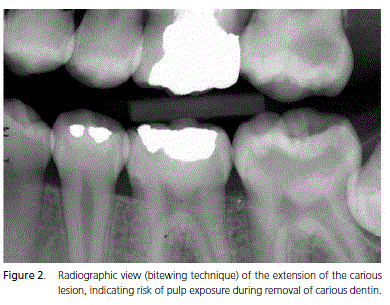
The patient, a fourteen year old boy, came to the Dentistry Graduation Course clinic at UnG complaining of non-spontaneous pain in tooth 37. In the clinical exam, the presence of an extensive cavity was noted (Figure 1) and through a radiographic exam, it was verified that the lesion extended well into the dentine with risk of pulp exposure during removal of carious tissue (Figure 2). Pulp vitality assessment with cold was performed, the diagnosis being reversible pulpitis.
Due to the patient's age and pulp condition, a stepwise excavation technique was chosen to avoid accidental pulp exposure that could lead to premature devitalization of a permanent tooth.
The absolute isolation of the operating area was carried out to avoid contamination, but without anesthesia, because pain reported during excavation indicates approximation to the pulp, in which case the excavation must be halted. Access to the cavity was performed with high speed spherical diamond tips. The carious dentin was removed with sharp curettes, starting with the dentine above the pulp wall, up until the moment that the patient reported pain, and the remaining carious dentin was left on the pulp wall. At this point, diagnosis between demineralized and contaminated dentin is critical; if only demineralized dentin is present, definitive restoration treatment with pulp protection with calcium hydroxide can be performed. However, if there is any doubt about the remaining dentin being contaminated, the ideal solution is to perform provisional sealing before definitive restoration.
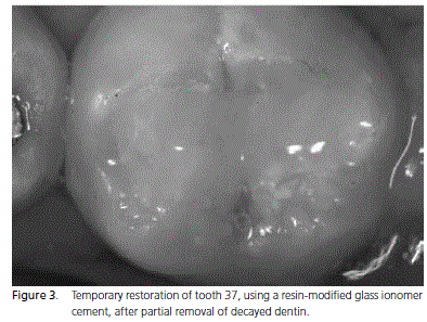
In this case, as there was a large amount of softened dentin, the cavity was cleaned with air/water jets and a layer of calcium hydroxide paste was applied to the entire pulp wall. The cavity was provisionally sealed with a hybrid material of glass ionomer cement and composite resin (Vitremer 3M ESPE - Figure 3) which is outstanding because it releases fluoride and is more resistant to occlusal forces.
A period of 60 to 90 days would have been sufficient for the stepwise excavation technique to result in dentin hardening and tertiary dentin deposition. It was noted that significant alterations occurred in the first 6 months4-8, but, as the painful symptoms disappeared, the patient did not return for treatment after 90 days and it was only after 12 months that it was possible to make contact and resume treatment. However, systematic observations of pulp vitality should be made during the waiting period, because the possibility exists of asymptomatic pulp necrosis developing6.
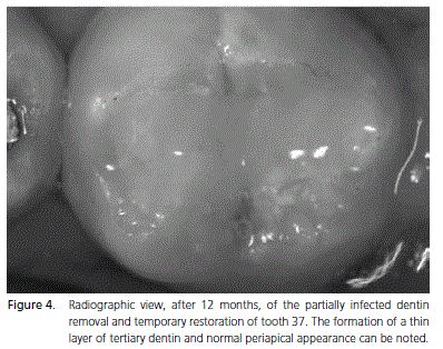
After 12 months, a radiographic exam was performed and the formation of a thin layer of tertiary dentin and normal periapical appearance was noted (Figure 4).
The tooth was isolated with a rubber dike and the temporary sealing was removed with high speed spherical diamond tips. Dentin presented a dark color, with a hard consistency on probing, with an appearance of dentin with arrested caries. Thus, the decision was taken not to remove this residual tissue.
Etching was performed with 37% phosphoric acid for 20 seconds on the enamel and 15 seconds on the dentin. The acid was removed with jets of water and the cavity was softly dried with jets of air, taking care to keep the dentin humid for the application of the adhesive system. Two layers of a water and alcohol-based single-bottle adhesive system (Single Bond, 3M ESPE) were applied for 5 seconds and the solvent evaporated with air jets. Next, the adhesive was photo-activated for 20 seconds.
A layer of resin-modified glass ionomer was applied as an indirect pulp protector (Vitremer, 3M ESPE) and it was photo-activated for 40 seconds. The restoration was performed with nanoparticle composite resin (Z350, 3M ESPE) inserted in layers using the incremental technique.
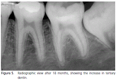
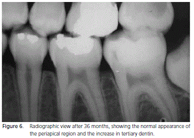
After 18 and 36 months of follow-up, the normal appearance of the apical region and an increase of tertiary dentin can be observed (Figures 5 and 6).

DISCUSSION
The clinician frequently considers the stepwise excavation technique to be a type of indirect protection of the pulp-dentin complex. However, in an ideal situation, an indirect protection should only be placed on sound or demineralized dentin and the stepwise technique consists of placing biocompatible materials on the carious dentin, in order to stimulate the formation of tertiary, sclerotic or remineralized dentin8.
However, it must be treated as a specific technique, developed to avoid pulp exposure during carious tissue removal from teeth with deep caries lesions2,8, since it consists of the application of a biomaterial which will remain active on the dentin for a period of at least 45 days and which, if necessary, will later be removed along with the remaining carious dentin. Since there is no evidence that the re-opening procedure is necessary, and provided that the patient does not present irreversible pulpitis, a thin layer of contaminated carious dentin can be left and the tooth can be definitively restored2.
However, non-reopening of the cavity characterizes a direct pulp capping and it is necessary to know how to clearly discern a direct pulp capping technique from a stepwise excavation technique, since the main difference between them is the characteristic and amount of remaining infected dentin, and the presence of infected dentin under a restoration. In a private practice, a direct pulp capping of an infected soft dentin cannot be accepted. In the stepwise excavation technique, the dentist chooses to provisory leave contaminated dentin in the cavity and, in a second intervention, the dentist must evaluate the final condition of the dentin before the definitive restoration is performed.
On the other hand, in the case of indirect pulp protection, the remaining dentin must be harder, i.e. only demineralized, and with the cavity restoration there will be inactivation of the bacteria, arrest of the lesion and tertiary dentin deposition. During a repeat intervention, there is a risk of pulp exposure in a cavity whose lesion will probably not progress7,9. But in this case, a constant follow-up is necessary to guarantee successful treatment.
The same effects of the biomaterial will also occur with contaminated dentin during the stepwise excavation technique. Associated with this, the temporary sealing will permit a change in cavity conditions, from one of a highly contaminated environment, with the possibility of rapid progression, to a condition of inactive lesion or of low progression. Moreover, it stimulates the pulp to recover, with the aim of removing all carious dentin in a subsequent intervention, in an effort to increase the longevity of the restorative treatment2,4 ,10.
At this time, there is nothing to prevent the demineralized dentin from being kept and indirect pulp capping being performed. Although the risk of pulp exposure after the stepwise excavation technique is lower, if it does happen, one can opt for direct pulp protection.
Thus, the stepwise excavation technique is recommended for teeth with deep caries lesions, where the removal of carious tissue in a single step would lead to pulp exposure2-4,8,10-11.
In the stepwise excavation technique, a positive response is expected from the pulp, with the formation of secondary and principally tertiary dentin. With the passing years, the pulp gradually becomes more fibrous with a reduction in volume due to the physiological production of dentin and reduction in blood supply and regeneration capacity. That is why the stepwise excavation technique is recommended more for young patients, whose pulp chamber is still broad, or even in teeth with incomplete apexes, because they have a better supply of nutrients and a greater potential for repair10.
Coincidentally, in teeth with dentin tubules of larger diameter and a broader pulp chamber, carious lesion progression is greater, as is the risk of accidentally hitting the pulp during carious tissue removal8.
Due to rapid lesion progression in young teeth, which are recommended for the stepwise excavation technique, the diagnosis of pulp condition is essential for the success of treatment, as it is necessary for the pulp to present as normal or with reversible inflammation, in order to obtain a potential healing response.
The removal of central cariogenic biomass of dentin and the superficial part of demineralized dentin already guarantees a lower degree of bacterial activity in the cavity, enabling the pulp to react better. However, removal of carious tissue in deep lesions does not need to be as rigorous as traditionally recommended, as there will be repeat intervention for the removal of this dentin3-4.
Removal of dentin associated with the application of calcium hydroxide can cause a potential dentin healing response. Calcium hydroxide has an anti-bacterial effect and it can act on the contaminated dentin, leading to a greater physiological reaction of the dentin organ3.
Studies have shown that cavity sealing alone is sufficient to promote hardening of carious lesion that indicates its arrest6,9,11. It should be noted that only cavity sealing of carious dentin causes a great reduction in the number of microorganisms5,9,11. Thus, it is supposed that, in order to guarantee success, all that is required is to use good biocompatible sealing material with the purpose ofpreventing the penetration of aggressive agents and their metabolic products, which affect the pulp through the dentinal tubules8.
Nevertheless, the procedure most recommended is to use material based on calcium hydroxide8, since clinically, pulp micro-exposure may occur before the stepwise excavation technique treatment is performed. Finally, cavity sealing must be performed with a temporary, but resistant, restorative material, as it may be left in place for at least 45 days8.
Ionomer cements are outstanding because they are biocompatible, they bond to the dental structure, have a thermal linear expansion coefficient similar to that of the dentin and they also release fluoride that enhances remineralization. It is reported that the action of fluoride from ionomeric materials is continuous, as this material also presents a sponge effect, incorporating the fluoride from the medium to release them in a situation of cariogenic challenge8. Among these materials, the resin-modified ionomers are excellent because, although they may cost more, they can guarantee better esthetics and mechanical resistance while waiting for pulp response.
During the waiting period, the professional must assess the pain symptoms and ascertain if the tooth remains vital and can receive definitive treatment8. It is suggested that the opening of the cavity for definitive restoration treatment must be performed at least 45 days after treatment with the stepwise excavation technique. This period is necessary for the pulp tissue to react to aggressions and for the inflammatory process to be inhibited.
A mineral gain occurs between 6-7 months and there is no need to wait longer than this to perform definitive treatment.2 However, it is important that, at the end of the waiting period, the responses to the pulp sensitivity and radiographic examinations are normal, so that the professional can proceed with definitive restorative treatment8.
Clinical studies have shown high success rates for the stepwise excavation technique (6-17% of pulp exposure4,12) when compared with total removal of carious dentin (40% of pulp exposure12), a success rate of 63% over 10 years of follow-up13, and after removal of provisional restoration an increase in the radiographic density of the remaining dentin is noted, which suggests an increase in mineral quantity with carious lesion arrest and tertiary dentin deposition2.
As in the present clinical case, longitudinal clinical assessments show that in cavities where the dentin was initially softened, this dentin presented with a darker coloring and harder consistency after reopening, which suggests that the carious process has been arrested3,6,11.
Many studies confirm the benefits of partial removal of carious dentin, the application of calcium hydroxide and cavity sealing, and at a later stage, complete removal of carious dentin (stepwise excavation technique) in contrast to the complete removal of carious tissue. Thus, the stepwise excavation technique treatment can be considered a safe therapy, scientifically corroborated, and with a high rate of success2-6,7-13.
CONCLUSION
It may be concluded that the stepwise excavation technique has the advantage of avoiding pulp exposure during carious tissue removal from teeth with deep caries lesions and also of changing the cavity conditions, promoting dentin formation and pulp repair.
Collaborators
V MONARI, YBO LIMA-ARSATI and JA RODRIGUES took part in all phases of the preparation of this article.
REFERENCES
1. Doméjean-Orliaguet S, Léger S, Auclair C, Gerbaud L, Tubert- Jeannin S. Caries management decision: influence of dentist and patient factors in the provision of dental services. J Dent. 2009;37(11):827-34. [ Links ]
2. Oliveira EF, Carminatti G, Fontanella V, Maltz M. The monitoring of deep caries lesions after incomplete dentine caries removal: results after 14-18 months. Clin Oral Invest. 2006;10(12):134-9.
3. Bjørndal L, Larsen T, Thylstrup A. A clinical and microbiological study of deep carious lesions during stepwise excavation using long treatment intervals. Caries Res. 1997;31(6):411-7.
4. Bjørndal L, Thylstrup A. A practice based study on stepwise excavation of deep carious lesions in permanent teeth: a 1 year follow up study. Community Dent Oral Epidemiol. 1998;26(2):122-8.
5. Bjørndal L. In deep cavities stepwise excavation of caries can preserve the pulp. Evid Based Dent. 2011;12(3):68.
6. Fisher FJ. The effect of a calcium hydroxide water paste on micro organisms in carious dentine. Br Dent J. 1972;133(1):19-21. 7. Kidd EAM. How clean must cavity be before restoration? Caries Res. 2004;38(10):305-13.
8. Orhan AI, Oz FT, Orhan K. Pulp exposure occurrence and outcomes after 1- or 2-visit indirect pulp therapy vs complete caries removal in primary and permanent molars. Pediatr Dent. 2010;32(4):347-55.
9. Weerheijm KL, De Soet JJ, van Amerongen WE, De Graaff J. The effect of glass-ionomer cement on carious dentine an in vivo study. Caries Res. 1993;27(5):417-23.
10. Ricketts D. Management of the deep carious lesion and the vital pulp dentine complex. Br Dent J. 2001;191(11):606-10.
11. Duque C, Negrini TC, Sacono NT, Spolidorio DM, de Souza Costa CA, Hebling J. Clinical and microbiological performance of resin-modified glass-ionomer liners after incomplete dentine caries removal. Clin Oral Investig. 2009;13(4):465-71.
12. Bjørndal L, Reit C, Bruun G, Markvart M, Kjaeldgaard M, Näsman P, et al. Treatment of deep caries lesions in adults: randomized clinical trials comparing stepwise vs. direct complete excavation, and direct pulp capping vs. partial pulpotomy. Eur J Oral Sci. 2010;118(3):290-7.
13. Maltz M, Alves LS, Jardim JJ, Moura MS, Oliveira EF. Incomplete caries removal in deep lesions: a 10-year prospective study. Am J Dent. 2011;24(4):211-4.
 Endereço para correspondência:
YBO LIMA-ARSATI
Universidade Estadual de Feira de Santana,
Departamento de Ciências Biológicas.
Av. Transnordestina, s/n., Novo Horizonte,
44036-900, Feira de Santana, BA, Brasil.
e-mail: ynaralima@yahoo.com
Recebido: 4/12/2010
Aceito:1/8/2011


