


Serviços Personalizados
Artigo
 pdf em Inglês
 Artigo em XML
 Referências do artigo
 Enviar este artigo por email
Links relacionados
Compartilhar

 Permalink
RGO.Revista Gaúcha de Odontologia (Online)
versão On-line ISSN 1981-8637
RGO, Rev. gaúch. odontol. (Online) vol.60 no.2 Porto Alegre Abr./Jun. 2012
ORIGINAL / ORIGINAL
Effect of dentin on the shear bond strength of different adhesive systems
Efeito do substrato dentinário na resistência de união ao cisalhamento de diferentes sistemas adesivos
Darlon Martins LIMA I; Maria Salete Machado CANDIDO, in memoriam II
I Universidade Federal do Maranhão, Curso de Odontologia, Programa de Pós-graduação em Odontologia, Av. dos Portugueses, s/n, Bacanga 65085-580, São Luís, MA, Brasil
II Universidade Estadual Paulista Júlio de Mesquita Filho, Faculdade de Odontologia. Araraquara, SP, Brasil
ABSTRACT
Objective
This study assessed the shear bond strength of total-etch and self-etch adhesive systems to sound and caries-affected human dentin.
Methods
The total-etch adhesive systems investigated were Scotchbond Multi Purpose Plus (3M ESPE, St. Paul, MN, USA) and Single Bond (3M ESPE, St. Paul, MN, USA), and the self-etch adhesive systems were Clearfill SE Bond (Kuraray, Osaka, Japan) and Adper Prompt L-Pop (3M ESPE, St. Paul, MN, USA). Eighty human teeth were used, 40 sound and 40 caries-affected (n=10). They were cut with a diamond disc to expose the dentinal surface and the adhesion area was standardized to 4 millimeters. Caries detector was used on the caries-affected4 teeth and the affected tissue was removed. The bond shear strength of different adhesives was tested on sound dentin (control group) and caries-affected dentin (experimental group). Z-100 composite resin cylinders were fabricated using a two-piece mold, and the specimens were stored in distilled water at 37°C for 24 hours, and submitted to the shear strength bond test using a crosshead speed of 0.5 mm/min.
Results
The data were treated by analysis of variance and Tukey test and the significance level was set at 5%. Caries-affected dentin had a significant negative impact on the shear bond strength of total-etch adhesive systems.
Conclusion
Shear bond strength depends on dentin status and type of adhesive system used.
Indexing terms: Dentin. Dentin adhesives. Shear bond strength.
RESUMO
Objetivo
Avaliar a resistência de união ao cisalhamento de sistemas adesivos de condicionamento ácido total e autocondicionantes em dentina humana hígida e afetada por cárie.
Métodos
Os sistemas adesivos com etapa prévia de condicionamento ácido da estrutura dental utilizados foram: Scotchbond Multi Purpose Plus (3M ESPE, St. Paul, MN, USA) e Single Bond (3M ESPE, St. Paul, MN, USA), e os sistemas adesivos autocondicionantes: Clearfill SE Bond (Kuraray, Osaka, Japan) e Adper Prompt L-Pop (3M ESPE, St. Paul, MN, USA). Foram utilizados 80 dentes humanos, sendo 40 hígidos e 40 cariados (n=10), cortados com disco de diamante, expondo a superfície de dentina, e a área de adesão padronizada, em 4 mm de diâmetro. Para os dentes cariados, após corte inicial, estes eram corados com solução evidenciadora de cárie, e o tecido corado removido. Nos Grupos controle, os sistemas adesivos eram aplicados em dentina hígida, e nos Grupos experimentais, aplicados em dentina afetada por cárie. Confeccionaram- -se cilindros de resina composta Z-100, com auxílio de matriz bipartida, os corpos-de-prova obtidos, armazenados em água destilada a 37°C por 24 horas, e submetidos ao ensaio de cisalhamento, a uma velocidade de 0,5 mm/min.
Resultados
Foram submetidos à Análise de Variância, e ao teste de Tukey, ao nível de 5% de significância. Observou-se influência do fator substrato para os valores de resistência de união, diminuídos significantemente quando sistemas adesivos de condicionamento ácido total eram aplicados em dentina afetada por cárie.
Conclusão
O presente estudo indicou que a resistência de união ao cisalhamento é dependente do tipo de dentina e do tipo de sistema adesivo utilizado.
Termos de indexação: Dentina. Adesivos dentinários. Resistência ao cisalhamento.
INTRODUCTION
One of the foundations of contemporary dentistry is the ability to bind certain materials to dental tissues. Adhesion principles allow the use of polymers in a wide range of clinical situations, ranging from minimally invasive preventive procedures to the replacement of significant crown tissue by direct or indirect techniques.
Most adhesion tests are done on sound dentin but the most common clinical situation involves what remains of this substrate after caries removal1-3, that is, two different layers of carious lesion on dentin with distinct hardness and stain susceptibility4-5. Microscopically, the topmost layer presents extensive demineralization6, denatured collagen fibrils7 and absence of viable odontoblastic processes. Consequently, the infected dentin does not remineralize4,8. The second layer, immediately below the topmost layer, is characterized by moderate demineralization, healthy collagen fibrils and viable odontoblastic processes, so it may recover on its own. This layer is called caries-affected dentin9-10.
The changes seen in caries-affected dentin significantly impact the performance of adhesive materials. Adhesion to this substrate is weaker than to sound dentin1,11-15. Therefore, there is no consensus on the effect that caries-affected dentin has on different adhesive systems.
The objective of this study is to assess the shear bond strength of different adhesive systems to sound and caries-affected dentin. The study will test the hypothesis that different dentinal substrates do not affect shear bond strength.
METHODS
The study was approved by the Research Ethics Committee of Paulista State University Júlio de Mesquita Filho's School of Dentistry of Araraquara, protocol number 16/07. Eighty permanent human molars obtained from the tooth bank of the said school were used: 40 sound and 40 affected by caries on the enamel and dentin of the occlusal surfaces. All teeth were examined under a stereo magnifying glass. The teeth were cleaned with periodontal curettes and a prophy brush, pumice and water, and stored in distilled water at 4°C until used. The teeth were mounted on wooden platforms (4.5 x 4.5 x 1 cm) and glued with a thermoplastic impression compound in stick (working temperature of roughly 55oC). The top occlusal third was then cut out by the ISOMET 1000 precision saw (Buehler Ltda., Lake Bluff, Illinois, EUA) to expose the amelodentinal junction.
The cut surfaces were then sanded with 100-grit sandpaper under plenty of cool running water to ensure that the entire area consisted of dentin. Sound and caries-affected dentin were worn to different depths. In sound teeth, all the occlusal enamel was sanded and in carious teeth, all the enamel and carious dentin, dyed by a caries detector, were sanded. Visible caries-affected dentin was confirmed by resistance to cutting by a manual dentin excavator. More specifically, a microbrush was used to administer a caries detector to the carious teeth, which was allowed to rest for 10 seconds and then rinsed for 30 seconds. The dyed tissue, which was very moist and soft, was then removed by a dentinal curette. The procedure was repeated when partly dyed tissue was still present. If the dyed dentin resisted cutting by a dental curette, it was not removed, since this was the parameter chosen for limiting dentinal removal. This parameter characterized the caries-affected dentin, that is, harder dentin with dentinal sclerosis. Teeth with exposed pulp chamber were discarded. The teeth were fixed on a glass plate with wax 07 to keep the chemically-activated acrylic resin from contaminating the dentinal surface and thereby hindering the adhesive procedures. Once the teeth were fixed, they were embedded in a chemicallyactivated acrylic resin contained inside a 4cm-long, 3cmwide polyvinyl chloride (PVC) cylinder finished with a lathe to ensure standard height and symmetry. The chemicallyactivated acrylic resin was poured inside the PVC cylinder with the occlusal surface of the tooth facing outward.
Standardization of the smear layer was the next step. The sets (tooth, PVC cylinder and chemically-activated acrylic resin) were sanded with a horizontal polisher (model DP - 10, Panambra Industrial e Técnica S.A., São Paulo, Brazil) for 30 seconds using 320- and 600-grit sandpaper and plenty of cooling. The dentinal surfaces were then rinsed with running water for at least 10 seconds to remove impurities that could compromise adhesion. After separation of the sound and caries-affected teeth, the specimens were randomly divided into the experimental groups and stored in distilled water until ready for use.
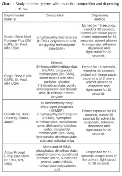
The shear bond strengths of 4 different adhesive systems of different compositions and dispensing methods were investigated. Two were total-etch and two were self-etch adhesive systems (Chart 1), which were then used on two different surfaces, sound and caries-affected dentin. Hence, 8 conditions were tested, each with 10 specimens, totaling 80 specimens.
The 80 teeth were divided into 8 groups. The control groups consisted of adhesive systems dispensed on sound dentin and the experimental groups consisted of the same adhesive systems dispensed on caries-affected dentin: G1 - Scotchbond Multipurpose Plus (3M ESPE, St. Paul, MN, USA) on sound dentin; G2 - Scotchbond Multipurpose Plus (3M ESPE, St. Paul, MN, USA) on cariesaffected dentin; G3 - Single Bond (3M ESPE, St. Paul, MN, USA) on sound dentin; G4 - Single Bond (3M ESPE, St. Paul, MN, USA) on caries-affected dentin; G5 - Clearfill SE Bond (Kuraray, Osaka, Japan) on sound dentin; G6 - Clearfill SE Bond (Kuraray, Osaka, Japan) on caries-affected dentin; G7 - Adper Prompt L-Pop (3M ESPE, St. Paul, MN, USA) on sound dentin; G8 - Adper Prompt L-Pop (3M ESPE, St. Paul, MN, USA) on caries-affected dentin.
Once the study design was established, the teeth were distributed into groups with the respective fabrication of the mounts. Adhesive tape (3M, Sumaré, Brazil) was used for limiting the exposed dentinal area to a circumference of 4mm, thereby standardizing the area submitted to the adhesive systems, as shown in Chart 1.
All adhesive systems were used according to the manufacturer's instructions. Composite resin cylinders were fabricated on stainless steel molds with a diameter of 4 cm and height of 4 mm, having a central hole with a diameter of 4mm. For firmness, a stainless steel ring with the same diameter as the PVC cylinder was fixed externally, placed over the PVC cylinder. Later, the composite resin was poured on the two-piece molds.
The molds were filled with two batches of the microhybrid composite resin Z-100 (3M ESPE, St. Paul, MN, USA). Each layer was 2mm thick and light-cured for 40 seconds using a halogen light mirror with a light intensity not less than 450 mw/cm2 verified by a calibrated radiometer. After the last layer was poured and cured, the polyester mold was placed on top of the metal mold. Once full, the external ring around the PVC was removed with a single movement, from bottom to top, the two pieces of the mold were removed, and the composite resin cylinder was light-cured for another 40 seconds.
Once all 80 specimens were ready (n=10), they were stored in individual flasks filled with distilled water for 24 hours in an incubator at 37°C. Each specimen was then coupled to an appropriate device and submitted to the shear bond strength test in a mechanical testing system (Material Test System 810, MTS Systems Corporation, Minneapolis, Minnesota, EUA) with a crosshead speed of 0.5 mm/minute, with a chisel-shaped tip. This tip was placed on the base of the composite resin cylinder, parallel to the dentinal surface. The machine was stopped as soon as the specimen broke or cracked. Data were collected by software (Test Works, Test Star 2 System, MTS Systems Corporation, Minneapolis, Minnesota, EUA) and the final bond strengths were calculated by dividing the maximum load in Newton (N) by the bond area in mm2, and expressed as MPa.
RESULTS
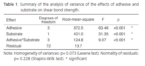
Two-way analysis of variance was used to assess the effect of the adhesive system and substrate on shear bond strength. This analysis was complemented by the Tukey multiple comparisons of means. The significance level was set at 5%.
Table 1 shows the summarized analyses of variance. The Levene and Shapiro-Wilk tests suggest acceptable homogeneity of variance and normality of residuals (p>0.05). Both variables, adhesive system and substrate, have a significant impact on shear bond strength (p<0.001), indicating that the performance of the adhesive system is affected by the substrate.
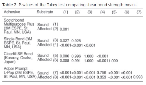
Pairs of bond strength means were compared by the Tukey test to unravel their dependence on the study variables. The strengths are shown in Table 2.
Table 3 shows the means and standard deviations of the experimental groups and the comparison of the means done by the Tukey test. Means followed by same letters are not significantly different at the 5% significance level. There is evidence that the shear bond strength means of total-etch systems to the caries-affected dentins are lower than those of total-etch systems to sound substrates, but the same was not observed with self-etch systems.
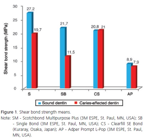
Figure 1 below shows the shear bond strength means.
Total-etch systems require etching of the dental structure by 37% phosphoric acid (Scotchbond Multi Purpose Plus (3M ESPE, St. Paul, MN, USA) and Single Bond 2 (3M ESPE, St. Paul, MN, USA)). Their shear bond strengths were significantly lower when used on cariesaffected dentin. Meanwhile, the self-etch systems Clearfill SE Bond (Kuraray, Osaka, Japan) and Adper Prompt L-Pop (3M ESPE, St. Paul, MN, USA) were not affected by dentin status.
The comparison of the data regarding single- and double-step, total-etch and self-etch adhesive systems commonly used in clinical practice shows that the totaletch system Scotchbond Multi Purpose Plus (3M ESPE, St. Paul, MN, USA) had a significantly higher shear bond strength than the Single Bond 2 (3M ESPE, St. Paul, MN, USA) on sound and caries-affected dentin. The same was observed on self-etch systems, that is, the shear bond strength of the double-step self-etch system Clearfill SE Bond (Kuraray, Osaka, Japan) was significantly higher than that of the single-step self-etch system Adper Prompt L-Pop (3M ESPE, St. Paul, MN, USA). When total-etch and self-etch systems used on sound dentin are compared, Scotchbond Multi Purpose Plus (3M ESPE, St. Paul, MN, USA) has a significantly higher shear bond strength than Single Bond 2 (3M ESPE, St. Paul, MN, USA) and Clearfill SE Bond (Kuraray, Osaka, Japan), and all these systems have a significantly higher shear bond strength than Adper Prompt L-Pop (3M ESPE, St. Paul, MN, USA). Therefore, (Scotchbond Multipurpose Plus (3M ESPE, St. Paul, MN, USA) > Single Bond (3M ESPE, St. Paul, MN, USA) = Clearfill SE Bond (Kuraray, Osaka, Japan) > Adper Prompt L-Pop (3M ESPE, St. Paul, MN, USA). However, the shear bond strength of these systems was significantly affected by caries-affected dentin. Scotchbond Multipurpose Plus (3M ESPE, St. Paul, MN, USA) was still better than Single Bond 2 (3M ESPE, St. Paul, MN, USA) and Adper Prompt L-Pop (3M ESPE, St. Paul, MN, USA), but similar to Clearfill SE Bond (Kuraray, Osaka, Japan). Likewise, on sound dentin, Single Bond 2 (3M ESPE, St. Paul, MN, USA)) was similar to Adper Prompt L-Pop (3M ESPE, St. Paul, MN, USA). Therefore, Scotchbond Multipurpose Plus (3M ESPE, St. Paul, MN, USA) = Clearfill SE Bond (Kuraray, Osaka, Japan) > Single Bond (3M ESPE, St. Paul, MN, USA) = Adper Prompt L-Pop (3M ESPE, St. Paul, MN, USA).

DISCUSSION
Many factors contribute to the bonding and curing of the adhesive and the formation of the hybrid layer. Scientists are continuously trying to understand the phenomenon of dental adhesion and comparing the performance of different types of adhesive systems. However, most studies study adhesion exclusively on sound dentin, and only a handful are on caries-affected dentin.
Since the most common clinical condition involves a caries-affected substrate, the present study assessed the effect of substrate on the shear bond strength of total-etch and self-etch systems.
The results of the present study showed that the shear bond strength of adhesive systems to sound and caries-affected dentin depends both on the adhesive and substrate. The shear bond strengths of the total-etch systems Scotchbond Multi Purpose Plus (3M ESPE, St. Paul, MN, USA) and Single Bond 2 (3M ESPE, St. Paul, MN, USA) were significantly affected by caries-affected dentin while the selfetch systems Clearfil SE Bond (Kuraray, Osaka, Japan) and Adper Prompt L-Pop (3M ESPE, St. Paul, MN, USA) were not.
These results are not entirely corroborated by those of other studies, that is, some aspects are and some are not. The results for the total-etch adhesive systems tested in the present study agree with other studies that compare shear bond strength to sound and cariesaffected dentin11,14,16 and disagree with the studies done by Nakajima et al.2 and Nakajima et al.11-12, who did not find significant shear bond strength differences of Scotchbond Multi Purpose Plus (3M ESPE, St. Paul, MN, USA) to cariesaffected dentin. The results of the present study are also in disagreement with those of Hosoya et al.17, who reported that caries-affected dentin did not affect the shear bond strength of Single Bond (3M ESPE, St. Paul, MN, USA). The method used in the latter17 was not the traditional method, that is, they used a jig that attached the samples strongly to the device, which resulted in low microtensile bond strengths. In addition to assessing different adhesive systems, they also assessed if mounting method for the microtensile test (dumbbell and stick) affected shear bond strength and found that it did not. They reported a mean shear bond strength of 9.0 MPa. Additionally, this study17 used deciduous dentin, which is physically and structurally different from permanent dentin.
The presence of pathological processes, such as caries and hypermineralization, can change the dentinal chemistry and structure. Since age, attrition, caries and restorative procedures can change dentinal composition, increase dentinal sclerosis and reduce its organic components, particularly collagen, they can also reduce the effectiveness of adhesive systems that rely on collagen for the formation of the hybrid layer18.
Dentinal tubules destroyed by sclerosis may also lower adhesion because of low dentinal fluid and high dentinal permeability. The constant presence of mineral deposits may also affect acid performance and infiltration of the resin19. The shear bond strength of the self-etch system Clearfill SE Bond (Kuraray, Osaka, Japan) may not have been significantly affected by caries-affected dentin because of its less rigorous dispensing requirements. The self-etch systems do not require rinsing and blotting but require dissolution in water, contrary to the adhesive systems that require etching with phosphoric acid, rinsing and blotting, and adhesion on a moist process. However, ideal rinsing and drying are difficult to obtain and very subjective. Self-etch systems such as Clearfil SE Bond (Kuraray, Osaka, Japan) tend to form a hybrid but thinner homogeneous layer with fewer imperfections20. Its mild formula does not dissolve the smear layer completely, which is then hybridized by the polymer. Partial dissociation of this structure allows the functional monomers of selfetch systems to interact with the hydroxyapatite crystals that remained around the partially exposed collagen fibrils. The acidic monomer 10-methacryloiloxydecyldihydrogen phosphate (10-MDP) in the Clearfil SE Bond (Kuraray, Osaka, Japan) system quickly binds to hydroxyapatite, forming very stable, water-insoluble bonds12. It is also speculated that the reason Clearfill SE Bond (Kuraray, Osaka, Japan) performed similarly in sound and cariesaffected dentin is because that study used a mechanical test to assess shear bond strength, and this type of bond requires a greater bonding area. Hence, because of the greater bonding area, it might not have favored the greater force needed for rupturing the bonded interface, since cohesive failures are common in specimens due to less uniform force distribution. Disagreements with other similar studies12-13,16,21 may be related to the different methodologies used for testing shear bond strength, since they used the micro-traction test. This test has a smaller bonding area making the test uniform, maximizing the concentration of forces and allowing maximum mechanical challenge.
Total or partial destruction of the dentinal tubules and the presence of intertubular dentin with mineral deposits may prevent a reliable adhesion of resinous restorative systems12,22 by reducing the number of resinous tags, elasticity model and cohesive dentinal strength14, corroborating reports22 that the deposition of betatricalcium phosphate crystals on carious lesions increases mineral content, which reduces dentinal permeability and thereby affects the shear bond strength of adhesive systems.
According to Nakajima et al.2, caries-affected dentin may contain certain substances, such as glycoproteins or mucopolysaccharides, that affect wettability and infiltration of the adhesive system through the dentinal micropores, and/or polymerization. Hence, collagen fibrils at the base of the hybrid layer will not be fully bonded by the primer and adhesive, resulting in a fragile area with low shear bond strength.
The results found for Adper Prompt L-Pop (3M ESPE, St. Paul, MN, USA) are in agreement with previous studies16,23 that reported low shear bond strengths and inconsistent results for this self-etch adhesive system, whose performance is similar in sound and caries-affected dentin. The low shear bond strength found for Adper Prompt L-Pop (3M ESPE, St. Paul, MN, USA) is directly associated with its high water sorption potential. Water in the adhesive and hybrid layer prevents adequate polymerization, resulting in hydrolysis of the hybrid layer and low shear bond strength, so this system does not have the requirements for a reliable adhesion to the dental tissue24.
The study of adhesion to different dentinal substrates is of utmost importance. In clinical practice, it is sometimes difficult to identify carious and infected tissue and remove all of it. When carious tissue remains on the tooth, adhesion is unstable and may promote microleakage and secondary caries, resulting in failed restorative treatment which is sometimes confused with aesthetic excellence because of the similarity between the composite resin and dental tissue. The present study makes evident that dentinal hybridization depends on many factors, such as the adhesive system and its formula and dentin status with its complex composition, and that these factors and the fact that restorative treatments are usually done to treat caries, which often result from high cavity configuration factor, could contribute to treatment failure. Therefore, it is extremely important for dental surgeons to be able to recognize infected dentin and eliminate it correctly from the hybridization site to avoid early treatment failure. In carious dentin, this aspect is controversial. Since caries-affected dentin is usually surrounded by sound dentin, the restorative procedure is more likely to succeed. From the biological point of view, maintenance of this dentin prevents overtreatment, which could expose the pulp. Once this altered dentin is sealed25, it may undergo reorganization, especially if glass ionomer cement is used. Recent evidence26-27 shows that there is no progression of the lesion when the dentin and enamel are correctly sealed, and even deposition of tertiary dentin is viable. Additionally, longitudinal clinical studies26-27 have shown that it is very important to seal the cavity properly to prevent the promotion of new caries.
Based on the premise that adhesive restorative procedures are done on all sorts of dentin, that is, sound, caries-affected, tertiary and sclerotic dentin, and that, invariably, the enamel is also involved, it is important to understand who adhesive systems work28 and their specific performance on different substrates, since this may affect treatment success and longevity.
CONCLUSION
The study methodology and results show that shear bond strength depends on the adhesive system used and the condition of the substrate.
Caries-affected dentin has a negative impact on the shear bond strength of total-etch systems but does not affect that of self-etch systems. Although the shear bond strength of the three-step total-etch system was negatively affected by caries-affected dentin, it was similar to that of the two-step self-etch system.
Collaborators
DM LIMA performed the experimental procedures from the pilot experiment to the study experiments, analyzed and interpreted the results and wrote the article. MSM CANDIDO conceived and designed the experiment, and supervised the different study stages and manuscript writing.
REFERENCES
1. Kimochi T, Yoshiyama M, Urayama A, Matsuo T. Adhesion of a new commercial self-etching/self-priming bonding resin to human caries-infected dentin. Dent Mater J. 1999;18(4):437- 43. doi: 10.4012/dmj.18.437. [ Links ]
2. Nakajima M, Sano H, Burrow MF, Tagami J, Yoshiyama M, Ebisu S, et al. Tensile bond strength and SEM evaluation of caries affected dentin using dentin adhesives. J Dent Res. 1995;74(10):1679-88. doi: 10.1177/00220345950740100901.
3. Yoshiyama M, Urayama A, Kimochi T, Matsuo T, Pashley DH. Comparison of a conventional vs self - etching adhesive bonds to caries-affected dentin. Oper Dent. 2000;25(3):163-9.
4. Fusayama T. Two layers of carious dentin: diagnosis and treatment. Oper Dent. 1979;4(2):63 -70.
5. Fusayama T, Nakamura M, Kurasaki N, Iwaku M. Non-pressure adhesion of a new adhesive restorative resin. J Dent Res. 1979;58(4):1364-70. doi: 10.1177/00220345790580041101.
6. Angker L, Nockolds C, Swain MV, Kilpatrick N. Correlating the mechanical properties to the mineral content of carious dentine: a comparative study using an ultra-micro indentation system (UMIS) and SEM-BSE signals. Arch Oral Biol. 2004; 49(5):369- 78. doi: 10.1016/j.archoralbio.2003.12.005.
7. Jackson RJ, Lim DV, Dao, ML. Identification and analysis of a collagenolytic activity in Streptococcus mutans. Curr Microbiol. 1997;34(1):49-54. doi: 10.1007/s002849900143.
8. Kuboki Y, Ohgushi K, Fusayama T. Collagen biochemistry of the two layers of carious dentin. J Dent Res. 1977;56(10):1233-7. doi: 10.1177/00220345770560102301.
9. Beeley KA, Yip HK, Stevenson AG. Chemo mechanical caries removal: a review of the techniques and latest developments. Ned Tijdschr Tandheelkd. 2001;108(7):277-81.
10. Sarnat H, Massler M. Microstructure of active and arrested dentinal caries. J Dent Res. 1965;44(6):1389-410. doi: 10.1177/00220345650440064601.
11. Nakajima M, Sano H, Zheng L, Tagami J, Pashley DH. Effect of moist vs. dry bonding to normal vs. caries affected dentin with Scotchbond multi-purpose plus. J Dent Res. 1999;78(7):1298- 303. doi: 10.1177/00220345990780070301.
12. Nakajima M, Ogata M, Okuda M, Tagami J, Sano H, Pashley DH. Bonding to caries-affected dentin using self - etching primers. Am J Dent. 1999;12(6):309-14.
13. Yoshiyama M, Tay FR, Doi J, Nishitani Y, Yamada T, Itou K, et al. Bonding of self-etch and total-etch adhesives to carious dentin. J Dent Res. 2002;81(8):556-60. doi: 10.1177/154405910208100811.
14. Arrais CA, Giannini M, Nakajima MT, Tagami J. Effects of additional and extended acid etching on bonding to cariesaffected dentine. Eur J Oral Sci. 2004;112(5):458-64.
15. Xie J, Flaitz CM, Hicks MJ, Powers JM. Bond strength of composite to sound and artificial carious dentin. Am J Dent. 1996;9(1):31-3.
16. Ceballos L, Camejo DG, Victoria Fuentes M, Osorio R, Toledano M, Carvalho RM, et al. Microtensile bond strength of total-etch and self-etching adhesives to caries-affected dentine. J Dent. 2003;31(7):469-77. doi: 10.1016/S0300-5712(03)00088-5.
17. Hosoya Y, Kawada E, Ushigome T, Oda Y, Garcia-Godoy F. Microtensile bond strength of sound and caries-affected primary tooth dentón measured with original designed jig. J Biomed Mater Res Part B: Appl Biomater. 2006;77:241-8.
18. Erickson RL. Surface interactions of dentine adhesive materials. Oper Dent. 1992;17(Suppl. 5):81-94.
19. Karakaya S, Unlu N, Say EC, Ozer F, Soyman M, Tagami J. Bond strengths of three different dentin adhesive systems to sclerotic dentin. Dent Mater J. 2008;27(3):471-9. doi: 10.4012/ dmj.27.471.
20. Tay FR, Carvalho R, Sano H, Pashley DH. Effect of smear layers on the bonding of a self-etching primer to dentin. J Adhes Dent. 2000;2(2):99-116.
21. Perdigao J, Swift Jr. EJ. Analysis of dental adhesives systems using scanning microscopy. Int Dent J. 1994;44(4):349-59.
22. Yoshiyama M, Tay FR, Torii Y, Nishitani Y, Doi J, Itou K, et al. Resin adhesion to carious dentin. Am J Dent. 2003;16(1):47-52.
23. Bouillaguet S, Gysi P, Wataha JC, Ciucchi B, Cattani M, Godin C, et al. Bond strength of composite to dentin using conventional, one-step, and self-etching adhesive systems. J Dent. 2001;29(1):55-61. doi: 10.1016/S0300-5712(00)00049-X.
24. Tanaka J, Ishikawa K, Yatani H, Yamashita A, Suzuki K. Correlation of dentin bond durability with water absorption of bonding layer. Dent Mater J. 1999;18(1):11-8. doi: 10.4012/ dmj.18.11.
25. Braga MM, Ekstrand KR, Martignon S, Imparato JC, Ricketts DN, Mendes FM. Clinical performance of two visual scoring systems in detecting and assessing activity status of occlusal caries in primary teeth. Caries Res. 2010;44(3):300-8. doi: 10.1159/000315616.
26. Mertz-Fairhust EJ, Curtis JW Jr, Erglew JW, Rueggeberg FA, Adair SM. Ultraconservative and cariostatic sealed restorations: results at year 10. J Am Dent Assoc. 1998;129(1):55-66.
27. Alves LS, Fontanella V, Damo AC, Ferreira de Oliveira E, Maltz M. Qualitative and quantitative radiographic assessment of sealed carious dentin: a 10-year prospective study. Oral Sur Oral Med Oral Pathol Oral Radiol Endod. 2010;109(1):135-41. doi: 10.1016/j.tripleo.2009.08.021.
28. Martins GC, Franco APGO, Godoy EP, Maluf DR, Gomes JC, Gomes OMM. Adesivos dentinários. RGO - Rev Gaúcha Odontol. 2008;56(4):429-36.
 Correspondence to:
DM LIMA
e-mail: darlonmartins@yahoo.com.br
Received on: 18/3/2010
Final version resubmitted on: 5/12/2010
Approved on: 3/4/2011
