


Serviços Personalizados
Artigo
 pdf em Inglês
 Artigo em XML
 Referências do artigo
 Enviar este artigo por email
Links relacionados
Compartilhar

 Permalink
RGO.Revista Gaúcha de Odontologia (Online)
versão On-line ISSN 1981-8637
RGO, Rev. gaúch. odontol. (Online) vol.60 no.3 Porto Alegre Jul./Set. 2012
ORIGINAL / ORIGINAL
Epidemiological survey of biopsy performed in a residency program in bucco maxillofacial surgery
Levantamento epidemiológico das biópsias realizadas em uma residência em cirurgia e traumatologia bucomaxilofacial
Marcos Rogério TAKASHIMA I; Adriana ETGES II
I Santa Casa de Pelotas, Residência em Cirurgia e Traumatologia Bucomaxilofacial. Praça Piratinino de Almeida, 53, 96015-290, Pelotas, RS, Brasil
II Universidade Federal de Pelotas, Faculdade de Odontologia, Centro de Diagnóstico das Doenças da Boca. Pelotas, RS, Brasil
ABSTRACT
Objective
To analyze the frequency of oral lesions and their characteristics in patients seen in the residency program at Santa Casa de Pelotas, Rio Grande do Sul, Brazil.
Methods
Reports of biopsies performed in the residency program at Santa Casa de Pelotas, during a period of three years, obtained from the Archives of the Center for Diagnosis of Diseases of the Mouth. From these, data were collected and analyzed with respect to histological diagnosis of the lesion, gender, age, ethnicity, lesion location, type of biopsy, and agreement between clinical and histopathology diagnosis.
Results
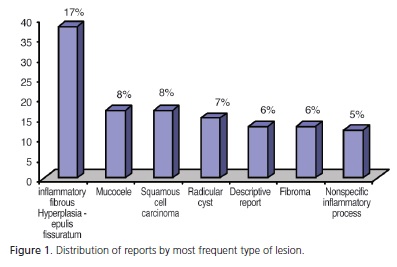
A total of 221 reports were analyzed, from which 55 different diagnoses were observed. Inflammatory fibrous hyperplasia was the lesion most commonly found, appearing in 38 reports (17%), followed by squamous cell carcinoma and mucocele in 17 reports, and radicular cyst in 15 reports. The main demographic characteristics shown were: Females in 59.09% of cases; the fifth decade of life in 29%, and caucasian ethnicity representing 85% of cases. As regards location, the lesions were concentrated mainly in the maxillary and mandibular soft tissues (29%). The number of excisional biopsies was higher, representing 91.4%. Agreement between clinical and histopathologic diagnosis was slightly higher (111 reports) than disagreements.
Conclusion
The results of this study were similar to those of the majority of the articles reviewed with regard to the most frequent oral lesions and their characteristics, in spite of the shortcomings found in the way requisition forms for histopathological examinations had been filled out.
Indexing terms: Biopsy. Epidemiology. Oral pathology.
RESUMO
Objetivo
Analisar a frequência das lesões bucais e suas características nos pacientes atendidos na residência em Cirurgia e Traumatologia Bucomaxilofacial da Santa Casa de Pelotas, Rio Grande do Sul.
Métodos
Foram coletados e analisados os dados dos laudos das biópsias realizadas na residência em Cirurgia e Traumatologia Bucomaxilofacial da Santa Casa de Pelotas durante um período de três anos, obtidos dos Arquivos do Centro de Diagnóstico das Doenças da Boca, em relação ao diagnóstico histológico da lesão, gênero, idade, etnia, localização da lesão, tipo de biópsia, e concordância entre o diagnóstico clínico e o histopatológico.
Resultados
Foram analisados 221 laudos, sendo observados 55 diagnósticos diferentes. A hiperplasia fibrosa inflamatória foi a lesão mais comumente encontrada com 38 laudos (17%), seguida de mucocele e carcinoma espinocelular com 17 laudos, e cisto radicular com 15 laudos. O gênero feminino com 59,09%, a quinta década de vida com 29% e a etnia leucoderma representando 85% foram as principais características demográficas. Com relação à localização, as lesões se concentraram mais nos tecidos moles da maxila e mandíbula (29%). As biópsias excisionais foram superiores e representaram um percentual de 91,4%. A concordância entre o diagnóstico clínico e o histopatológico foi levemente superior (111 laudos) em relação à discordância.
Conclusão
Os resultados obtidos neste estudo são semelhantes aos da maioria dos artigos revisados no que se refere às lesões bucais mais frequentes e suas características, apesar das falhas verificadas no preenchimento das fichas de requisição dos exames histopatológicos.
Termos de indexação: Biópsia. Epidemiologia. Patologia bucal.
INTRODUCTION
Epidemiological survey studies of lesions have revealed their prevalence and incidence, characterizing the population and environment in which they are conducted, and have contributed to improving the diagnosis and treatment of the lesions most commonly found. Therefore, it is important to have a reliable data collection method that generates more precise results, which can be used with confidence.
When lesions related to the bucco maxillofacial complex were verified, the majority of epidemiological studies have shown inflammatory fibrous hyperplasia to be the most frequently occurring lesion1-7. In pediatric population, mucocele has been reported more frequently8-10. With respect to gender, the female has been found more frequently in studies1,4-6,11-15. The age-range of patients affected by lesions varies a great deal according to the studies, and have been observed to occur mostly in the fourth13,15 and fifth decades of life4,7,11,16. In pediatric patients, the age-ranged comprised between 8 and 15 years is the most affected, according to the large majority of studies reviewed9-10,17-18. Furthermore, with regard to pediatric populations, the larger portion of studies9,18-19 found no statistically significant differences in distribution by gender.
The aim of this study was to analyze the characteristics of the most frequently occurring oral lesions, by means of collecting the data and results of biopsies performed in patients cared for in the residency program in Surgery and Bucco Maxillofacial Traumatology ("Cirurgia e Traumatologia Bucomaxilofacial - CTBMF") of "Hospital Santa Casa de Misericórdia" in Pelotas, between the years 2006 and 2008, and compare this information with the literature reviewed.
METHODS
An observational, retrospective, descriptive study was conducted, in which secondary data were collected from January 2006 to December 2008 from the reports of the Center for Diagnosis of Oral Diseases ("Centro de Diagnóstico das Doenças da Boca - CDDB") of the School of Dentistry, Federal University of Pelotas, which served as reference for forwarding the biopsies performed in the Hospital Residency Program in Surgery and Bucco Maxillofacial Traumatology, at "Santa Casa de Pelotas" (RS), and for the Southern Region of the State.
The data collected were analyzed according to the independent variables: histologic diagnosis of the lesion, gender, age (divided into decades), ethnicity (leukoderma, melanoderma or xeroderma), location of the lesion in the oral cavity [soft tissues related to the maxilla and mandible (gingiva, alveolar mucosa, alveolar ridge, palate), maxilla, mandible, tongue, floor of the mouth, jugal mucosa, lip, and other regions], type of biopsy (incisional or excisional), and agreement between clinical and histopathological diagnosis (equal or different). These variables were analyzed in relation to the set of all the exams.
This study was conducted in compliance with all the demands, rules and guidelines of the Research Ethics Committee of the School of Dentistry, Federal University of Pelotas, after obtaining a report in favor of its implementation under Protocol No.054/2008.
RESULTS
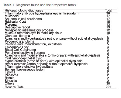
A total of 221 reports were analyzed, with reference to patients cared for between January 2006 to December 2008, by the Hospital Residency program in CTBMF, at Santa Casa de Pelotas (RS). The data of the survey of diagnoses found and their respective totals are shown in Table 1. Of the reports checked in this study, there were histological results that presented only a diagnosis, therefore, in the table, they were grouped in a set denominated "others", which comprised: pleomorphic adenoma, unicystic ameloblastoma, angiofibroma, candidiasis, carcinoma in situ, verrucous carcinoma, actinic keratosis, cystadenocarcinoma, papillary cystadenoma, dentigerous cyst, paradental cyst, compatible with acuminate condyloma, focal cemento-osseous dysplasia, moderate epithelial dysplasia, lichenoid dysplasia, central ossifying fibroma, ectopic salivary gland, hemangioma, reactive lymphoid hyperplasia, vascular leiomyoma (angiomyoma), reactive lymph node, neurofibroma, pigmented cell nevus, compound odontoma, chronic granulomatous infectious process, bone sequestration, suggestive of benign fibro-osseous lesion, suggestive of lichen planus, keratocystic odontogenic tumor, granular cell tumor. These, associated with those listed in the table total 55 different histopathological results.
The main lesions found and their percentages are represented in Figure 1.
There was greater demand for care by women, presenting a ratio of men/women of 0.69, corresponding to 59.09% (130) for women, and 40.91% (90) for men. One of the biopsy charts did not contain this information.
With regard to age, five patients (2%) were in the first decade of life; 22 (10%) in the second; 20 (9%) in the third; 22 (10%) in the fourth; 64 (29%) in the fifth; 47 (21%) in the sixth; and 31 (14%) were in the age-range of over 60 years. In 10 charts (5%) this datum was not filled out.
Leukoderma ethnicity represented 85% of the patients; 12% melanodermas and in seven reports (3%) this information was not filled out. No patient was classified as xeroderma.
The lesions were mostly concentrated in the soft tissues related to the maxilla and mandible - represented by the gingiva, alveolar mucosa, alveolar ridge and palate (29%). In sequence, the other locations observed were the maxilla (15%), lip (14%), jugal mucosa (12%), tongue (11%), mandible (9%), others regions (5%) and floor of the mouth (3%). The description of the lesion site was not filled out in four of the charts (2%).
There were a larger number of excisional biopsies compared with the incisional type. Of a total of 221 biopsied lesions, 202 were performed by the excisional technique (91.4%) and 16 by the incisional technique (7.2%). In three charts (1.4%) this datum was not filled out.
Agreement between the clinical and histopathological diagnosis occurred in 111 cases (50%). In 102 cases (46%) there was no agreement between the diagnoses. In eight charts (4%) this datum was not filled out.
DISCUSSION
Bucco maxillofacial Surgery and Traumatology is a specialty that differs from the majority of the others in dentistry because it presents more activity at hospital level, and by the more frequent interaction with other medical specialties. Because it concerns a reference services, and is free of charge at times, the data in these institutions may serve as a parameter for other studies and dental care services, allowing an estimate of the relative magnitude of the oral health problems in the population cared for, which makes it easier to establish the priority of preventive and therapeutic actions.
As was the case in this survey, a large portion of the studies reviewed1-7 presented inflammatory fibrous hyperplasia as the predominant lesion. With the exception of the studies of Vier et al.4 and Cruz et al.5 who analyzed charts from the pathology services within a hospital, all the others data from Oral Pathology Laboratories, a fact that ratifies inflammatory fibrous hyperplasia as a very common lesion. Another important factor is that the larger portion of the population cared for was concentrated in the more advanced age-ranges, in which there is greater use of prosthetic devices, which may cause a larger number of hyperplastic and inflammatory lesions.
In this study, mucocele was the second most frequently found lesion and was also among the most frequent type in the articles of Tay2, Vier et al.4, Cruz et al.5, Nascimento et al.6 and Weir et al.11, demonstrating that it was a frequent finding in analyses of samples of varying age ranges. But, it is known that it is frequently predominant in populations of youngsters and children, as specific studies in pediatric patients have shown8-10.
Radicular cyst, in turn, was also among the most commonly found lesions in this and other studes1-2,11-12,14. Because it is a pathology that is difficult to resolve by conservative endodontic therapy, it is still frequently removed surgically and consequently sent for histopathological analysis.
In this study, the outstanding finding was the expressive quantity of squamous cell carcinomas (a total of 17 - 8%). This fact may be explained, because the Bucco Maxillofacial Surgery and Traumatology Service of Santa Casa de Misericórdia of Pelotas is the reference in the region for various treatments, which cares for patients who present malignant neoplasias and seek diagnosis for oral manifestations. Annually, the residency program in CTBMF holds an oral cancer prevention week, in addition to providing continuous care for the population throughout the year. In studies that cite the significant presence of carcinomas, squamous cell o carcinoma4,20 is the most frequent. Corroborating the data of this study, Leite Segundo et al.20 and Vier et al.4 also found expressive percentages of squamous cell carcinomas of 10% and 12% respectively. Furthermore, according to Leite Segundo et al.20 the laboratory at which the data of this study were analyzed, is the reference in its region, in the Service of biopsy of lesions of the bucco maxillofacial complex, which may have contributed to the high index of carcinomas.
With regard to the "descriptive report", it is mentioned when it was not possible to give the specific name of a lesion, by virtue of the poor manner in which the biopsy chart was filled out, and/or the biopsied material was removed from a site that was unsuitable for definition of the diagnosis, and was incompatible with the presumptive diagnosis of the surgeon. The descriptive report may also indicate only the presence of normal tissues in the biopsied area. The factor that may have contributed to the high rate of this requirement was the incomplete filling out of the biopsy charts, considering that a total of 110 were in this incompletely filled out condition, representing approximately 50% of the total number of biopsies examined. The item that was most frequently incompletely filled out was with reference to the size of the lesion, and for this reason this feature was left out of the results and discussion. Others authors12-14 have also found incorrectly filled out charts, but in lower quantity (1% to 21% of the total analyzed).
In the majority of studies similar to the present one, a greater predominance of the female gender was also found1,4-6,11-15. Of the six articles8-10,17-19 that analyzed pediatric populations included in this study, half of them9,18-19 found no statistically significant differences in the distribution of lesions by gender. These two abovementioned facts may be represented by the greater care women take of their and their children's health. As regards the malignant neoplasias in this study, the male gender presented greater prevalence, and this datum has also been verified in studies that analyzed the specific prevalence of malignant neoplasias16,21. More drinking and smoking habits in men in comparison with women may justify this datum, and once again, the greater care taken by women as regards their health, between the genders.
In the literature, in spite of the great variation shown in the most affected age-ranges, the fifth decade of life has generally been the most representative4,7,11,16, as it was in this study. Onofre et al.21 analyzing oral neoplasias, verified that the age-range from 61 to 70 years of age was the time of greatest occurrence. Lesions such as inflammatory fibrous hyperplasia and carcinomas are related to a population at a more advanced age, and they were among the most found in this study, which may have resulted in the predominance of a higher age-range.
As was the case in this study, the majority of studies1,8-9,13-14,16 also showed evidence of greater incidence of lesions in leukoderma patients, when ethnicity was analyzed. The fact that over 80% of the population of the State of Rio Grande Sul is white, according to the Brazilian Institute of Geography and Statistics22, may explain this demographic characteristic. Moreover, access to information and health, in this case, continue to be greater in the white population.
As occurred in various studies5,8,15-16,18,21 the site of lesions was predominantly situated in the soft tissues related to the maxilla and mandible, most probably due to the large quantity of reactive and inflammatory lesions present in the major portion of these tissues. It is worth noting that in these studies, the options for the classification of anatomic locations were more specific, such as: gingiva, alveolar mucosa, alveolar ridge, palate; and frequently they were not similar among them. For this reason, and also for the purpose of comparison with this study, they were included in generalized locations in the item "soft tissues of the maxilla and mandible".
In this study, the majority of the biopsies were excisional (90%), reflecting the fact that these biopsied lesions were of a small size, as are a large portion of oral lesions, and which in many cases corresponds to treatment of the lesion. Studies that mentioned this type of datum4,6, also presented a higher number of excisional than incisional biopsies at a percentage higher than 70%, being justified in the same way.
Agreement between the clinical and histopathological diagnosis was slightly higher than disagreement in this study. Vier et al4, in turn, found a coincident percentage of 79.9% in their study. The agreement rates may be the result of the fact that the lesions that are relatively easy to diagnose clinically, such as inflammatory fibrous hyperplasia, mucocele, fibroma, were among the most biopsied type in these studies.
CONCLUSION
From analysis of the results obtained in this study, it could be concluded that the oral lesions most frequently found and their characteristics are similar to those in the majority of the articles reviewed, and so are the demographic data of the population attended, in spite of the failures that occurred with regard to the lack of data about the lesion on the exam requisition forms, when forwarding the material sent to the Histopathological Services.
ACKNOWLEDGEMENTS
The authors thank the Center for the Diagnosis of Diseases of the Mouth of the School of Dentistry, Federal University of Pelotas, its team of professionals, and the Coordinator of the Residency Program in Bucco Maxillofacial Surgery and Traumatology, of Santa Casa in Pelotas, Dr. Ronaldo Lemes da Silva.
Collaborators
MR TAKASHIMA and A ETGES were responsible for the entire elaboration of the article.
REFERENCES
1. Marin HJI, Da Silveira MMF, De Souza GFM, Pereira JRD. Lesões bucais: concordância diagnóstica na Faculdade de Odontologia de Pernambuco. Odontologia Clín-Científ. 2007;6(4):315-8. [ Links ]
2. Tay ABG. A 5-year survey of oral biopsies in an oral surgical unit in Singapore: 1993-1997. Ann Acad Med Singapore. 1999;28(5):665-71.
3. Leonel ECF, Vieira EH, Gabrielli MAC. Análise retrospectiva da incidência, diagnóstico e tratamento das lesöes bucais encontradas no Serviço de Cirurgia e Traumatologia Bucomaxilofacial da Faculdade de Odontologia de Araraquara - UNESP. Rev Paul Odontol. 2002;24(3):18-22.
4. Vier FV, Rockenbach MIB, Gabriel JG, Yurgel LS, Cherubini K, Figueiredo MAZ. Diagnósticos histopatológicos do Laboratório de Patologia do Serviço de Estomatologia da PUCRS, nos anos de 2000 a 2002 e sua relação com o diagnóstico clínico. Rev Odonto Ciênc. 2004;19(46):382-8.
5. Cruz MCFN, Almeida KGB, Lopes FF, Bastos EG, Freitas RA. Levantamento e biópsias da cavidade oral realizadas no Hospital Universitário - Unidade Presidente Dutra/UFMA, da cidade de São Luís (MA), no período de 1992 a 2002. Rev Bras Patol Oral. 2005;4(3):185-8.
6. Nascimento GJF, Paraíso DP, Góes PSA, Sobral APV. Estudo epidemiológico de 2.147 casos de lesões bucomaxilofaciais. Rev Bras Patol Oral. 2005;4(2):82-9.
7. Bertoja IC, Tomazini JG, Braosi APR, Zielak JC, Reis LFG, Giovanini AF. Prevalência de lesões bucais diagnosticadas pelo Laboratório de Histopatologia do UnicenP. Rev Sul-Bras Odontol. 2007;4(2):41-6.
8. Das S, Das AK. A review of pediatric oral biopsies from a surgical pathology service in a dental school. Pediatr Dent J. 1993;15(3):208-11.
9. Sousa FB, Etges A, Corrêa L, Mesquita RA, de Araújo NS. Pediatric oral lesions: a 15-year review from São Paulo, Brazil. J Clin Pediatr Dent. 2002;26(4):413-8.
10. Lima GS, Fontes ST, Araújo LMA, Etges A, Tarquinio SBC, Gomes APN. A survey of oral and maxillofacial biopsies in children. A single-center retrospective study of 20 years in Pelotas-Brazil. J Appl Oral Sci. 2008;16(6):397-402. doi: 10.1590/S1678- 77572008000600008.
11. Weir JC, Davenport WD, Skinner RL. A diagnostic and epidemiologic survey of 15,783 oral lesions. J Am Dent Assoc. 1987;115(3):439-42. 12. Ovalle CJW. Prevalencia de lesiones histopatológicas bucales en la Zona del Bajío. Rev ADM. 2000;57(4):132-6.
13. Grandi G, Maito FDM, Sant'ana Filho M. Estudo epidemiológico das lesões ósseas diagnosticadas no serviço de patologia bucal da PUCRS [citado 2010 Maio 6]. Disponível em: <www. odontologia.com.br>.
14. Deboni MCZ, Traina AA, Trindade IK, Rocha EMV, Teixeira VCB, Takahashi A. Levantamento retrospectivo dos resultados dos exames anatomopatológicos da disciplina de cirurgia da FOUSPSP. RPG Rev Pós Grad. 2005;12(2):229-33.
15. Bataineh A, Al-Dwairi ZN. A survey of localized lesions of oral tissues: a clinicopathological study. J Contemp Dent Pract. 2005;6(3):30-9.
16. Amorim AG, Amorim RFB, Freitas RA. Estudo epidemiológico do carcinoma epidermóide oral: análise de 85 casos. Odontol Clin- Cient. 2002;1(1):41-5.
17. Cavalcante ASR, Marsilio AL, Kühne SS, Carvalho YR. Lesões bucais de tecido mole e ósseo em crianças e adolescentes. PGR Pós-Grad Rev Fac Odontol Säo José dos Campos. 1999;2(1):67- 75.
18. Gültelkin SE, Tokman B, Türkseven MR. A review of paediatric oral biopsies in Turkey. Int Dent J. 2003;53(1):26-32.
19. Dhanuthai K, Banrai M, Limpanaputtajak S. A retrospective study of paediatric oral lesions from Thailand. Int J Paediatr Dent. 2007;17(4):248-53. doi: 10.1111/j.1365-263X.2007.00828.x.
20. Leite Segundo AV, Da Silva UH, Martelli PJL. Estudo retrospectivo de exames anatomopatológicos do Laboratório de Anatomia Patológica da Faculdade de Odontologia de Caruaru / PE. Odontol Clin-Cient. 2003;2(1):15-20.
21. Onofre MA, Sposto MR, Simões ME, Scaf G, Ferreira LA, Turatti E. Prevalência de câncer bucal: no serviço de medicina bucal da Faculdade de Odontologia de Araraquara/UNESP: 1989-1995. RGO - Rev Gaúcha Odontol. 1997;45(2):101-4.
22. Instituto Brasileiro de Geografia e Estatística. Censo 2000 [citado 2010 Maio 6]. Disponível em: <http://www.ibge.gov.br/home/estatistica/populacao/defaulttab_amostra.shtm>.
 Correspondence to:
MR TAKASHIMA
e-mail: mrtakashima@yahoo.com.br
Received on: 18/8/2010
Final version resubmitted on: 18/11/2010
Approved on: 16/3/2011
