


Serviços Personalizados
Artigo
 pdf em Inglês
 Artigo em XML
 Referências do artigo
 Enviar este artigo por email
Links relacionados
Compartilhar

 Permalink
RGO.Revista Gaúcha de Odontologia (Online)
versão On-line ISSN 1981-8637
RGO, Rev. gaúch. odontol. (Online) vol.60 no.4 Porto Alegre Out./Dez. 2012
ORIGINAL / ORIGINAL
Prevalence of dental anomalies of number, size, shape and structure
Prevalência de anomalias dentais de número, tamanho, forma e estrutura
Deborah Queiroz de FREITAS I; Richard Yuiti TSUMURAI II; Denilson Newton São Pedro MACHADO FILHO II
I Universidade Estadual de Campinas, Faculdade de Odontologia, Departamento de Radiologia Odontológica. Piracicaba, SP
II Centro Universitário de Rio Preto, Curso de Odontologia. São José do Rio Preto, SP, Brasil
ABSTRACT
Objective
To evaluate the prevalence of dental anomalies (of number, form, size and structure) using panoramic images, in São José do Rio Preto, São Paulo, Brazil.
Methods
512 panoramic radiographies were evaluated from orthodontic patients between the ages of 6 and 20; only suitable images were examined. We considered all permanent teeth; the analysis of third molars was included from 12 years of age onwards. A chi-square test was used to determine the difference in prevalence of dental anomalies between the genders.
Results
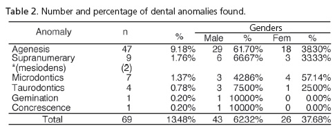
Dental anomalies were observed in 69 (13.48%) radiographies; 47 (9.18%) were agenesis, 9 (1.76%) were supernumerary teeth, 7 (1.37%) were microdontic teeth, 4 (0.78%) were taurodontism, 1 (0.2%) was gemination and 1 (0.2%) was concrescence. There was a higher prevalence of anomalies in males (p=0.013). Other dental anomalies were not observed.
Conclusion
The dental anomalies show a significant rate of incidence; therefore, the professional should be present in the planning of dental treatment, especially orthodontic planning; in addition, radiographic evaluation is essential in such cases.
Indexing terms: Anodontia. Epidemiology. Panoramic radiography. Tooth abnormalities.
RESUMO
Objetivo
Avaliar a prevalência das anomalias dentais (de número, tamanho, forma e estrutura) pelo exame de radiografias panorâmicas, na cidade de São José do Rio Preto, São Paulo.
Métodos
Foram avaliadas 512 radiografias panorâmicas, de indivíduos do sexo masculino e feminino, com idade entre 6 a 20 anos, realizadas para início de tratamento ortodôntico. Apenas radiografias com qualidade adequada foram incluídas na amostra. Foram considerados todos os dentes permanentes; a avaliação dos terceiros molares foi incluída a partir dos 12 anos de idade. O teste qui-quadrado foi utilizado para determinar diferença na prevalência de anomalias dentais entre os sexos.
Resultados
Anomalias dentais foram encontradas em 69 (13,48%) radiografias panorâmicas avaliadas, sendo 47 (9,18%) agenesias, 9 (1,76%) dentes supranumerários, 7 (1,37%) microdontias, 4 (0,78%) taurodontias, 1 (0,2%) geminação e 1 (0,2%) concrescência. Houve maior prevalência de anomalias no sexo masculino (p=0,013). Outras anomalias de forma e anomalias de estrutura não foram observadas.
Conclusão
As anomalias dentais apresentam uma importante incidência, por isso o profissional deve considerar sua presença no plano de tratamento, especialmente nos ortodônticos, para favorecer a estética e a função desse paciente; também é importante ressaltar o papel do exame radiográfico na sua detecção.
Termos de indexação: Anodontia. Epidemiologia. Radiografia panorâmica. Anormalidades dentárias.
INTRODUCTION
Generally speaking, anomalies can be classified as hereditary, congenital or acquired. With hereditary anomalies, etiological factors acted on the genetic information phase, causing alterations in cell differentiation that cause structural modifications either before or after birth. With congenital anomalies, etiological factors acted on the intra-uterine formation phase, altering the composition and/or function of the affected organ, which also takes place with acquired anomalies; with the latter, however, the etiological factors acted in the post-natal formation phase. The main causes of congenital and acquired anomalies are infections, traumas, nutritional variations and temperature as well as intoxication from chemical substances.
Dental anomalies may be classified as to number, size, shape and structure of the afflicted tooth or teeth.
According to Yamada1, 5% of the population is born with some hereditary anomaly and around 60% of these anomalies involve the teeth, the upper jaw or the face.
Certain anomalies of form, such as lacerations, supernumerary root, invaginated tooth and taurodontism, do not cause significant alterations to a patient's oral health, his treatment needs or changes in treatment plans. On the other hand, other anomalies of form, shape, number, size or structure, generally require evaluation and intervention by a professional or must at least be considered during dental treatment, particularly in the planning of orthodontic treatment.
Many anomalies can only be detected or confirmed by radiographic examination and indeed panoramic radiography is considered to be very important to this evaluation, as it is an extremely comprehensive examination which makes it possible to evaluate all dental elements in just one image; moreover, they are easily obtainable and use low doses of radiation2-3.
Due to the high clinical importance, there are many studies about the prevalence of dental anomalies in the literature in various regions of the country and also in other countries2-20. There are a number of differences in these studies, in terms of the frequency of the anomaly and the teeth involved, which have been attributed to differences in the regions and the examined population.
The aim of this study is to evaluate the prevalence of dental anomalies of number, size, form and structure, by means of the panoramic radiography of individuals between the ages of 6 and 20, in the city of São José do Rio Preto, in the state of São Paulo, Brazil.
METHODS
A total of 512 panoramic radiographies were evaluated of patients in the 6 to 20 age range, from the records of three Dental Clinics in the city of São José do Rio Preto.
Patient age was the first criterion for inclusion in the sample. In addition, the quality was analyzed of the panoramic radiography taken prior to commencement of dental treatment. Only those of good quality were selected, with a minimum of distortion and adequate density and contrast, these being considered ideal for evaluation. Cases where it was not possible to determine if the absence of one or more dental elements was a result of agenesis or extraction, were excluded.
The area used for the radiographic interpretation possessed ideal lighting conditions, that is, a dark room whose only light came from the negatoscope, and with the assistance of a magnifying glass.
The analysis of the radiographies was carried out by a duly qualified examiner who evaluated the number and morphology of all the permanent teeth present. The presence of all anomalies was taken into consideration (number, size, form and structure), with the exception of supranumerary roots and lacerations. The evaluation of third molars was only conducted in patients aged 12 or above.
The data were noted on a record card specially developed for this case study and were subsequently tabulated. The chi-square test was used to assess the existence of differences in the prevalence of dental anomalies between the genders, with a 5% level of significance.
The study was approved by the Ethics in Research Committee at the Faculty of Dentistry in Piracicaba (protocol no. 040/2007), and all the requirements and standards of the National Health Board's, Resolution 196 of June 13, 1996 were observed during execution as were the main ethical principles contained in the Declaration of Helsinki (2000).
RESULTS
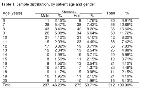
Table 1 shows the sample distribution, by patient age and gender. A total of 46.29% were male while 53.71% were female, characterizing a sample with a homogenous distribution between the genders. The majority of the sample (63.89%) fell within the 7 to 12 age group, with an average age of 11.2 years.
Table 2 shows the number and percentage of dental anomalies found, by gender. There was a greater prevalence of anomalies in the male gender (62.32%). The chi-square test showed a statistically significant difference between the genders for the anomalies (p=0.013).
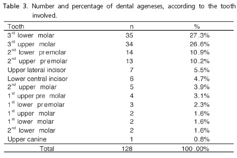
Table 3 exhibits the frequency of agenesis, according to the tooth involved.
DISCUSSION
Normally, anomalies in dental development are detected by routine clinical examinations and periapical and/or panoramic radiographic exams2-3. Their diagnosis, carried out at the start of the mixed dentition at around 6 years of age, permits the dental surgeon to adopt a preventive attitude towards the possible esthetic and functional problems which could in the future exist in the normal development pattern. Early diagnosis benefits normal occlusion since permanent tooth eruption can be planned21.
Panoramic radiography is very important in the study of the prevalence of dental anomalies and increases the possibility of early detection, as it is an examination that shows all the upper and lower teeth, it is easy to perform, comfortable for the patient and uses low doses of radiation2-3. These characteristics led to it being chosen for the evaluation of anomalies in the present research study.
Several authors recommend the performance of the radiographic exam at school age to enable the planning of preventive therapy2,5,7.
The most frequent occurrence of anomalies was detected in the 7 to 12 age range, a result which is in agreement with the majority of authors14,22. In the present study, it is possible to attribute this to the fact that the majority of the sample was in this age group, coinciding with the age at which the patients seek dental treatment, since the radiographies were selected from Orthodontic Clinics.
Dental anomalies were found in 13.48% of the radiographies examined. Out of the grand total of anomalies, a larger percentage of dental anomalies was noted in the male gender (62.32%) compared with females (37.68%), an outcome different from that found by Watanabe et al.14, who found a greater prevalence of anomalies in the female gender, and that found by Paula & Ferrer3 and Tallón-Walton et al.20, who all observed a greater prevalence of agenesis in the female gender. Bearing in mind that the samples of the aforementioned authors were composed of a higher percentage of female patients and that the sample in the current study has a similar number of patients of both sexes, it may be stated that the outcome found here better reflects the distribution of anomalies by gender. The differences in the populations studied also have to be taken into consideration as the authors evaluated patients in Piracicaba, Goiânia and the Spanish hinterland, respectively. Grieco et al.18 and Gomes et al.19 found a greater prevalence of agenesis in the female gender in samples with a homogeneous distribution between the genders, however there was no statistically significant difference in their studies.
The most prevalent anomalies were those of number (10.94%), with the majority of them (9.18%) relating to dental agenesis. Similar values were found by Ciamponi & Frassei11 (9.31%), by Ledesma-Montes et al.12 (8.3%) and by Tallón-Walton et al.20 (9.48%). However, Marques et al.15, Barbosa & Kroll16, Paula & Ferrer3, Grieco et al.18 and Gomes et al.19 all found lower prevalence of agenesis than that of this study: 6%, 4.54%, 2.9%, 6.89% and 6.3%, respectively. On the other hand, Castilho et al.9 observed 24.37% prevalence for ageneses.
The order of teeth afflicted in relation to dental agenesis is in agreement with the studies of several authors2,5,21-22 who found the following sequence: upper and lower second premolars, upper lateral incisor, lower central incisor, upper first premolar, lower lateral incisor, upper and lower second molars (third molars were not included in these samples). It was ascertained that there was only a difference in respect of the lower lateral incisors, as no instance of this was found in the present study. Paula & Ferrer3, Castilho et al.9, Borba et al.13 and Barbosa & Kroll16, who did consider third molars in their evaluations, found similar data to those in the present study, where lower and upper third molars demonstrated higher values of agenesis. It is also in agreement with the studies of Dahlberg23 and his classification of teeth into stable and variable.
Here, the absence of third molars was only computed after 12 years of age since it may not be possible to discern them in radiographies of patients younger than this, which does not mean that these teeth were not in the process of formation and would not appear in subsequent radiographies. This could lead to overestimated values for agenesis as this was not a longitudinal study. Castilho et al.9 also used a starting age of 12 as a parameter in their study of agenesis of third molars.
Of the total of ageneses, 51.6% occurred in the maxilla and 48.4% in the mandible, which showed similar values for upper and lower teeth afflictions, as also occurred in the study of Paula & Ferrer3.
Differences in the frequency of agenesis, and in afflicted teeth in the various studies, may be explained by the influence of racial variations, socio-economic variations and geographic location, factors which should not be overlooked until such time as evidence is found that they do not impact the congenital absence of teeth8.
The prevalence of supranumerary teeth was 1.76%, which was similar to that of McKibben & Brearley4, who obtained 1.53% and Lecco Berrocal et al.17, who found 1.05% prevalence, and was higher than that of Tallón-Walton et al.20, whose study pointed to 0.39% of supranumerary cases.
These results could be evidence that the human dentition today has a tendency towards a reduced number of teeth, rather than the opposite. Modern anthropological science states that the number of teeth in human beings shows a tendency to diminish in accordance with the way we chew today14.
As regards size anomalies, microdontia was found in 1.37% of cases, a similar value to that of Ledesma-Montes et al.12 (1.2%), but lower, however, than that of Carvalho & Tamburús6, who found 4.83%, Carvalho et al.10, who obtained 3.96% and Tallón-Walton et al.20, who noted 5.5% in their study. Macrodontia was not observed in this sample.
Dental anomalies of form comprised 0.2% gemination and 0.2% concrescence, results close to those in the studies of McKibben & Brearley4; there was also 0.78% taurodontia, data similar to those of Carvalho & Tamburús6, who found 0.52% and to those of Carvalho et al.10, who found a prevalence of 1%.
In the present study, numbers related to supranumerary roots and lacerations were not included. Taking into consideration that previous studies6,10,12-14,20 did not evaluate the aforementioned anomalies, a comparison with other studies of prevalence found here, would not be possible. Accordingly, it was decided not to record them. These authors did not provide justification for these exclusions, but it is believed that, as they relate to the more commonly observed alterations, their evaluation could have led to an overestimated number of anomalies. Moreover, they are alterations which do not occasion the need for treatment, as do those evaluated here.
Other form and structure anomalies were not observed.
CONCLUSION
The prevalence values obtained were similar to those of several studies, but differed from others, demonstrating the difference between populations in different regions. Despite this, it should be considered that the anomalies show a significant relative incidence: the professional must be alert to its presence for the performance of early diagnosis and a plan of treatment which benefits patient esthetics and function. It should also be emphasized that, in the majority of cases, the radiographic exam is essential to its detection.
Collaborators
DQ FREITAS was responsible for the supervision of all the stages of research, performance of the statistical analysis, corrections and composition of the finished article. RY TSUMURAI was responsible for the conception, evaluation of panoramic radiographies and composition of the article. DNSP MACHADO FILHO was responsible for a review of the literature and the composition of the article.
REFERENCES
1. Yamada N. Oral radiographic abnormalities in genetic diseases. Dent. Outl. 1983;62(1):71-8. [ Links ]
2. Buenviaje TM, Rapp R. Dental anomalies in children: a clinical and radiographic survey. ASDC J Dent Child. 1984;51(1):42-6.
3. Paula AFB, Ferrer KJN. Prevalência de agenesia em uma clínica ortodôntica de Goiânia. RGO - Rev Gaúcha Odontol. 2007;55(2):149-53.
4. McKibben DR, Brearley LJ. Radiographic determination of the prevalence of selected dental anomalies in children. ASDC J Dent Child.1971;28(6):390-8.
5. Davis PJ. Hypodontia and hyperdontia of permanent teeth in Hong Kong school children. Community Dent Oral Epidemiol. 1987;15(4):218-20.
6. Carvalho FR, Tamburús JR. Estudo radiográfico da incidência de anomalias dentais - contribuição ao estudo de algumas anomalias. Rev Assoc Paul Cir Dent. 1988;42(3):217-9.
7. Neal JJ, Bowden DE. The diagnostic value of panoramic radiographs in children aged nine to ten years. Br J Orthod. 1988;15(3):193-7.
8. Glen FF. A consecutive six year study of the prevalence of congenitally missing teeth in private pedodontic practice of two geographically separated areas. J Dent Child. 1964;31(3):264-70.
9. Castilho JCM, Nicodemo RA, Bazzarella CB. Prevalência de anodontia entre estudantes do 2º grau da cidade de São José dos Campos: correlação dessa anomalia entre terceiros molares e outros órgãos dentários. Rev Odontol UNESP. 1990;19(1):269-76.
10. Carvalho PL, Simi R, Abdalla CM, Ferrero CA, Oliveira RJ. Estudo da prevalência das anomalias dentais por meio das radiografias panorâmicas. Rev Odontol Univ Santo Amaro. 1997;2(3):28-30.
11. Ciamponi AL, Frassei VAS. Anodontias parciais de dentes permanentes: estudo da prevalência em crianças residentes na cidade de São Paulo. RPG Rev Pos-Grad. 1999;6(3):213-7.
12. Ledesma-Montes C, Salcido-García JF, Hernández-Flores F, Garcés-Ortíz M. Pathological findings in a sample of Mexican pediatric patients. Clinical and radiographic survey. Minerva Stomatol. 2012;61(5):205-12.
13. Borba GVC, Borba Júnior JC, Pereira KFS, Silva PG. Levantamento da prevalência de agenesias dentais em pacientes com idade entre 7 e 16 anos. RGO - Rev Gaúcha Odontol. 2010;58(1):35-9.
14. Watanabe PCA, Souza JG, Almeida SM, Montebello Filho A. Estudo Radiográfico (Ortopantomográfico) da incidência das anomalias dentais de número na região de Piracicaba - SP. ROBRAC. 1997;6(21):32-8.
15. Marques LS, Souki BQ, Mazzieiro ET. Desenvolvimento dentário: um estudo radiográfico. JBP J Bras Odontopediatr Odontol Bebê. 2002;5(28):464-9.
16. Barbosa SRC, Kroll LB. Contribuição ao estudo da prevalência das agenesias dentais da cidade de Iporá - GO. Rev ABRO. 2004;5(2):72-4.
17. Leco Berrocal MI, Martín Morales JF, Martínez González JM. An observational study of the frequency of supernumerary teeth in a population of 2000 patients. Med Oral Patol Oral Cir Bucal. 2007;12(2):134-8.
18. Grieco FAD, Carvalho PEG, Guedes-Pinto E, Garib DG, Valle- Corrotti KM. Prevalência de agenesia dentária em pacientes ortodônticos da cidade de São Paulo. RPG Rev Pós Grad. 2007;13(4):312-7.
19. Gomes RR, da Fonseca JA, Paula LM, Faber J, Acevedo AC. Prevalence of hypodontia in orthodontic patients in Brasilia, Brazil. Eur J Orthod. 2010;32(3):302-6. doi: doi: 10.1093/ejo/ cjp107.
20. Tallón-Walton V, Nieminen P, Arte S, Carvalho-Lobato P, Ustrell- Torrent JM, Manzanares-Céspedes MC. An epidemiological study of dental agenesis in a primary health area in Spain: estimated prevalence and associated factors. Med Oral Patol Oral Cir Bucal. 2010;15(4):e569-74. doi: doi:10.4317/medoral.15.e569.
21. Montebello Filho A, Possobon RF, Freitas DQ, Pistóia GD. Agenesia bilateral de primeiro molar permanente: revisão de literatura e relato de casos. Rev ABRO. 2003;4(1):15-9.
22. Salinas CF. Orodental findings and genetic disorders. Birth Defects Orig Artic Ser. 1982;18(1):79-120.
23. Dahlberg AA. Concepts of occlusion in physical anthropology and comparative anatomy. J Am Dent Assoc. 1953;46(5):530-5.
 Correspondence to:
DQ FREITAS
e-mail: deborahqf@hotmail.com
Received on: 18/5/2010
Final version resubmitted on: 14/11/2010
Approved on: 12/1/2011
