


Serviços Personalizados
Artigo
 pdf em Inglês
 Artigo em XML
 Referências do artigo
 Enviar este artigo por email
Links relacionados
Compartilhar

 Permalink
RGO.Revista Gaúcha de Odontologia (Online)
versão On-line ISSN 1981-8637
RGO, Rev. gaúch. odontol. (Online) vol.60 no.4 Porto Alegre Out./Dez. 2012
ORIGINAL / ORIGINAL
Influence of calcium hydroxide remains on marginal leakage of filled standard root sections
Influência dos resíduos de hidróxido de cálcio na infiltração marginal de corpos de prova obturados
Sara Teodoro MARRA I; Maria Antonieta Veloso Carvalho de OLIVEIRA I; João Carlos Gabrielli BIFfiI
I Universidade Federal de Uberlândia, Faculdade de Odontologia. Uberlândia, MG, Brasil
ABSTRACT
Objective
To evaluate the influence of calcium hydroxide on the degree of marginal leakage in roots filled with two different types of sealers.
Methods
Roots of single-rooted bovine teeth (n=54) were instrumented and divided into four groups of 12 samples each: G1 and G2 were filled with zinc oxide-eugenol sealer, G3 and G4 filled with resin calcium hydroxide based-sealer. The G1 and G3 received calcium hydroxide intracanal medication for seven days. After removal of the medication, samples of the four groups were filled using the lateral condensation technique. The six remaining roots underwent biomechanical preparation, but received no medication or filling, serving as a control group. The apical third and part of the middle third were cut, leaving just 6mm in total length. The specimens were immersed in India ink for 72 hours. Cross sections were made to determine the length and depth of penetration of the dye and were quantified macroscopically with the software ImageTool 3.00.
Results
The results showed no statistical difference (p <0.05) with the groups that were filled with resin calcium hydroxide based-sealer, regardless of previous use of intracanal medication or not. However when the filling was made with zinc oxide-eugenol sealer, dye penetration was higher in the group that received no intracanal medication.
Conclusion
The presence of residues of calcium hydroxide only had an influence on leakage when the sealer was made with zinc oxide-eugenol.
Indexing terms: Calcium hydroxide. Coloring agents. Dental leakage.
RESUMO
Objetivo
Avaliar a influência do hidróxido de cálcio sobre o grau de infiltração marginal em raízes obturadas com dois tipos de cimentos.
Métodos
Raízes de dentes bovinos unirradiculares (n=54) foram instrumentadas e divididas em quatro grupos com 12 amostras: G1 e G2 obturados com cimento à base de óxido de zinco e eugenol; G3 e G4 obturados com cimento resinoso com hidróxido de cálcio. Os grupos G1 e G3 receberam medicação intracanal de hidróxido de cálcio por sete dias. Após a remoção da medicação, os quatro grupos foram obturados pela técnica de condensação lateral. As seis raízes restantes passaram pelo preparo biomecânico, mas não receberam medicação intracanal nem obturação, serviram como grupo controle. O terço cervical e parte do terço médio das raízes foram seccionados, permanecendo somente 6,0 mm do comprimento total. Foram imersas no corante tinta India por 72 horas. Secções transversais foram feitas para verificação da extensão e profundidade de penetração do corante e quantificadas macroscopicamente com o software ImageTool 3.00.
Resultados
Os resultados não apresentaram diferença estatística (p<0,05) com relação aos grupos obturados com cimento resinoso, independente do uso prévio ou não da medicação intracanal. Entretanto quando o cimento à base de óxido de zinco e eugenol foi usado a penetração do corante foi maior no grupo que não recebeu medicação intracanal.
Conclusão
A presença dos resíduos do hidróxido de cálcio influenciou na infiltração marginal, somente quando o cimento usado foi à base de óxido de zinco e eugenol.
Termos de indexação: Hidróxido de cálcio. Corantes. Infiltração dentária.
INTRODUCTION
The permanence of residue from calcium hydroxide (Ca(OH)2) intracanal medication, even after its clinical removal, and regardless of the technique or material used, is a fact proven in the literature1-3. There have been various studies and explanations to answer the question concerning the potential impact of these Ca(OH)2 residues on the root canal filling1,3-5.
In the opinion of some authors, the remaining Ca(OH)2 would be incorporated into the cement of the filling, causing a decrease in the permeability of this filling1. Others have proposed that these residues would determine an acceleration in the setting time and mainly the expansion of certain cements, which would provide a better adaptation to the walls of the root canal4. However, several studies have demonstrated that Ca(OH)2, in contact with the filling cement, has a negative impact on the latter's adhesion to the wall of the root canal2-3. There also exists the possibility of the Ca(OH)2 reacting with the tissue fluids forming calcium carbonate, which is reabsorbable and, in the long run, could create spaces in the interface between the filling and the wall of the root canal1.
Normally, the ability of filling materials to provide a seal is evaluated by means of marginal leakage tests, as they are easy to perform and do not require sophisticated materials. Their results, however, are questionable due to the numerous factors that may have an impact on these studies, such as operator experience, the period between the filling and immersion, the time the samples remain in the dye, the type of dye, the presence or not of the smear layer, the presence of air or liquid inside the canal after filling, the linear measurement of the leakage6. When extracted teeth are used in these studies, several other factors may have an impact on the outcome, such as dental anatomy, the morphology of the root canal system, the dentin structure, the efficiency of root canal preparation, the irrigation procedure, the type of cement and how it is mixed and the filling materials7.
The lack of sample standardization in the leakage studies is an important factor, mainly dictated by the impossibility of anatomic standardization, as dental anatomy is extremely diversified. In an attempt to minimize the anatomical variances, the majority of studies use the same dental group, with similar characteristics. Others, besides belonging to the same dental group, also remove the crowns and the root tips of the teeth8-9. The use of cylindrical test specimens made from bovine teeth10 is also common.
Many studies have evaluated the marginal leakage of dyes, using as a model simulated root canals in resin blocks11, in glass tubes7-12 or in plastic blocks13. These models permit the standardization of the apical diameter and length of the canal, enabling comparisons of preparation, irrigation and filling in experimental conditions without the variability of the biological material in samples11.
Based on questions such as these, it seemed to us important to carry out a leakage study by employing standardized test specimens, with the aim of evaluating the influence of calcium hydroxide on the degree of marginal leakage in filled roots with two different types of cement, one having a zinc oxide-eugenol base (Endofill) and the other a resin base with Ca(OH)2 (Sealer 26).
METHODS
Fifty-four lower bovine teeth from adult animals with completely formed roots and single canals were selected for the present research study. After the teeth were extracted, they were stored in 10% formaldehyde (Oxidial, Mar Del Plata, Argentina) and the soft tissue adhering to the root was removed with the aid of a scalpel blade no. 15 (Golgran, São Paulo, Brazil).
With the aim of facilitating the instrumentation of the canals, the dental crowns were sectioned in the cervical region with a double-sided diamond disk (Buehler, Wawkegan, USA) mounted on a precision cutter (Buehler, Wawkegan, USA), at a speed of 350 rpm. The removal of pulp tissue was carried out using a barbed broach no. 30 (8000 Munich 70, Germany), and only those teeth that had an anatomic diameter of the apical foramen compatible with a Kerr file no. 40 (Sybron Kerr, Orange, USA) remained in the study.
The instrumentation technique used was the stepback technique, establishing the apical stop at 1 mm (19 mm) using as the memory instrument file type K 55 and step-back as far as the file type K 80. During the entire instrumentation, irrigation was conducted with a solution of 1% sodium hypochlorite (Farma, Serrana, Brazil), with around 2 ml of solution at each instrument change. Vacuuming was carried out with the aid of a vacuum cannula connected to a micropump (Nevoni, Barueri, Brazil). The final cleaning of the foramen was performed with a file type K 15, ensuring that it was not obstructed, followed by irrigation with 5 ml of saline solution (Farmax, São Paulo, Brazil). After the instrumentation, the root canals were dried with absorbent paper cones (Tanari, Manacapuru, Brazil) and calibrated with the diameter of the memory instrument.
The teeth were divided into groups according to whether or not intracanal medication was used and to the type of cement employed: Group 1 (n=12) - with intracanal medication and filled with a zinc oxide-eugenol (ZOE) based cement; Group 2 (n=12) – no intracanal medication and filled with an ZOE based cement; Group 3 (n=12) – with intracanal medication and filled with a resin cement with Ca(OH)2; Group 4 (n=12) - no intracanal medication and filled with a resin cement with Ca(OH)2; Control group (n=6) - no intracanal medication and without filling.
The roots of groups G1 and G3 were secured, alternately, in a bench vice (Western, São Paulo, Brazil) to help with administering the intracanal medication. The canals were filled with calcium hydroxide paste P.A. (Biodinâmica, Ibiporã, Brazil) associated with the saline solution and prepared on a glass dish until a creamy consistency was achieved. This paste was applied to the root canals using a disposable syringe with a 25 x 0.7 hypodermic needle (Embramac, Barranquilla, Colombia), along the whole extension of the working length (19 mm), by injecting the paste until seen to be seeping out of the apex.
The temporary sealing of the cervical portion was performed using ZOE based cement (IRM, Dentsply, Petrópolis, Brazil). The samples were kept for a period of seven days in an oven at 37°C and an environment with 100% humidity. To maintain humidity, sponges were adapted to the lids of the jars where the roots were kept, and periodically moistened with saline solution.
The medication was removed with abundant irrigation of saline solution (5 ml) and gentle filing with the memory instrument along the working length. The irrigation was carried out until the reflux came out clear. A vacuum micropump was used for vacuuming and no. 55 absorbent paper cones (Tanari, Manacapuru, Brazil) were used for the final drying of the root canals.
The filling was performed following the technique of lateral condensation, with a ZOE based cement (Endofill, Dentsply, Petrópolis, Brazil) or a resin cement with Ca(OH)2 (Sealer 26, Dentsply, Petrópolis, Brazil). Three roots were filled at a time, previously secured in a bench vice. The cements were transported to the inside of the canals with the aid of digital spacer no. 25 (8000 Munich 70, Germany). The main gutta-percha cones (nº 55) (Tanari, Manacapuru/AM, Brazil) whose diameters were previously checked by means of a gauge ruler (Endo calibradora GT, Maillefer, Ballaigues, Switzerland), were introduced as far as their adaptation to the apical stop. Active lateral condensation was carried out using a no. 25 digital spacer, which was pressed apically and laterally for the seating of the cone and opening of space for the FF accessory cones (Tanari, Manacapuru, Brazil), placing the necessary number of accessory cones so that the spacer only penetrated the cervical third of the root segments. The excess of filling material was removed with heated Paiva tampers (Golgran, São Paulo, Brazil) and the vertical condensation was carried out. After temporary sealing with ZOE cement (IRM, Dentsply, Petrópolis, Brazil), the roots remained in the oven at 37°C and 100% humidity for 72 hours.
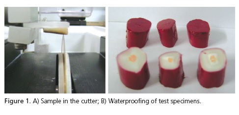
The roots had the apical third and a part of the middle third removed using the precision cutter (Buehler, Wawkegan, USA), leaving only 6 mm of length in total for leakage with dye (Figure 1A). The samples were fully waterproofed with the filling material remaining exposed on the side facing the apical third (Figure 1B), with two layers of nail varnish (Colorama, Procosa, São Paulo, Brazil) allowed to dry for one hour.
They were then placed for 10 minutes in a vacuum pump (Primar, São Paulo, Brazil) at a pressure of 56 KPa. After being fully immersed in the India ink dye (Faber Castell, Germany), they were placed once again in the vacuum pump for 10 minutes. This vacuum environment was used to eliminate possible bubbles that might be present inside the root canal fillings. After 72 hours stored in an environment at 37°C and 100% humidity, the roots were washed in running water for an hour to remove the excess dye solution. The varnish coating was removed with the aid of scalpel blades no. 15.
The six roots that underwent biomechanical preparation, but which did not have intracanal medication or fillings, were separated into two groups, after removal of the apical third and a part of the middle third, to check the leakage stage. The positive control group (n=3), did not have its roots waterproofed in order to test the effectiveness of the India ink in penetrating the root canal. The negative control group (n=3) had its roots fully waterproofed in order to test the effectiveness of the varnish in preventing penetration of the dye into the root canal.
Cross-sections 1.5 mm thick were produced for all samples using a precision cutter. The number of sections obtained in each test specimen was determined by the presence of dye leakage into the root canal walls, seen with the aid of a stereoscopic magnifier (Biosan, Belo Horizonte, Brazil).
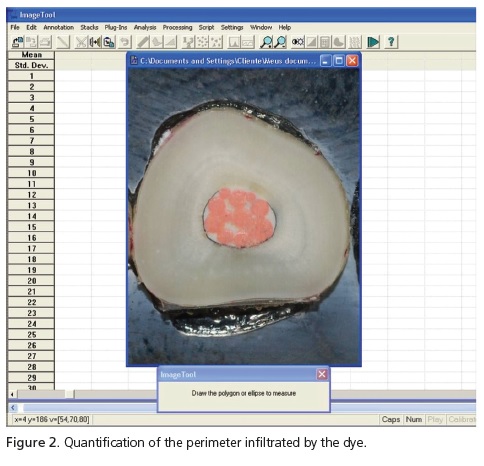
Immediately after the cutting, each root section was placed in contact with absorbent paper and kept in a natural drying process in the environment for fifteen minutes. The previously identified cross-sections of each root were glued with cyanoacrylate (Super Bonder, Henkel, São Paulo, Brazil) to glass slides (Sommer, São Paulo, Brazil), with the apical third of each root remaining visible. Images were captured of all the root sections with a digital camera (Nikon D60, Tokyo, Japan). The quantification of dye leakage was carried out with the assistance of the software application known as Image Tool 3.00 (The University Texas Health Science Center in San Antonio) (Figure 2).
The dye leakage in the samples of the four experimental groups was analyzed in length and depth. The analysis of the length was done using the infiltrated perimeter in the inside of the root canal of the crosssections of each sample. The depth analysis of the dye was performed by counting the number of cross sections of each sample that revealed the presence of India ink.
With the objective of ascertaining the existence or otherwise of statistically significant differences between the percentages of infiltrated perimeter obtained between the groups, the Mann-Whitney U-test was applied to the values obtained in the four sections and to those values related to depth. The level of significance was established at 0.05 in a two-tailed test.
RESULTS
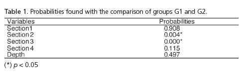
Groups G1 and G2, filled with ZOE based cement, showed statistically significant differences between the values of sections 2 and 3 (Table 1), and the highest leakage values were obtained with those roots that did not receive intracanal medication (Group G2).
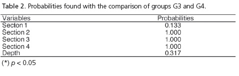
As for groups G3 and G4, filled with a resin cement with Ca(OH)2, the results did not show statistically significant differences between the values of all the comparisons made between the samples, regardless of whether or not intracanal medication was used (Table 2).
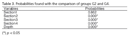
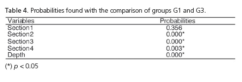
When comparing the groups that did not receive intracanal medication and which were filled with different cements (Table 3), the results showed statistically significant differences between the values of sections 2, 3 and 4 and between the measurements of depth, and the highest leakage values were obtained with the samples that used ZOE based cement (G2). When the groups were compared that received intercanal medication and which were filled with different cements (G1 and G3), the results showed statistically significant differences between the values of sections 2, 3 and 4, and between the depth measurements, and the highest leakage values were obtained in group (G1), filled with ZOE based cement (Table 4).
As for the roots that served as a control for the leakage stage, there was no penetration of the dye in the negative control group while in the positive control group, there was leakage along the whole length and depth of the root canal.
DISCUSSION
There have been few dye leakage studies like ours, that have evaluated the influence of Ca(OH)2 in filled teeth using two types of endodontic cement, one ZOE based and the other resin based with Ca(OH)2 4-5. Our results were similar to those of Çaliskan et al.5 in respect of the leakage of dye into the filled teeth using ZOE based cement, which showed the highest values in the samples that did not receive intracanal medication. Meanwhile, Holland & Murata4 found contradictory results, though this is due to the fact that they used methylene blue dye and not India ink as in the present research study and that of Çaliskan et al.5. This methylene blue dye becomes discolored in contact with intracanal medication of Ca(OH)2 due to its incompatibility with alkaline substances9, producing unreliable results.
With regard to the roots filled with resin cement with Ca(OH)2, there was no statistically significant difference between the samples that did or did not receive intracanal medication, such results being in agreement with previous studies4-5.
The use of standardized test specimens, i.e. of roots having the apical and middle thirds removed, provided a reduction in the possible effects of the apical ramification of the root canals8. This apical area could accumulate dye and thus interfere with the measurement of leakage occurring inside the root canal1. Moreover, the roots of bovine teeth maintain the same characteristics as human dentin with their dentinal tubules and humidity11. The natural porosity of dentin allows room for air to be displaced by the dye during leakage12, which does not occur with the samples of glass capillary tubes and resin or plastic blocks. The impermeable resin walls of the canals simulated in the blocks as they have humidifying characteristics different from dentin, could affect the distribution and effectiveness of the cement11, while the plastic blocks, being a highly hydrophobic material, do not easily permit the passage of water and dye like the tooth. Moreover, they exhibit a greater underfilling and overfilling than the teeth, because they do not represent the anatomical configuration of the root canal system, with the presence of constriction of the foramen13. Experimental samples of resin blocks and glass capillary tubes are only recommended for studies that compare dye leakage methods and for investigation of variations inside the samples7-11.
Variations in sealing ability between the experimental groups in the present study may have been caused by the chemical interactions or reactions between the Ca(OH)2 paste and the type of cement. Several studies have testified to the impacts of Ca(OH)2 residues on the mechanical properties of different types of cement and on the apical sealing ability after the filling of the canal2-5. In the study by Hosoya et al.3, the resin cement with Ca(OH)2 had its sealing ability enhanced when the Ca(OH)2 medication was used in advance, which did not happen with the ZOE based cement. However, the authors stress that the improvement observed in sealing ability cannot be interpreted as a clinically relevant result as it is an in vitro study and does not reflect a clinical scenario (5). Due to the complex interrelationship between the multiple factors that affect endodontic treatment, it is impossible to study the correlation between in vitro dye coloration and in vivo treatment failure7-8.
The smaller leakage of India ink in the samples in the present research study, which received medication using Ca(OH)2 and which were filled with ZOE based cement, and in the samples either with or without medication, filled using resin cement and Ca(OH)2, in our opinion does not signify that there was an increased capacity to seal of the filling. Much less does it signify that the presence of Ca(OH)2 residues in the root canal caused discoloration in the India ink, as in the case of methylene blue, as there are no studies which confirm this. We believe that the India ink was actually prevented from penetrating when the Ca(OH)2 was present inside the root canal, perhaps via some chemical interaction or due to the formation of a physical barrier. Further studies will be needed to corroborate our hypothesis. Further studies should also be conducted with the aim of developing vehicles linked to Ca(OH)2 that can help with its removal.
CONCLUSION
In accordance with the experimental conditions employed and the results obtained, we conclude that the presence of calcium hydroxide residues only had an impact on marginal leakage when the cement used was ZOE based. The remains of the calcium hydroxide intracanal medication significantly reduced the marginal leakage of the dye, in length as well as depth, when the cement used for the filling was ZOE based (Endofill). When the cement used was resinous and with Ca(OH)2 (Sealer 26), there was no difference in dye leakage, irrespective of whether or not intracanal medication had been previously applied.
Collaborators
ST MARRA was responsible for the composition of the article and for carrying out all of the experimental part, except for the endodontic treatment. MAVC OLIVEIRA jointly directed and, being a specialist in the area, carried out part of the endodontic treatment, as well as helping with the performance of other phases and the composition of the article. JCG BIFfidirected the research study and took part in the composition of the article.
REFERENCES
1. Porkaew P, Retief H, Barfield RD, Lacefield WR, Soong SJ. Effects of calcium hydroxide paste as an intracanal medicament on apical seal. J Endod. 1990;16(8):369-74. doi: 10.1016/S0099- 2399(06)81908-4. [ Links ]
2. Margelos J, Eliades G, Verdelis C, Palaghias G. Interaction of calcium hydroxide with zinc oxide-eugenol type sealers: a potential clinical problem. J Endod. 1997;23(1):43-8. doi: 10.1016/S0099-2399(97)80206-3.
3. Hosoya N, Kurayama F, Lino F, Arai T. Effects of calcium hydroxide on physical and sealing properties of canal sealer. Int Endod J. 2004;37(3):178-84. doi: 10.1111/j.0143-2885.2004.00781.x.
4. Holland R, Murata SS. Efeito do hidróxido de cálcio como curativo de demora no selamento marginal após a obturação do canal. Rev Assoc Paul Cirur Dent. 1993;47(6):1203-7.
5. Çaliskan MK, Turkun M, Turkun S. Effect of calcium hydroxide as an intracanal dressing on apical leakage. Int Endod J. 1998;31(3):173-7. doi: 10.1046/j.1365-2591.1998.00145.x.
6. Wu MK, Wesselink PR. Endodontic leakage studies reconsidered. Part I. Methodology, application and relevance. Int Endod J. 1993;26(1):37-43. doi: 10.1111/j.1365-2591.1993.tb00540.x.
7. Plotino G, Grande NM, Manzulli N, Chiaradia G, La Torre G, Somma F. Influence of reduced air pressure methods on dye penetration in standardized voids. Oral Surg Oral Med Oral Pathol Oral Radiol Endod. 2007;103(2):289-94. doi: 10.1016/j. tripleo.2006.05.018.
8. Kazemi RB, Spandberg LSW. Effect of reduced air pressure on dye penetration in standardized voids. Oral Surg Oral Med Oral Pathol Oral Radiol Endod. 1995;80(6):720-5. doi: 10.1016/ S1079-2104(05)80257-4.
9. Kontakiotis EG, Wu MK, Wesselink PR. Effect of calcium hydroxide dressing on seal of permanent root filling. Endod Dent Traumatol. 1997;13(6):281-4.
10. Wu MK, De Gee AJ, Wesselink PR. Leakage of four root canal sealers at different thicknesses. Int Endod J. 1994;27(6):304-30. doi: 10.1111/j.1365-2591.1994.tb00273.x.
11. Patomvanich S, Edmunds DH. Variation in the microleakage produced by four different techniques in root fillings in simulated root canal model. Int Endod J. 1996;29(3):156-62.
12. Masters J, Higa R, Torabinejad M. Effects of vacuuming on dye penetration patterns in root canals and glass tubes. J Endod. 1995;21(6):332-4. doi: 10.1016/S0099-2399(06)81011-3.
13. Chohayed AA. Comparison of conventional root canal obturation techniques with thermafil obturators. J Endod. 1992;18(1):10-2. doi: 10.1016/S0099-2399(06)81135-0.
 Correspondence to:
ST MARRA
e-mail: sarateodoromarra@yahoo.com.br
Received on: 7/6/2010
Final version resubmitted on: 11/11/2010
Approved on: 17/1/2011
