


Serviços Personalizados
Artigo
 pdf em Inglês
 Artigo em XML
 Referências do artigo
 Enviar este artigo por email
Links relacionados
Compartilhar

 Permalink
RGO.Revista Gaúcha de Odontologia (Online)
versão On-line ISSN 1981-8637
RGO, Rev. gaúch. odontol. (Online) vol.60 no.4 Porto Alegre Out./Dez. 2012
ORIGINAL / ORIGINAL
Characteristics of the primary dentition of children who attended a Mother and Child Dental Care Program
Características da dentição decídua em crianças que freqüentaram um Programa Odontológico de Atenção Materno-Infantil
Lúcia de Fátima Almeida de Deus MOURA I; Marcoeli Silva de MOURA I; Marina de Deus Moura de LIMA II; Marcos Antônio da Mota ARAÚJO III; Wagner Leal de MOURA I
I Universidade Federal do Piauí, Faculdade de Odontologia, Departamento de Patologia e Clínica Odontológica. Teresina, PI, Brasil
II Universidade de São Paulo, Faculdade de Odontologia, Departamento de Patologia Bucal. São Paulo, SP, Brasil
III Fundação Municipal de Saúde. Teresina, PI, Brasil
ABSTRACT
Objective
To assess the type of occlusion prevalent in children who attended a Mother and Child Dental Care Program in the city of Teresina.
Methods
The clinical records of children who had participated in the Preventive Program for Expectant Mothers and Babies were chosen randomly and a letter was sent to 1,801 mothers. 343 (19%) responded by attending a consultation, and 261 of these were selected. They were aged 3 to 6, of both sexes and with complete deciduous dentiti on. The data were transcribed to individual clinical record cards and then tabulated and analyzed statistically using EPI-INFO software, version 6.04. An alpha error of 5.0% was considered for false rejection hypothesis.
Results
A prevalence of type II arch was observed, in both the upper and lower jaw. The chi-square test showed there was no significant difference between the types of dental arch with regard to age. The presence of primate space in the upper arch was found in 71.2% of the sample, while in the lower arch the frequency was 44.8%. Age was not a determining factor of changes with regard to the presence or absence of primate space. The vertical terminal plane between second primary was prevalent in all ages and the situation showed no difference with regard to the dental arches. Regarding the presence of overjet, overbite and anterior open bite, there was a statistically significant decrease in their frequency with the increase in age.
Conclusion
The children examined exhibited predominantly type II arch, with primate space in the upper arch and vertical terminal plane between second primary molars.
Indexing terms: Dental occlusion. Dentition primary. Orthodontics preventive.
RESUMO
Objetivo
Avaliar o tipo de oclusão prevalente em crianças que freqð?entaram um Programa Odontológico de Atenção Materno-Infantil na cidade de Teresina.
Métodos
Realizou-se uma seleção aleatória de fichas clinicas de crianças que haviam participado do PPGB e, através destas fichas foram enviadas cartas a 1801 mães. Das cartas enviadas houve um retorno de 343 crianças (19%) e destas, foram selecionadas 261 com dentadura decídua completa, de ambos os gêneros, na faixa etária de 3 a 6 anos. Os dados obtidos foram transcritos para fichas clínicas individuais e posteriormente tabulados e analisados estatisticamente através do programa EPI-INFO, versão 6.04. Considerou-se 5,0% de erro alfa para hipótese de falsa rejeição.
Resultados
Foi observada prevalência de arco do tipo II, t anto para a arcada superior quanto para a inferior. O teste do qui-quadrado não demonstrou haver diferença significativa entre os tipos de arco dentário com relação à idade. A presença de espaço primata na arcada superior representou 71,2% da amostra avaliada, enquanto na arcada inferior a freqð?ência foi de 44,8%. A idade não foi fator desencadeante de alterações com relação à presença ou ausência de espaço primata. A relação terminal de segundos molares decíduos em plano/reto foi prevalente em todas as idades e a situação não apresentou diferenças quanto aos hemi-arcos dentários. Quanto à presença de trespasses vertical e horizontal e mordida aberta anterior com relação a idade, observou-se que com o aumento da idade houve diminuição das situações avaliadas com significância estatística.
Conclusão
As crianças examinadas exibiram predominantemente arco tipo II, com espaço primata na arcada superior e relação terminal dos segundos molares em plano terminal reto.
Termos de indexação: Dentição decídua. Oclusão dentária. Ortodontia preventiva.
INTRODUCTION
The Preventive Program for Expectant Mothers and Babies (PPGB) is an extension activity of the Dentistry course at the Federal University of Piauí (UFPI), implemented in 1997, whose practices take place in the Institute of Social Perinatology of Piauí (IPSP) and which focus on a preventive/promotional perspective1.
The goal of the PPGB is to recover and maintain the oral health of expectant mothers and children up to the age of 36 months. The program's activities are undertaken by students of the Dentistry course, under the supervision and guidance of lecturers at UFPI2-3.
The first studies related to the occlusion of the primary dentition were carried out by Baume4. The author classified the deciduous dental arches according to the presence or absence of generalized spaces in the anterior region as type I - when there were spaces, and type II - when there were no spaces. Baume4 found that 70% of the children evaluated suffered from type I arches and 30% from type II, in the maxilla; as for the mandible, the frequency was 63% of type I arches and 37% type II. The presence was also noted in this article of specific diastemas that were independent of the type of arch: in the upper arch they were located between the lateral incisors and the canines, while in the lower arch they were between the canines and first molars, known as primate diastemas.
The types of dental arch and the presence of primate gaps are characteristics that are independent of the primary dentition; they may or may not be present, connected or isolated on the mandible and/or maxilla4-5.
Baume4 also analyzed the upper and lower deciduous arches via the distal surfaces of the second molars and noted that they may have three different positions: in plane or straight (76%), in mesial step (14%) or in distal step (10%). The observations used the mandible as a reference.
In the relationship of deciduous second molars, with a distal step formation, the occlusion has a tendency toward the development of a probable skeletal imbalance which could result in a Class II (Angle) malocclusion in the permanent dentition. Terminal plane or mesial step relationships are considered to be the most favorable, enabling the permanent first molars to experience normal occlusion development6.
Relevant literature on the subject is scant though there is a consensus amongst authors that the eruption of the permanent first molars is guided by the distal surface of the deciduous second molars. Barbosa et al.7 studied the prevalence of terminal relationships of the deciduous second molars and the type of dental arch, by evaluating study models of 27 children of both sexes aged between 4 and 6 yearsold. The authors concluded that there was a predominance of terminal relationships in mesial step, followed by straight and distal. Type I arches were the most frequent and were independent of terminal relationships of the second molars.
Fernandes et al.8 analyzed the primary dentition of 354 children from the city of Niterói (in the state of Rio de Janeiro) and observed that the terminal relationship frequency of deciduous second molars in plane with canines in class I was 41.52%, distal step and canines in class II 29.66% and mesial step accompanied by canines in class III 1.69%. In addition, the most prevalent malocclusions were the crossbite and deep overbite.
The genetic factor is thought to be predominant in the growth and development patterns of the dental arches, although this could also be heavily influenced by environmental factor such as oral habits, quality of nutrition and personal health. These statements may be explained using Moss's functional matrix hypothesis according to which facial growth occurs in response to functional needs, which means conceptually that the soft tissue in the mouth, during development, has an impact on structures such as bone and cartilage9.
The aim of the present study was to determine the type of occlusion in the children attending the university extension program Preventive Program for Expectant Mothers and Babies (PPGB) at the Federal University of Piauí (UFPI) and to observe potential alterations in the spacing between teeth and terminal relationships of the deciduous second molars with increasing age.
METHODS
Following approval by the Ethics in Research Committee at the Federal University of Piauí (UFPI) (opinion 004/2003), the study began. Those responsible signed a free and informed consent form observing resolution 196/96 of the Brazilian National Health Board (CNS), which regulates policies and standards of research involving human beings.
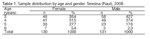
The study was developed using 261 children of both sexes, aged between 3 and 6, and with complete primary dentitions, who lived in the metropolitan region of Teresina (state of Piauí). All the subjects in the study attended the UFPI Extension Project - "Preventive Program for Expectant Mothers and Babies (PPGB)" and were eliminated if they had passed the age limit of 36 months (Table 1).
The data were collected in the months of December 2003 and January, February and July in 2004. The children were examined by the authors of the study and assisted by note-takers. Intra-examiner error was evaluated using the kappa calculation, whose congruity was considered to be excellent (k=0.9). For the validation of the diagnosis criteria, the exams were repeated for 10% of the evaluated sample.
The children were examined in dental consulting rooms under the direct light of a reflector bulb and were instructed to close the mouth in normal maximum intercuspation and to remain in this position through to the end of the examination. The types of dental arches4, terminal relationship of deciduous second molars and the presence or absence of primate diastemas were observed, as well horizontal and vertical encroachment.
The types of arch were evaluated for the presence or absence of interdental spaces4. In order to determine the presence of interdental spaces, a 0.25mm brass wire was used and when this was able to pass freely between the teeth, these were regarded as spaces. Children with arch type I, those who had generalized dental spaces in the anterior region, should have at least three spaced teeth.
Those children who presented with a horizontal encroachment greater than or equal to 3mm were considered to be suffering from overjet. The values were obtained with the assistance of a millimetric ruler placed on the incisal ridge of the upper anterior teeth.
Vertical encroachment was considered to be excessive when the upper teeth overlapped a third or more of the crown of the lower incisors.
Anterior open bites were characterized by the absence of contact between anterior teeth when the posterior teeth were in normal maximum intercuspation3.
The data obtained were transcribed to the individual clinical record cards and subsequently tabulated and statistically analyzed using the software application EPI-INFO, version 6.04. An alpha error rate of 5% was assumed for the hypothesis of false rejection.
RESULTS
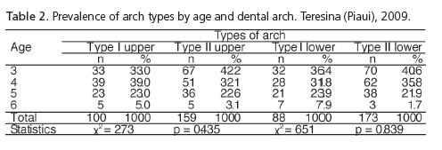
The results of Table 2 showed a prevalence of the type II arch, for both the upper and lower arches. The chisquare test showed no significant difference between the types of dental arch with regard to age.
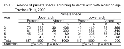
The presence of primate space in the upper arch equated to 71.2% of the sample tested while in the lower arch, the frequency was 44.8%. Age was not a factor which triggered alterations with regard to the presence or absence of primate space (Table 3).
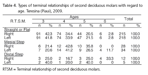
The terminal relationship of second deciduous molars in flat/straight was prevalent at all ages and the situation showed no difference with regard to dental hemiarches. In 2.3% of children, it was not possible to determine the terminal relationship of second deciduous molars due to the lack of cooperation by the children. Amongst the variables studied, there was no significance (Table 4).
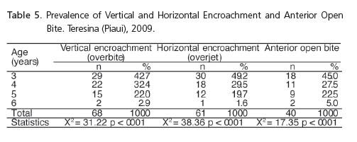
The proportion test was applied between ages and each variable studied. It was noted that, in all situations, statistical significance existed (Table 5).
DISCUSSION
The results presented in this study confirm the prevalence of dental arches without generalized spaces, classified by Baume4 as a type II arch (Table 2). Some of the authors researched presented different results from those found here, in other words, a prevalence of type I arches was detected in the two dental arches4,7,9-10. As for the results obtained by Usberti et al.5 they were partially in disagreement, seeing that the authors noted a prevalence of type II arch in the mandible and type I in the maxilla.
Baume4 states that the deciduous dental arches which present with or without spaces do not change spontaneously in the age range between 3 and 5½ years, and that they are not acquired through the influences of the environment, but rather are congenital. Looking at Table 2, it was concluded that age was not a determining factor in alterations in the dental arches, results which go against those presented by Soviero et al.11. In relation to the variables arch type and gender of the children surveyed, it was noted that the percentages were quite similar, and there was no statistical significance between them, and similar results were found by other authors5,12.
It can be seen from the analysis in Table 3 that there was a prevalence of primate spaces in the upper arch (71.2%) while in the lower arch the frequency was 44.8%. Age was not a determining factor of the presence or absence of primate spaces. Of the 261 children examined, the stratum which presented the greatest frequency of primate space was the one before 4 years of age, and then the ages of 4 and 5, however no significant statistical association was observed between the variables analyzed. When making comparisons with data presented by Moura et al.13, whose analyzed sample comprised children from the state of Piaui who were also residents of the city of Teresina, the authors found similar results to those found in this study and which also agree with other authors researched9,14, however a number of others presented results which were not in agreement5,7.
Through the results shown in Table 4, it can be seen that there was a prevalence of terminal relationship of second deciduous molars in-plane/straight. Age was not a factor that triggered alterations with regard to the terminal relationship of second deciduous molars.
Baume4 states that there are no dimensional changes in the deciduous dental arches between the ages of 3 and 5½ and that the aforementioned arches only suffer dimensional changes in the sagittal and transverse direction, when suffering adverse environmental influences. The author also observed that the vertical growth of the alveolar processes occurs simultaneously with the development of germs in the permanent successor teeth.
According to Baume4, the presence of interdental spaces is determined by genetic patterns and, after the complete eruption of the tooth and stabilization of the primary dentition, new spaces are not created spontaneously and any diastemas present do not undergo spontaneous alteration11.
The primary dentition presents with normal occlusal characteristics, horizontal encroachment of less than 2mm and vertical encroachment between 1mm and 2mm16. Alterations in the characteristics described are directly related to the presence of non-nutritive sucking habits such as the use of pacifiers and thumb sucking15-19. Katz et al.16 found no association between facial morphology and malocclusions.
The increased overjet values of 23.4%, found in the present study, and considering the sample as a whole, are lower than those of Mendes et al.15 who noted a prevalence of 67.8%, Bezerra & Cavalcanti17 49.1% and Medeiros et al.19 49.1% and are close to those found by Katz et al.16 at 29.7%. Similarly, the prevalence of anterior open bite of 15.3%, found in this study, and considering the sample as a whole, is less than that observed by Bezerra & Cavalcanti17 who found a prevalence of 45.3%, Katz et al.16 36.4%, Sousa et al.18 37.8%. The differences between the data in the present study and the literature may be down to the instructions emitted by the PPGB3, as the aforementioned alterations are heavily associated with the deleterious habits of sucking which, when the children paid return visits to the program, were actively discouraged.
The results presented in Table 5 demonstrate that, with increased age, there is a significant reduction in vertical and horizontal encroachment and in anterior open bite. The reduction in vertical encroachment (overbite) with increased age may perhaps be attributed to the growth of the vertical dimensions prompted by the eruption of the first permanent molars and the reduction in horizontal encroachment and anterior open bite to the abandonment of the habit of non-nutritional sucking. Castro et al.9 presented results that did not agree with those found in this study in which the authors observed an increased frequency in overbite with the passing of time.
Comparing the results of the present study with others found in the literature, it can be seen that environmental factors exert an influence over the development of deciduous occlusion and that gender was not seen to be a significant element in such alterations5,10,12.
CONCLUSION
Type II arches (Baume) were prevalent in both sexes and dental arches. Age was not a determining factor of alterations with regard to the type of arch. As for the primate diastemas, these were present in the majority of the children, in both the upper arch (71.2%) and the lower arch (44.8%).
The relationship of canines in neutral occlusion was the most prevalent. Age was not a determining factor of alterations. Terminal relationships with second deciduous molars were plane or straight, followed by mesial step then distal step. No significant statistical differences were found between gender, and age was not a determining factor in the alterations.
Collaborators
LFAD MOURA participated in the conception, outline, data collection, analysis, interpretation of data and composition of the article. MS MOURA and MDM LIMA participated in the data collection and composition of the article. WL MOURA participated in the critical review and composition of the article. MAM ARAÚJO performed the statistical analysis and participated in the composition of the article.
REFERENCES
1. Moura LFAD, Moura MS, Toledo OA. Dental caries in children that participated in a dental program providing mother and child care. J Applied Sci. 2006;14(1):53-60. doi: 10.1590/S1678- 77572006000100011. [ Links ]
2. Moura LFAD, Lira DMMP, Moura MS, Lopes TSP, Leopoldino VD, Moura MDM. Apresentação do Programa Preventivo para Gestantes e Bebês. J Bras de Odontopediatr Odontol Bebê. 2001;4(17):10-4.
3. Moura LFAD, Moura MS, Toledo AO. Conhecimentos e práticas em saúde bucal de mães que freqüentaram um programa odontológico de atenção materno infantil. Ciênc Saúde Coletiva. 2007;12(4):1079-86. doi: 10.1590/S1413- 81232007000400029.
4. Baume LJ. Physiological tooth migration and its significance for the development of occlusion. I - The biogenetic course of the deciduous dentition. J Dent Res. 1950;29(2):123-32. doi: 10.1177/00220345500290020301.
5. Usberti AC, Cunha JCM. Freqüência de arcos tipo I e II de Baume e espaço primata. RGO - Rev Gaúcha Odontol. 1987;35(6):474- 8.
6. Burdi AR, Moyers RE. Desenvolvimento da dentição e da oclusão. In: Moyers RE. Ortodontia. 4ª ed. Rio de Janeiro: Guanabara Koogan; 1991.
7. Barbosa CS, Di Nicolo R, Ursi WJS. Estudo da prevalência dos tipos de planos terminais dos segundos molares decíduos. PGR Pós-Grad Rev Fac Odontol São José dos Campos. 2000;3(1):41- 8.
8. Fernandes KP, Amaral MAT, Monico MA. Ocorrência de maloclusão e necessidade de tratamento ortodôntico na dentição decídua. RGO - Rev Gaúcha Odontol. 2007;55(3):223- 7.
9. Castro LA, Modesto A, Vianna R, Soviero VLM. Estudo transversal da evolução da dentição decídua: forma dos arcos, sobressaliência e sobremordida. Pesqui Odontol Bras. 2002;16(4):367-73. doi: 10.1590/S1517-74912002000400015.
10. Alexander S, Prabhu NT. Profiles, occlusal plane relationships and spacing of teeth in the dentitions of 3 to 4 year old children. J Clin Peditr Dent. 1998;22(4):329-34.
11. Soviero VM, Bastos EPS, Souza IPR. Dentição decídua: estudo da prevalência dos espaços interproximais em crianças brasileiras. Rev Odontol Univ São Paulo. 1999;13(2):159-65.
12. Ferreira RI, Barreira AK, Soares CD, Alves AC. Prevalência de características da oclusão normal na dentição decídua. Pesqui Odontol Bras. 2001;15(1):23-8. doi: 10.1590/S1517- 74912001000100005.
13. Moura MS, Simplício AHM, Moura LFAD, Moura WL Alterações na relação molar entre as dentaduras decídua e mista. Rev. ABO Nac. 1994;2(5):333-9.
14. Shimizu RH, Michaelis G, Liu J, Shimizu IA, Ignácio SA. Estudos das características da dentição decídua em crianças entre 3 e 6 anos de idade. J Bras Ortodon Ortop Facial. 2003;8(44):124-31.
15. Mendes AC, Valença AMG, Lima CCM. Associação entre aleitamento materno, hábitos de sucção não nutritivos e maloclusões em crianças de 3 a 5 anos. Cienc Odontotol Bras. 2008;11(1):67-75.
16. Katz CRT, Rosenblat AR, Gondim PPC. Non-nutritive sucking habits in Brazilian children: Effects on deciduous dentition and relationship with facial morphology. Am J Orthod Orthop. 2004; 126(1):53-57. doi: 10.1016/j.ajodo.2003.06.011.
17. Bezerra PKM, Cavalcanti AL. Características e distribuição das maloclusões em pré-escolares. Rev Cien Med Biol. 2006; 5(2):117-23.
18. Souza FRN, Taveira GS, Almeida RVD, Padilha WWN. O aleitamento materno e sua relação com hábitos deletérios e maloclusaão dentária. Pesqui Bras Odontopediatria Clin Integr. 2004;4(3):211-6.
19. Medeiros PKB, Cavalcanti AL, Bezerra PM, Moura C. Maloclusões, Tipos de aleitamento e hábitos bucais deletérios em pré-escolares: um estudo de associação. Pesqui Bras Odontopediatria Clin Integr. 2005;5(3):267-74.
 Correspondence to:
LFAD MOURA
e-mail: mouraiso@uol.com.br
Received on: 20/3/2009
Final version resubmitted on: 26/6/2009
Approved on: 17/8/2009
