Serviços Personalizados
Artigo
Links relacionados
Compartilhar
RGO.Revista Gaúcha de Odontologia (Online)
versão On-line ISSN 1981-8637
RGO, Rev. gaúch. odontol. (Online) vol.60 no.4 Porto Alegre Out./Dez. 2012
ORIGINAL / ORIGINAL
Radiographic analysis of pixel intensity with aluminum step-wedge and different spatial resolutions
Análise radiográfica da intensidade de pixel com a utilização de penetrômetro de alumínio e diferentes resoluções espaciais
Adele Verônica Tonietto GOMES I; Juliana Zavala BAZZI I; Rafaela Elvira ROZZA I; Soraya de Azambuja BERTI I; Paulo Henrique COUTO SOUZA I; Fernando Henrique WESTPHALEN I; Ana Lúcia TOLAZZI I
I Pontifícia Universidade Católica do Paraná, Curso de Odontologia. Curitiba, PR, Brasil
ABSTRACT
Objective
To evaluate the pixel intensity and to compare it with different spatial resolutions, using an aluminum step-wedge.
Methods
The sample consisted of three bone chips of two dry pig mandibles. Then, each bone chip and the aluminum step-wedge were placed on periapical radiographs in order to take the images with Ekta Speed film, with an exposure time of 0.2 seconds and focal length of 25 cm. All radiographs were scanned with spatial resolutions of 150 and 300 dpi and saved as JPEG files. The images were measured using the histogram tool provided by the Image Tool program (UTHSCSA, Texas, USA) selecting specific areas on the bone chips and on the aluminum step-wedge.
Results
The data were analyzed by ANOVA and Tukey statistical tests which showed that there was no significant difference in pixel intensity between bone chips and step 3 (3.6 mmEq/Al) of the aluminum step-wedge (p>0.05). It was also observed that there were no significant differences in pixel intensity value measured on step-wedges 2 (3.3 mmEq/Al), 3 (3.6 mmEq/Al), 4 (3.9 mmEq/Al) and on the bone chips, between spatial resolutions of 150 and 300 dpi.
Conclusion
Small differences in spatial resolution did not interfere with the evaluation of pixel intensity. It is recommended to measure pixel intensity on digitalized radiographs using an aluminum step-wedge as a reference density value.
Indexing terms: Evaluation. Radiography dental digital. Radiographic image enhancement.
RESUMO
Objetivo
Avaliar a intensidade de pixels de fragmentos de tecido ósseo mandibular, bem como compará-la em diferentes resoluções espaciais, utilizando um penetrômetro de alumínio.
Métodos
A amostra consistiu de 3 fragmentos ósseos, provenientes de 2 mandíbulas secas de suínos. Em seguida, cada fragmento ósseo, juntamente com o penetrômetro de alumínio, foram posicionados sobre filmes radiográficos periapicais. As radiografias foram realizadas com filme Ekta Speed, tempo de exposição de 0,20 segundos e distância focal de 25 cm. Todas as radiografias foram digitalizadas com resoluções espaciais de 150 e 300 dpi e, em seguida, arquivadas em JPEG. As imagens foram medidas por meio da ferramenta histograma do programa Image Tool (UTHSCSA, Texas, USA) selecionando-se áreas nos fragmentos ósseos e no penetrômetro de alumínio.
Resultados
A análise dos resultados pelos testes estatísticos ANOVA e Tukey mostrou que não houve diferença significativa de intensidade de pixel entre os fragmentos ósseos e o degrau 3 do penetrômetro de alumínio (3,6 mmEq/Al) (p>0,05). Observou-se ainda que não houve diferenças significativas dos valores da intensidade de pixel dos degraus 2 (3,3 mmEq/Al), 3 (3,6 mmEq/Al) e 4 (3,9 mmEq/Al) e os fragmentos ósseos, entre as resoluções espaciais avaliadas nesse estudo.
Conclusão
Pequenas diferenças de resolução espacial não interferiram nos valores de intensidade de pixel. É recomendável medir esta intensidade com a utilização de penetrômetro de alumínio como referência para valor de densidade.
Termos de indexação: Avaliação. Radiografia dentária digital. Intensificação de Imagem Radiográfica.
INTRODUCTION
The discovery of x-rays was undeniably one of the greatest contributions to humanity. However after a few years in use, harmful effects began to appear, caused by inadvertent use. In order to minimize these effects, studies were conducted to develop new equipment and accessories that would contribute to reducing the dose of x-rays to which individuals were exposed1-2.
These studies resulted in the introduction of radiographic film of different sensitivities which reduced radiation doses and exposure time. Another huge contribution came from the new image diagnosis systems such as digital radiography1-2. These were described for the first time in 1988 and introduced into the United Kingdom after the publication by Mouyen et al.3, who described the RVG system (radiovisiography). This system made it possible to obtain directly digital dental radiographic images.
In the beginning there were three types of digital imaging systems, classified in accordance with the capture of the image. The first was Digital Dental Radiography (DDR), represented by systems that have sensors connected via cable to a computer. The second was Computed Radiography (CR) which uses an image capture board similar to the size of a radiographic film. Finally, there was the indirect digital dental image obtained by means of the digital scanning of a conventional x-ray4-5.
This classification still prevails today, though many more items of equipment have surfaced with the aim of improving direct digital systems. One example of this is wireless technology6 which, according to Haiter Neto et al.7, demonstrates significantly greater sensitivity when compared to its predecessors.
According to several studies, the digitized radiographic image made possible the evaluation of the maxilla and mandibular dental complex by considering the possibility of using resources such as the change in brilliance and contrast, image inversion, the application of high and low relief and the magnification of images in specific regions2,8-11, as well as a reduction in exposure time by as much as 90%12-13.
Studies such as those of Berti et al.14, Berti et al.15 and Kirsten et al.16 evaluated the pixel intensity in the mandible by means of technically standardized intraoral x-rays adapted to an aluminum step-wedge. This intensity, obtained in levels of gray, can be transformed into millimeters aluminum equivalent (mm eq Al), as Ruijter et al.17 stated, thus becoming a measurable value.
As for the scanning of x-rays, the literature reveals the use of different spatial resolution standards, amongst which the 300 dpi standard is the most frequently employed18. As there is no standardization in terms of the type of spatial resolution when pixel intensity is being considered, the possibility exists that small differences in this spatial resolution could have an impact on the measurement of pixel intensity, by using an aluminum step-wedge as the reference measurement. Therefore, the aim of the current study is to evaluate the influence of different spatial resolutions on pixel intensity values in digital periapical x-rays.
METHODS
The present study was approved by the Ethics in Research Committee at the Pontifical Catholic University of Paraná (PUC - PR), filed as record no. 320.
In the study, three bone chips from the vestibular cortical region of two dry pig mandibles were prepared in advance.
To begin with, areas in the mandibles were demarcated of 1.2 cm2 and then, using a low-speed no. 4 spherical drill bit, the osteotomy was carried out until total separation of the bone chips was achieved. Each chip was rendered uniform using sandpapers of varying grits in order to obtain areas of 1 cm2 and thicknesses of 2 mm, in accordance with the methodology used by Kirsten et al.16.
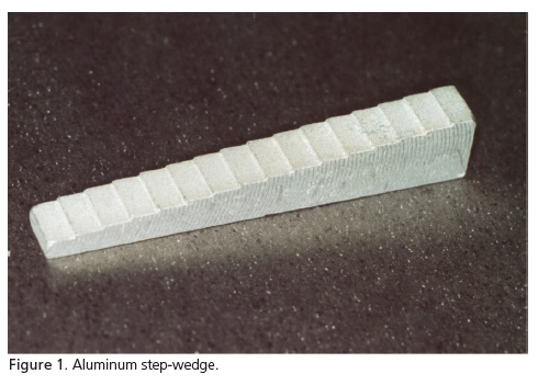
For the measurement of pixel intensity, an aluminum step-wedge was fabricated (Aluminum alloy 272/domestic, Mechanical Engineering Laboratory at PUC-PR), with a length of 40 mm and a width of 4 mm, composed of 16 step-wedges, with a thickness of 0.3 mm between each step-wedge (Figure 1).
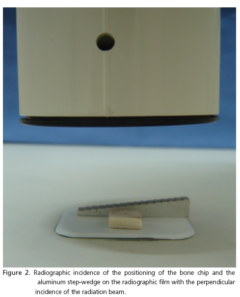
The radiographs were taken using an x-ray machine (Sirona, Heliodent, 70 kVp, 10 mA-120 V, SP), and Ekta Speed film (Eastman Kodak Company, Rochester, USA), with an exposure of 0.2 seconds and focal length of 25 cm. The chip was placed in the center of the film and the aluminum step-wedge in increasing order of stepwedges, from left to right (Figure 2).
Each chip, along with the step-wedge, was x-rayed three times, within the determined exposure time, amounting to 9 x-rays in total. The chemical processing of all the x-rays was carried out simultaneously by the time and temperature method in a darkroom, using the same developing solution (GBX developer and replenisher - KODAK Brasileira Com. e Ind. Ltda.) and fixer (GBX fixer and replenisher - KODAK Brasileira Com. e Ind. Ltda.). The time taken to develop the x-rays was 4 minutes and 30 seconds, and for the fixation, 10 minutes. In this stage, procedure gloves were used to avoid getting fingerprints on the x-rays, thus avoiding potential alteration during measurement. The drying of the x-rays was performed in an automatic machine (EMB - Indústria Brasileira, SP).
For digitalization, the x-rays were placed on the scanner (ScanMaker, 9800XL, 48 bits, 3200X1600 dpi; 12"X17" tabloid size, USB & SCSI-2, Microtek, USA), inverted and juxtaposed. A paper mask was then placed alongside the full set of x-rays. The spatial resolution standard of 150 dpi and 300 dpi was used with a dynamic range of 8 bytes. The images were saved as JPEG files.
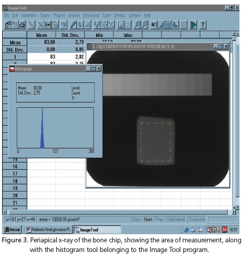
The digitized images were measured by means of the histogram tool from the application Image Tool, version 3.0 (UTHSCSA, USA), whose software was developed and made available free of charge by the University of Santo Antonio, Texas, USA (UTHSCSA, USA). Firstly, the images were manipulated by way of the automatic brilliance and contrast alteration function. Then, in order to take the measurements of the bone chips, areas of 50X50 pixels were selected for 150 dpi x-rays and 100X100 pixels for 300 dpi x-rays. The step-wedges of the aluminum step-wedge were selected for measurement, in accordance with similarities in the shades of gray, when compared with the aforementioned region of bone. In the selected step, areas of 10X20 pixels were measured for the 150 dpi x-rays and 20X50 pixels for the 300 dpi x-rays, using the same tool (Figure 3).
The results were obtained using the following statistical tests: Variance Analysis (ANOVA) to compare the independent continuous variables and the Tukey test, used to test for any differences between the means of the groups. For both tests, a level of significance of p<0.05 was assumed.
RESULTS
The step-wedges of numbers 2 (3.3 mmEq/Al), 3 (3.6 mmEq/Al) and 4 (3.9 mmEq/Al) of the aluminum step-wedge were selected according to similarities in the shades of gray between these step-wedges and the bone chips analyzed in this study. Then the pixel intensity of the bone chips was compared to the intensity obtained at each step on the step-wedge, considering the spatial resolutions analyzed.
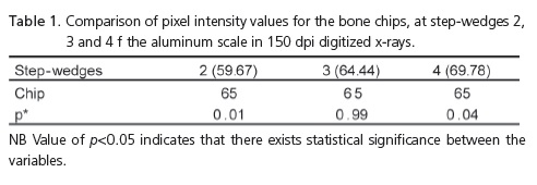
Table 1 shows the comparisons of the pixel intensity values obtained at step-wedges 2 (3.3 mmEq/Al), 3 (3.6 mmEq/Al) and 4 (3.9 mmEq/Al) of the aluminum step-wedge, with those of the bone chips in the 150 dpi digitized x-rays.
According to the results presented, it could be noted that there were no statistically significant differences in the pixel intensity values of the bone chips only with step 3 (3.6 mmEq/Al) on the aluminum scale, for p>0.05.
Table 2 shows the comparisons of pixel intensity values obtained at step-wedges 2 (3.3 mmEq/Al), 3 (3.6 mmEq/Al) and 4 (3.9 mmEq/Al) of the aluminum stepwedge, with those obtained from the bone chips, in the 300 dpi digitized x-rays.
It was observed that there were no statistically significant differences for the pixel intensity values of the bone chips only with step 3 (3.6 mmEq/Al) of the aluminum scale, for p>0.05.
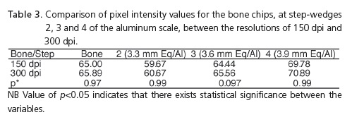
Table 3 shows the comparisons of the pixel intensity values obtained at steps 2 (3.3 mmEq/Al), 3 (3.6 mmEq/Al) and 4 (3.9 mmEq/Al) and the bone chips, between the two resolutions of 150 dpi and 300 dpi.
It was found that there were no statistically significant differences between the pixel intensity values of step-wedges 2 (3.3 mEq/Al), 3 (3.6 mmEq/Al) and 4 (3.9 mmEq/Al) of the step-wedge and the bone chips, in the spatial resolutions of 150 dpi and 300 dpi (p=0.05).

DISCUSSION
For the evaluation of pixel intensity, studies such as those of Souza et al.19, Puppin et al.20, Pereira et al.21 and Southard & Southard22 used periapical radiographic film of different sensitivities such as Ekta-speed, Ultra-speed and also D-Speed film. Zlataric & Celebic18, on the other hand, used panoramic x-rays to observe bone quality and the presence of the signs of reabsorption. All of these studies, however, measured bone density with images digitized by way of pixel intensity.
The evaluation of bone density can be performed with the aid of aluminum step-wedge, as shown by Berti et al.15, Koparal & Akdeniz23 and Shrout et al.24. These authors determined pixel intensity by selecting regions of interest, in digitized periapical x-rays, to determine the mean values of the gray levels and values in equivalent millimeters of aluminum.
Almeida et al.10, Puppin et al.20, Rawlinson et al.9, Ruijter et al.17 and Southard & Southard22 considered that aluminum possesses a density similar to that of bone. There are in the literature, however, studies like those of Zlataric & Celebic18 which evaluated the loss of bone mineral density in the mandibles of patients with total prostheses and removable partial prostheses, by way of digitized panoramic x-rays, using to this end a copper step-wedge. Corroborating the aforementioned studies, the present study also used an aluminum step-wedge, however this was composed of 16 step-wedges, with a gap of 0.3 mm between each. As for Puppin et al.20, they used a step-wedge of the same material, but with 8 stepwedges and gaps of 1 mm. Zlataric & Celebic18, contrary to these two research studies, used a 5-step copper stepwedge with gaps of 0.1 mm. It should be emphasized that the step-wedge need to have a sufficient number of stepwedges with different nuances in order to evaluate more accurately the different regions of bone. Nevertheless, the nuances cannot be too smooth to the point where it is difficult to view the different shades of gray as well as the pixel intensity measurements.
The sample in the present study comprised bone chips from the vestibular cortical of dry pig mandibles, as described also by Christgau et al.25. The periapical film was Ekta-speed (Eastman Kodak Company, Rochester, USA), the same as that used by Souza et al.19, and also an x-ray machine (Sirona, Heliodent, 70 kVp, 10 mA-120 V, SP), which reproduced the studies of Almeida et al.10, Kirsten et al.16, Pereira et al.21 and Southard & Southard22.
In the present study, the digitalization of x-rays was performed using a scanner (ScanMaker, 9800XL, 48 bits, 3200X1600 dpi; 12"X17" tabloid size, USB & SCSI-2, Microtek, USA) in spatial resolutions of 150 dpi and 300 dpi. On the other hand, the study conducted by Berti et al.15 and Schulze et al.26 only used a resolution of 300 dpi. As for Koparal & Akdeniz23 and Scarfe et al.27, they used a spatial resolution of 600 dpi. In view of the fact that both the resolution of 150 dpi and that of 300 dpi in JPEG format, do not take up a lot of space for computer storage, the comparison performed in the present study showed that it is possible to use either of the resolutions, without any great impact on results in terms of the measurement of pixel intensity in digitized periapical x-rays. It should be emphasized, however, that to obtain a standardization for the purposes of carrying out fresh measurements, as well as for greater reliability of results, the same type of radiographic film, the same exposure, the same processing technique and also the same aluminum scale should all be used.
Also to be taken into account, the difference in resolution investigated in this work was small, which did not interfere with the results obtained. Normally, there is no spatial resolution standard which is used during the process of scanning radiographic images, as far as pixel intensity measurement is concerned. Apparently, in accordance with the preliminary results of this study, small differences in spatial resolution do not interfere with the measurement of pixel intensity in digitized periapical x-rays. In addition, the images at 150 dpi or 300 dpi are not so cumbersome in terms of file size, enabling their transmission via the Internet. Nevertheless, bigger differences in spatial resolution with the analysis of pixel intensity in digitized periapical x-rays should be investigated in future studies.
CONCLUSION
With the results obtained in the present study, it was possible to conclude that the pixel intensity value found at step 3 (3.6 mmEq/Al) of the aluminum scale was the one which was closest to the value obtained in the bone chip. Moreover, it was ascertained that there was no significant difference between the storage size of spatial resolutions of 150 dpi and 300 dpi during the evaluation of pixel intensity. With the aid of the aluminum step-wedge, it was possible to establish a measurable vale of this intensity in the digitized periapical x-rays.
Collaborators
AVT GOMES and JZ BAZZI were responsible for the performance of the study, from the adaptation of the project design to the submission to the Ethics Committee, the preparation and processing of the x-rays, analysis of material by means of the Image Tool program and finally the composition of the article. RE ROZZA and SA BERTI assisted with the performance of the study, guidance and a search of literary reviews, the bibliographical adaptation and update and the composition of the article. PHC SOUZA was responsible for directing the research study, the monitoring of the performance of the study, in terms of the treatment of the mandible osteotomy, chemical processing and use of the x-ray machine, scanner and Image Tool software, and participated in the composition of the article. FH WESTPHALEN and AL TOLAZZI contributed with the interpretation of the statistical results, manipulation of the Image Tool software and the composition of the article.
REFERENCES
1. Freitas A, Rosa JE, Souza IF. Radiologia odontológica. São Paulo: Artes Médicas; 2000. [ Links ]
2. Schweitzer DM, Berg RW. A digital radiographic artifact: a clinical report. J Prosthet Dent. 2010;103(6):326-9. doi: 10.1016/S0022-3913(10)00082-X.
3. Mouyen F, Benz C, Sonnabend E, Lodter JP. Presentation and physical evaluation of radiovisiography. Oral Surg Oral Med Oral Pathol. 1989;68(2):238-42. doi: 10.1016/0030-4220(89)90200-4.
4. Khademi JA. Digital images & sound. J Dent Educ. 1996;60(1):41- 6.
5. Parks ET. Digital radiographic imaging. Is the dental practice ready? J Am Dent Assoc. 2008;139(4):477-81.
6. Tsuchida R, Araki K, Endo A, Funahashi I, Okano T. Physical properties and ease of operation of a wireless intraoral x-ray sensor. Oral Surg Oral Med Oral Pathol Oral Radiol Endod. 2005;100(5):603-8. doi: 10.1016/j.tripleo.2005.05.059.
7. Haiter-Neto F, dos Anjos Pontual A, Frydenberg M, Wenzel A. A comparison of older and newer versions of intraoral digital radiography systems: diagnosing non cavitated proximal carious lesions. J Am Dent Assoc. 2007;138(10):1353-9.
8. Frommer HH. Radiology for dental auxiliaries. 7th ed. St. Louis: Mosby; 2000.
9. Rawlinson A, Ellwood RP, Davies RM. An in vitro evaluation of a dental subtraction radiography system using bone chips on dried human mandibles. J Clin Periodont. 1999;26(3):138-42. doi: 10.1034/j.1600-051X.1999.260302.x.
10. Almeida SM, Oliveira AEF, Ferreira RI, Boscolo FN. Image quality in digital radiographic systems. Braz Dent J. 2003;14(2):136-41. doi: 10.1590/S0103-64402003000200012.
11. Fenyo- Pereira M. Radiologia odontológica. In Freitas A, Rosa JE, Souza IF. Radiografias digitais. São Paulo: Artes Médicas; 2004. p. 673-80.
12. Vandenberghe B, Jacobs R, Bosmans H. Modern Dental Image: a review of a current technology and clinical applications in dental practice. Eur Radiol. 2010;20(11):2637-55. doi: 10.1007/ s00330-010-1836-1.
13. Bardauil MRRS, Netto CM, Moura AAM. Evaluation of the maxillary premolar roots dissociation using radiographic holders with conventional and digital radiographic. Braz Oral Res. 2010;24(3):284-9. doi: 10.1590/S1806-83242010000300005.
14. Berti SA, Souza PHC, Westphalen FH, Westphalen VPD, Martins WD, Ignácio SA. Estudo radiográfico da densidade óssea mandibular em pixels e milímetros equivalentes de alumínio. Rev Odonto Ciênc. 2005;20(49):251-6.
15. Berti SA, Souza PHC, Westphalen FH, Tolazzi AL, Ignácio AS. Estudo radiográfico da densidade óssea relativa da região retromolar em pixels e milímetros equivalentes de alumínio. Rev Fac Odontol Porto Alegre. 2006;47(1):30-3.
16. Kirsten GA, Berti AS, Brancher JA, Westphalen FH, Ignácio SA, Souza PHC. Estudo radiográfico da densidade óssea, utilizando diferentes penetrômetros de alumínio. Rev Odonto Ciênc. 2007;22(58):299-304.
17. Ruijter JM, Verhoeven JW, van der Linden JM, Terlou M. Image processing and analysis program for measurement of bone density changes in reference and follow-up standardized extraoral oblique lateral cephalometric radiographs of the mandible. Dentomaxilofac Radiol. 2003;32(6):379-84.
18. Zlataric DK, Celebic A. Clinical bone densitometric evaluation of the mandible in removable denture wearers dependent on the morphology of the mandibular cortex. J Prosthet Dent. 2003;90(1):86-91. doi: 10.1016/S0022-3913(03)00171-9.
19. Souza PHC, Costa NP, Veeck EB. Influência dos tecidos moles nos níveis de cinza da região retromolar mandibular, utilizando filmes Ektaspeed plus e o programa de imagens digitalizadas Digora. Rev Odonto Ciênc. 2001;16(34):268-74.
20. Puppin AAC, Costa NP, Garcia RS. Análise da densidade óssea pela leitura óptica na região de molares em mandíbulas secas. Rev Odonto Ciênc. 1998;13(25):151-65.
21. Pereira BR, Souza PHC, Westphalen FH. Estudo dos níveis de cinza da densidade óssea mandibular mediante radiografias convencionais digitalizadas. Rev Odonto Ciênc. 2004;19(44):176-9.
22. Southard TE, Southard K. Detection of stimulated osteoporosis in maxillae using radiographic texture analysis. IEEE Trans Biomed Eng. 1996;43(2):123-32. doi: 10.1109/10.481981.
23. Koparal E, Akdeniz BG. Qualification of the lamina dura and dentin density in children. J Dent Child. 2001;68:335-8.
24. Shrout MK, Jett S, Mailhot JM, Potter BJ, Borke JL, Hildebolt CF. Digital Image analyses of cadaver mandibular trabecular bone patterns. J Periodontol. 2003;74(9):1342-7. doi: 10.1902/ jop.2003.74.9.1342.
25. Christgau M, Hiller FA, Schmalz G, Kolbeck C, Wenzel A. Accuracy of quantitative digital substraction radiography for determine changes in calcium mass in mandibular bone: an in vitro study. J Periodontol Res. 1998;33(3):138-49.
26. Schulze RK, Rosing ST, D'Hoedt B. Contrast perception in digitized panoramic radiographs compared with their filmbased origin. Oral Surg Oral Med Oral Pathol Oral Radiol Endod. 2002;94(3):388-94. doi: 10.1067/moe.2002.126450.
27. Scaf G, Morihisa O, Loffredo LC. Comparison between inverted and unprocessed digitalized radiographic imaging periodontal bone loss measurements. J Appl Oral Sci. 2007;15(6):492-4. doi: 10.1590/S1678-77572007000600007.
 Correspondence to:
PH COUTO SOUZA
e-mail: adelegomes@hotmail.com
Received on: 16/5/2011
Final version resubmitted on: 19/8/2011
Approved on: 2/4/2012


