


Serviços Personalizados
Artigo
 pdf em Inglês
 Artigo em XML
 Referências do artigo
 Enviar este artigo por email
Links relacionados
Compartilhar

 Permalink
RGO.Revista Gaúcha de Odontologia (Online)
versão On-line ISSN 1981-8637
RGO, Rev. gaúch. odontol. (Online) vol.60 no.4 Porto Alegre Out./Dez. 2012
CLÍNICO / CLINICAL
Forensic importance of panoramic radiographs for human identification
Importância pericial das radiografias panorâmicas para a identificação humana
Rhonan Ferreira da SILVA I; Fernando Gomes NUNES II; Jadir Camilo de FARIA NETO II; Inara Carneiro Costa REGE II; Eduardo DARUGE JÚNIOR III
I Instituto Médico-Legal, Seção de Antropologia Forense e Odontologia Legal. Goiânia, GO, Brasil
II Universidade Paulista, Campus Flamboyant. Rodovia BR, 153, Km 503, Fazenda Botafogo, Goiânia, GO, Brasil
III Universidade Estadual de Campinas, Faculdade de Odontologia. Piracicaba, SP, Brasil
ABSTRACT
Human identification is a procedure of great importance for the proceedings instituted in the various spheres of law, and is also required by the community for cultural or religious reasons. Among the processes of identification, the technique of forensic dentistry is a comparative methodology in which it is necessary for the person to be identified to have his dental characteristics recorded in some kind of documentation produced during his lifetime. In this context, panoramic radiographs are extra-oral images often requested by dentists due to the broad view structures of the oral-maxillofacial complex, which assist in obtaining the diagnosis and definition of the treatment plan. In this study, a case of human identification was reported using a panoramic x-ray, produced more than seven years earlier to support the planning of dental treatment. The radiographic comparison was made possible because a post-mortem panoramic x-ray was produced, allowing the viewing of anatomical and reconstructive features with a quality and quantity sufficient to safely establish a positive correlation between the skeletonized corpse and the missing person.
Indexing terms: Forensic anthropology. Forensic dentistry. Panoramic radiography.
RESUMO
A identificação humana constitui procedimento de grande importância para os processos instaurados nas várias esferas do Direito, também sendo exigida pela sociedade por questões culturais/religiosas. Dentre os processos de identificação pode-se citar a identificação odontolegal, uma metodologia comparativa na qual torna-se necessário que o indivíduo a ser identificado possua as suas particularidades odontológicas registradas em algum tipo de documento produzido em vida. Neste contexto, as radiografias panorâmicas são exames imaginológicos extrabucais freqüentemente solicitados pelos Cirurgiões-dentistas devido à ampla visualização de estruturas do complexo bucomaxilofacial, que auxiliam na obtenção do diagnóstico e delimitação do plano de tratamento. No presente trabalho, foi relatado um caso de identificação humana utilizando-se uma radiografia panorâmica, produzida há mais de sete anos para subsidiar um planejamento ortodôntico. O confronto radiográfico foi viabilizado realizando-se uma radiografia panorâmica pós-morte que permitiu a visualização de particularidades anatômicas e reabilitadoras com qualidade e quantidade suficientes para estabelecer uma correlação positiva, com segurança, entre o corpo esqueletizado como pertencente à pessoa desaparecida.
Termos de indexação: Antropologia forense. Odontologia legal. Radiografia panorâmica.
INTRODUCTION
In Brazil, a person's death may have ramifications in various spheres of Law, mainly in terms of the resolution of civil issues (division of assets and inheritance), criminal issues (victims of accidents, homicides or suicides), welfare and/or insurance issues. Also linked to this legal question, Brazilian society also demands that their family members are properly identified, for cultural/religious reasons, so that the post mortem rituals for the deceased persons may be carried out.
Regardless of the legal or socio-cultural motives related to the death of a human being, it is vital that the deceased is identified using reliable, objective methods supported by technical/scientific aspects so that there is no doubt as to the identity of the person being examined.
Given this context, the identification of human beings should be dictated by accuracy, being one of the most routine procedures performed in the various departments or Coroners Offices in our country. The methods that can be used to this end are diverse, and they may be applied in isolation or in conjunction with one another, one of the main factors in the choice of the identification process being the stage of conservation / decomposition of the corpses1.
For a corpse that presents with the soft tissue undamaged, especially the fingertips, papilloscopy (of the fingerprints) is the method of choice. However, if this method cannot be applied, it becomes necessary to use other techniques that also provide reliable results and, preferably low cost. Therefore, when confronted with bodies judged to be "unrecognizable", such as those bodies that are skeletonized, burnt, decomposed or mutilated, forensic examination of the dental arches becomes the method of first choice, as teeth, bones and dental materials are extremely resistant to environmental agents, particularly to the action of heat and fire2-3.
The technique of forensic dental identification in particular is classified as a comparative methodology and for this to furnish satisfactory results, it is necessary for the individual being examined to have his dental particularities recorded on some type of document, created while alive. These records are normally present amongst the various items that make up dental documentation (dental records, plaster molds, photographs, dental x-rays, etc.) which, depending on the quality and quantity of the information available, may provide adequate support in the resolution of legal questions3-5.
Knowing that x-rays are mainly produced with a clinical purpose, and with the aim of providing support for the dental diagnoses and treatment plans, but that in certain circumstances, these imagiological examinations may also be used for legal ends, the aim of the present study is to demonstrate the importance of dental x-rays for human identification, particularly panoramic x-rays, by means of a forensic case. Moreover, aspects of professional liability are stressed in terms of the accurate production and archiving of these complementary examinations together with the dental records, thereby permitting them to be used when faced with legal issues.
CASE REPORT
In October 2008, a skeletonized body was found in undergrowth in the interior of the state of Goias and, after carrying out forensic exams in situ, these human remains were sent to the Coroner's Office in Goiânia (Goias state) to establish the cause of death, the instrument used to perpetrate the homicide and the victim's identity.
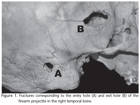
To this end, the bone structure and the dental remains of the body were suitably cleaned and subsequently subjected to a detailed necroscopy that permitted the identification of four fractures located in different regions of the skull, all a result of violence. One of the fractures had a circular format and was situated in the petrous region of the right temporal bone, while another had an abnormal format, situated in the squamous part of the ipsilateral temporal bone. These two fractures may be classified as sharp-edged/blunt weapon lesions, produced by the entry and exit, respectively, of a projectile from a firearm, fired at different moments (Figure 1). The other two fractures were located at the base of the skull, in the greater wing of the sphenoid on the right-hand side and in the region lateral to the left occipital condyle. Therefore, the cause of the victim's death was related to the cranioencephalic trauma perpetrated by a sharp/blunt instrument (firearm projectiles).
As regards the identification of the corpse, anthropological and forensic dental analyses were conducted as the soft tissue, which made up the fingertips, was found to be destroyed by the decomposition process. During the forensic dental exam, it was found that the victim had the following: old tooth loss, whose remaining alveolar ridges had been remodeled; teeth lost after death, whose dental cavities were empty and the interdental septa were preserved; a crown fracture which occurred after death (tooth 12); restorations in compound resin (cingulum of the canine teeth); restorations with silver amalgam; rotations; and diastemas.
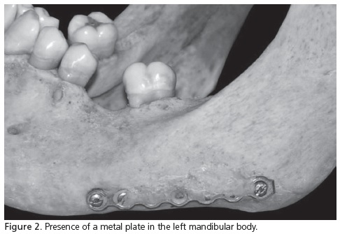
Two other dental peculiarities attracted attention: the first was related to the presence of an orthodontic band fixed to the crown in tooth 17, which indicated that the victim would have been undergoing dental treatment; the second peculiarity related to the presence of a rectilinear metal plate, fixed to the left mandibular body by means of 6 screws. This plate had areas of bony tissue covering its outer surface, indicating that it was used in the reduction of an old jaw fracture (Figure 2).
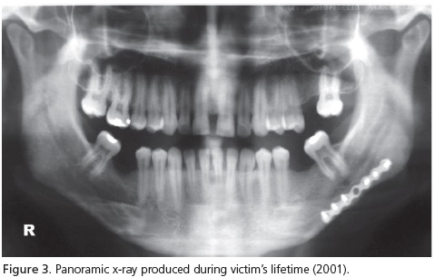
While the forensic exams were being carried out, the police investigation was progressing and the name of the supposed victim came up in the context, relating to an adult male who had disappeared some 30 days prior to the date the skeletonized body was found. So the supposed family members were instructed to look for any form of documentation produced as a result of medical or dental treatment, the result of this search culminating in the finding of a dental document dated 2001, comprising extra and intraoral photographs as well as a panoramic x-ray (Figure 3).
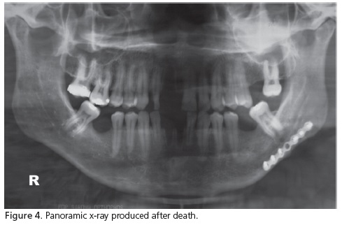
As the panoramic x-ray was the imagiological exam that permitted the disclosure of a larger number of anatomical and therapeutic peculiarities situated in the oral-maxillofacial complex, a post-mortem panoramic x-ray was performed (Figure 4), with the jaw being fixed to the skull using thermoplastic glue, with the anterior teeth in the top-to-top position.
A comparison of the dental characteristics, present in the radiographs taken before and after death, allowed us to positively connect the examined corpse to the missing person, bearing in mind the following details: the old tooth absences; the amount, location and type of restoration; the contour of the maxillary septum in the second molar region as well as a metal plate in the left mandibular body. The other forensic exams that were performed also provided results consistent with those obtained in the radiographic comparison, though in a lesser quantity and quality than those evidenced in the panoramic x-rays.
In order to carry out the present study, the ethical principles contained in the Declaration of Helsinki (2000) were followed and compliance with specific Brazilian legislation was also observed.
DISCUSSION
The panoramic x-ray is an imagiological exam produced by a technique that exhibits images of the facial structure present in the upper and lower dental arches, as well as the support structures. This exam is used clinically in the diagnosis of problems that require a broad view of the oral-maxillofacial complex, being commonly requested in the assessment of traumatisms, localization of third molars, extensive lesions, dental development, tooth retention and growth anomalies6.
Due to the fact that this type of imagiological exam requires the use of high-cost apparatus, its production is basically limited to radiological clinics. Accordingly, it is up to the dental surgeon to request one when he believes it relevant to the treatment he will be carrying out, mainly given the need to observe the oral-maxillofacial complex in greater magnification, as with the performance of orthodontic, implant, surgical and/or rehabilitation treatments.
In the current case, the initial panoramic x-ray was produced with the aim of providing support for an orthodontic treatment whose therapy was begun and evidenced by the presence of an orthodontic band, though with little work having been done, considering that the malocclusions present at the start of treatment were still there at the time of the post-mortem examination. Even though the orthodontic treatment had not continued as initially planned, the care taken by the professional in archiving the documentation produced for over seven years was evident, demonstrating compliance with the ethical recommendation of Clause VIII of Article 5 of the Dental Code of Ethics7.
It is understood that patients have the right of access to all dental documents produced as a result of their clinical treatment8, the Dental Surgeon being the guardian of this documentation for an indefinite period of time. This lack of definition concerning how long to keep dental documentation is mainly due to the possibility of the dental treatment having a defect which is difficult to prove (latent defect), a factor which extends the statutory retention period of 90 days to the moment in which the defect is evidenced, as set out in Article 26, §3 of the Consumer Protection Code9. In addition to the possibility of questions arising concerning professional conduct in the creation of products and performance of services, the archiving of dental documentation for an indefinite period also allows for the resolution of other legal issues as in cases of human identification where the requested dental documentation was produced more than ten years previously10.
It is up to the radiologist to produce the imagiological exams within the technical standards established in literature and to process the film so that the radiographic image enables an adequate view of the various anatomic and reconstructive peculiarities potentially present in the oral-maxillofacial complex. The care taken to make technical recommendations relevant to the carrying out of the panoramic x-rays also demonstrates an ethical care to the patient, who shall be exposed to the minimum amount of X-rays necessary to produce this radiograph and, as a consequence, ensures the quality of this complementary exam both from a clinical perspective and also where faced with legal questions11.
CONCLUSION
In the context of forensic investigations, forensic dental literature reports more cases of human identification using periapical and interproximal x-rays1,12-14, when compared with extra-oral x-rays. This can be explained by the greater production of these two types of x-ray with the aim of providing support for basic dental treatment, such as the study of caries, endodontic and periodontal treatments, and also by the greater ease of acquiring equipment for intra-oral dental x-rays and the low cost of production and ease of execution of the technique, when compared to the production of panoramic radiographs.
On the other hand, panoramic x-rays are being requested more and more with the aim of providing support for planning and diagnosis, mainly in the areas of Orthodontics, surgery and oral rehabilitation10. Consequently, these complementary exams also come to provide important support in cases of human identification1,15.
Finally, it should be stressed that, besides the factors inherent to dental documentation (good-quality panoramic x-rays) and to the corpse (integrity of the oral-maxillofacial complex), the presence of forensic dentists working together with forensic experts from the Coroner's Office in Brazil also represents an important factor in obtaining a satisfactory outcome in cases of human identification. Legally, according to clauses I and IX of Article 6 of Law 5.081/6616, the Dental Surgeon is the only professional with the legal capacity and specific academic training to analyze the dental peculiarities in the context of forensic examination.
Collaborators
RF SILVA carried out the forensic examination and the structuring and composition of the article. FG NUNES participated in the forensic examination, performed the bibliographical survey and the composition of the article. JC FARIA NETO took part in the post-mortem radiological exam; performed a bibliographical survey and composition of the article. ICC REGE conducted the radiographic comparison and the composition of the article. E DARUGE JÚNIOR guided the discussion of the results obtained, structuring and composition of the article.
REFERENCES
1. Silva RF, Daruge Júnior E, Pereira SDR, Almeida SM, Oliveira RN. Identificação de cadáver carbonizado utilizando documentação odontológica. Rev Odonto Ciênc. 2008;23(1):90-3. [ Links ]
2. Muller M, Berytrand MF, Quatrehomme G, Bolla M, Rocca JP. Macroscopic and microscopic aspects of incinerated teeth. J Forensic Odontostomatol. 1998;16(1):1-7.
3. Silva RF, Ramos DIA, Pereira SDR, Daruge E, Daruge Júnior E. Modelos de gesso: importância pericial e orientações odontolegais para arquivamento. Rev Assoc Paul Cir Dent. 2007;61(5):381-4.
4. Silva RF, Pereira SDR, Prado FB, Daruge Júnior E, Daruge E. Forensic odontology identification using smile photograph analysis: case reports. J Forensic Odontostomatol. 2008;27(1):12-7.
5. Ramos DIA, Daruge Júnior E, Daruge E, Antunes FCM, Meléndez BVC, Francesquini Junior L, et al. Transposición dental y sus implicaciones eticas y legais. Rev ADM. 2005;62(5):185-90.
6. White SC, Pharoah MJ. Radiologia oral: fundamentos e interpretação. 5ª ed. Rio de Janeiro: Elsevier; 2007.
7. Brasil. Conselho Federal de Odontologia. Resolução n. 42, de 20 de maio de 2003. Aprova o Código de Ética em Odontologia [texto na Internet]. Diário Oficial da República Federativa do Brasil, Brasília (DF); 2003 [citado 2008 Fev 10]. Disponível em: < http://www.forp.usp.br/restauradora/etica/c_etica/ceo_05_03. html>.
8. Severo AFR, Franco F, Kraether Neto L, Costa NP, Veeck EB. Radiografias odontológicas pertencem ao profissional ou ao paciente? Odontologia Clín Científ. 2002;2(1):97-102.
9. Brasil. Lei n. 8078, de 11 de setembro de 2011. Dispõe sobre a proteção do consumidor e dá outras providências [texto na Internet]. Diário Oficial da República Federativa do Brasil, Brasília (DF); 1990 Set 12 [citado 2008 Fev 10]. Disponível em: <https://legislacao.planalto.gov.br/legisla/legislacao.nsf/web search?openagent&tipo=LEI&codigo=8.078&ementa=2&da ta=19900911>.
10. Silva RF, Pereira SDR, Mendes SDSC, Pereira MMAF, Daruge E, Daruge Júnior E. Importância dos registros odontológicos para a identificação de corpo esqueletizado: relato de caso pericial. RCO Rev Curso Odontol Unievangélica. 2007;9(1):63-6.
11. Silva AE, Larentis NL, Fontanella V. Avaliação da freqüência dos erros na aquisição de radiografias panorâmicas num serviço de radiologia odontológica. RFO UPF. 2007;12(1):32-6.
12. Silva RF, De la Cruz BVM, Daruge Júnior E, Daruge E, Francesquini Júnior E. La importancia de la documentación odontológica en la identificación humana: relato de un caso. Acta Odontol Venez. 2005;43(2):159-64.
13. Silva RF, Pereira SDR, Mendes SDSC, Marinho DEA, Daruge Júnior E. Radiografias odontológicas: fonte de informação para a identificação humana. Odontologia Clín Científ. 2006;5(3):239- 42.
14. Barbosa A, Costa LRS, Barros LV, Rabbi R. Importância dos registros odontológicos na identificação odonto-legal: relato de caso. UFES - Rev Odontol. 1999;1(2):78-83.
15. Paranhos LR, Caldas JCF, Iwashita AR, Scanavini MA, Daruge Júnior E. A importância da documentação ortodôntica nas perícias de identificação humana. Ortodontia. 2008;41(Ed Esp):297-301.
16. Brasil. Lei n. 5081, de 24 de agosto de 1966. Regulariza o exercício da profissão de dentista no Brasil [texto na Internet]. Diário Oficial da República Federativa do Brasil, Brasília (DF); 25 Agosto 1966 [citado 2008 Fev 10]. Disponível em: <https://legislacao. planalto.gov.br/legisla/legislacao.nsf/websearch?openagent&tip o=LEI&codigo=5.081&ementa=2&data=19660824>.
 Correspondence to:
RF SILVA
e-mail: rhonanfs@terra.com.br
Received on: 17/11/2008
Final version resubmitted on: 4/2/2009
Approved on: 22/4/2009
