


Serviços Personalizados
Artigo
 pdf em Inglês
 Artigo em XML
 Referências do artigo
 Enviar este artigo por email
Links relacionados
Compartilhar

 Permalink
RGO.Revista Gaúcha de Odontologia (Online)
versão On-line ISSN 1981-8637
RGO, Rev. gaúch. odontol. (Online) vol.61 no.2 Porto Alegre Abr./Jun. 2013
ORIGINAL / ORIGINAL
Effect of chronic cortisone use in rats strain susceptible to ligature-induced periodontitis
Efeito do uso crônico da cortisona em ratas de linhagem susceptível à periodontite induzida por ligadura
Alex Semenoff SEGUNDOI; Samyra Lopes BuzelleII; Alessandra Nogueira PORTOII; Álvaro Henrique BORGESI; Fabio Luís Miranda PEDROI; Tereza Aparecida Delle Vedove SEMENOFFII
I Universidade de Cuiabá, Faculdade de Odontologia. Rua Manoel Ferreira Mendonça, 149, Bandeirantes, 78010-160, Cuiabá, MT, Brasil.
II Universidade de Cuiabá, Programa de Mestrado em Ciências Odontológicas Integradas. Cuiabá, MT, Brasil.
ABSTRACT
Objective
The present study assessed the effect of prolonged betamethasone use in ligature-induced periodontitis in adult Fischer-344 rats.
Methods
Thirty-six Fisher rats were randomly assigned to three groups: group B (betamethasone); group S (sham) and group C (control). The animals in group B were given intramuscular betamethasone daily, those in group S were given saline injections daily, and those in group C were not submitted to any intervention. The interventions lasted 60 days, and all groups were submitted to the same environmental conditions. Ten days after starting the injections, periodontal disease was induced in the animals from groups B and S by tying a sterile silk suture thread around the upper right second molar. Fifty days later, all animals were sacrificed and the connective tissue attachment level (Jac-E) and bone loss (Jac-O) were measured.
Results
Jac-E and Jac-O of groups B and S were not significantly different, but they differed significantly from those of group C.
Conclusion
Prolonged use of betamethasone did not affect the progression of induced periodontitis in rats.
Indexing terms: Betamethasone. Periodontitis. Rats, Inbred F344.
RESUMO
Objetivo
Avaliar o efeito do uso prolongado da betametasona na periodontite induzida por ligadura, em ratas adultas da linhagem Fischer-344.
Métodos
Selecionou-se trinta e seis ratas Fisher, as quais foram divididas aleatoriamente em três grupos: grupo B (betametasona); grupo S (soro) e grupo C (controle). Os animais do grupo B receberam injeções diárias intramuscular de betametasona, o grupo S recebeu injeções diárias de soro fisiológico e o grupo C foi mantido sem nenhuma intervenção. Estes procedimentos duraram 60 dias. Os três grupos foram mantidos nas mesmas condições ambientais. Decorridos dez dias a partir do início das injeções, os animais dos grupos B e S foram submetidos a indução da doença periodontal, através da colocação do fio de seda em volta do segundo molar superior direito. Ao final do período experimental (50 dias) os animais foram submetidos à eutanásia. O nível de inserção histológica (Jac-E) e perda óssea histológica (Jac-O) foram mensurados.
Resultados
Entre os grupos B e S, não houve diferenças estatisticamente significativas para Jac-E e Jac-O. Entretanto, notaram-se diferenças significativas em ambos os parâmetros avaliados quando o grupo C foi comparado aos grupos B e S.
Conclusão
Pode-se concluir que o uso prolongado de betametasona não modificou a progressão de periodontite induzida.
Termos de indexação: Betametasona. Periodontite. Ratos Endogâmicos F344.
INTRODUCTION
The relationship between the central nervous system and the immune system has been well established. However, much is yet unknown about the ways in which they connect and communicate1,2.
The hypothalamic-pituitary-adrenal (HPA) axis is responsible for many body functions in mammals. Exogenous or endogenous stimuli may affect the HPA response, changing an animal's mood, body temperature, appetite, hematopoietic organs, and leucocyte activity, among others, and facilitating the onset of infectious diseases, such as periodontitis3-4.
Inbred Fischer rats have greater HPA activation. The activation begins in the paraventricular nucleus (PVN) of the hypothalamus. Once excited, the PVN produces the neuropeptide corticotropin-releasing factor (CRF), which travels through the hypophyseal portal system and stimulates the adrenal and pituitary glands to produce many neurotransmitters. The most relevant hormones for the neuroendocrine axis are the adrenocorticotropic hormone (ACTH) and ß-endorphins, which stimulate the secretion of various endogenous substances, such as glucocorticoids, produced in the adrenal cortex, and noradrenaline and adrenaline, produced in the adrenal medulla and nerve endings5-7.
Inbred Fischer rats produce greater amounts of the abovementioned substances, which has a direct effect on the nervous, connective, and epithelial tissues of the periodontium, facilitating the progression of ligatureinduced periodontitis7-9.
The use of pharmaceutical drugs that suppress the inflammatory response in individuals with chronic diseases, such as arthritis, is a good option for improving their quality of life. Therefore, one of the alternatives is to use drugs that inhibit the inflammatory cascade. However, prolonged used of these drugs causes weight gain, gastric ulcers, and changes in blood pressure and the urinary system, among others10-11.
Corticosteroids are widely used in medical practice because of their numerous indications, such as the control of allergies, arthritis, changes in hard and soft tissues, skin problems, and neoplasms, and are usually recommended after surgeries. Sometimes prolonged use of these substances is necessary.
The present study measured histological parameters to assess the effect of prolonged betamethasone use in Fischer-344 rats.
METHODS
Thirty-six female Fischer rats aged two months and weighing 186.00±12.35 grams were selected for this study. They were given one week to adapt to the new environment which consisted of five or six animals housed per polyethylene cage (16x40x30 cm) under a 12- hour light/12-hour dark cycle at a temperature of 23°C and approximate air moisture of 40%. All animals had free access to a standard chow and water. The study was approved by the Animal Research Ethics Committee under protocol number CEEA74-05. The study strictly followed all the ethical principles set forth by the Declaration of Helsinki (2000).
Study design
On day 1 the rats were divided randomly by a technician into three groups as follows: a) Group B: ligature + betamethasone (n=12); b) Group S: ligature + saline (n=12); and c) Group C: negative control (n=12).
Group B was given daily intramuscular injections containing 1mg/kg of betamethasone (Duoflam, betamethasone dipropionate + betamethasone sodium phosphate, Cristália, Itapira, Brazil). The animals were weighed weekly to adjust the dosage. Group S was given daily injections containing 1ml of saline at 0.9%. These injections were given for 60 days. Group C was not given any injection.
Induced periodontal disease
Ten days after the injections began, the animals in groups B and S were anesthetized by the intramuscular administration of 0.1ml of ketamine hydrochloride (Dopalen, Agribrands, Saúde Animal, Paulínea, Brazil) at 50mg/ml and 0.05ml of xylazine hydrochloride (Rompun, Bayer, Saúde Animal, São Paulo, SP, Brazil) at 2g/100ml for every 100g of body weight.
After the anesthesia, a sterile silk suture thread number 4 (Ethicon, Johnson & Johnson, São Paulo, Brazil) was tied around the upper right second molar, and left there for 50 days12.
Histological analysis
After the intervention, the animals were sacrificed by anesthetic overdose and the right and left hemimaxillas were removed and fixed in 10% aqueous formaldehyde for 48 hours. After this period, the pieces were placed in ethylenediaminetetraacetic acid (EDTA) for five weeks for decalcification, then rinsed, dehydrated, cleared and embedded in paraffin blocks in a way that allowed cutting along the mesiodistal axes of the teeth. The 6 μm cuts were stained with hematoxylin and eosin in alcohol.
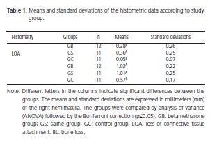
Ten serial slides were selected for the analysis. All slides showed the upper first and second molars and the following structures: a) coronal pulp; b) radicular pulp; c) evident cementoenamel junction of the mesial portion of the second molar; d) interproximal bone crest; and e) level of the connective tissue attachment. Histometry was done by capturing images and using the software ImageLab 2000 (Diracon Bio Informática Ltda., Vargem Grande do Sul, Brazil) to measure the study features in millimeters. The study features were: a) the distance between the cementoenamel junction in the mesial surface of the second molar and the apical portion of the connective epithelial tissue (LOA), representing the loss of attachment; and b) the distance between the cementoenamel junction and the alveolar crest (LOA), representing bone loss.
The slides were numbered and analyzed by different individuals who were not aware of their original groups. To ensure data reliability, intraindividual calibration was done during analysis of the slides by having the first slide of a lot reanalyzed at every ten slides. The differences between the standard error of the means for the attachment level and bone loss were 0.006mm and 0.005mm, respectively.
Result analysis
The mean weights of the different groups in the last week of the study were calculated and compared. The histometric data were grouped as either LOA or BL parameter and compared.
The independent samples were treated by one-way analysis of variance (one-way ANOVA) and post hoc tests, the Bonferroni correction and the t-test. The significance level for all tests was set at 5%.
RESULTS
The loss of attachment (LOA) and bone loss (BL) of the groups B and S submitted to ligature-induced periodontitis did not differ significantly (p>0.05) (Table 1).
The control group, that is, the group that had not been submitted to ligature-induced periodontitis or injections, had significantly less loss of attachment (LOA) and bone loss (BL) than groups B and S (p<0.05) (Table 1).
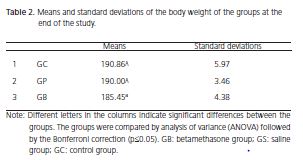
The mean body weights of the three groups did not differ significantly at the end of the experiment (Table 2).
DISCUSSION
The study results evidence that the study variables of groups S and B progress at the same rate, confirming the results of other studies. A study that used betamethasone in young rats observed that this drug prevented the progression of induced periodontitis in adult rats 13. Another study compared kidney transplant patients on high dosages of immunosuppressive drugs for long periods of time with patients without systemic conditions and found that said drugs did not promote bone loss or increase pocket depth14.
Although these findings are supported by many studies, some studies have opposite findings. Cavagni et al.15 found greater periodontal destruction in Wistar rats submitted to betamethasone and ligature-induced periodontitis for 40 days. A Lewis rat strain not susceptible to periodontal disease treated with pelleted corticosteroids also presented greater loss of connective tissue attachment than the control group8. A factor that may explain the conflicting results is the rat strains used by these studies. Lewis and Wistar rats have less intense HPA axis responses than the Fischer strain, which directly affects susceptibility to infectious diseases, such as periodontitis9,16.
A limitation of this study is that serum cortisone levels were not measured during or at the end of the study. However, the weights of the hematopoietic organs and parameters changed, as did the biochemical parameters (data not shown), proving that the study drug was active.
The duration of immunosuppressive treatment is a factor that may affect the progression of periodontitis. Fischer & Kingle17 used probing to analyze periodontal destruction at 14 and 28 days, and found more destruction at 14 days in the experimental group than in the control group, but equal destruction at 28 days.
Fischer rats were chosen for the present study because they are more susceptible to ligature-induced periodontitis. This is due to the greater genetic reactivity of their HPA axis, which produces greater amounts of glucocorticoids, adrenaline, and noradrenaline, which in turn are capable of interfering in the etiopathogenesis of periodontitis7,9.
The corticosteroid dosage used in the present study followed the manufacturer's recommendation and was based on the animal's body weight. An animal's weight is an indication of its health18, and the mean weights of the study groups were not significantly different.
Other important methodological procedures regard intraindividual calibration and maintaining the analyst unaware of the slide groups (blind analysis)19. This study performed both procedures, which renders good reliability to the results and avoids measuring bias.
Sex is a factor that may protect against the progression of periodontitis – only females were used in this study. However, a study has reported that induced periodontitis progresses at similar rates in male and female rats8.
Rat studies are an important tool for the testing of hypotheses, but what remains to be established is if the prolonged use of immunosuppressants affects the progression of periodontitis in humans.
CONCLUSION
The prolonged used of betamethasone did not affect the progression rate of ligature-induced periodontitis in rats strain susceptible to periodontal disease.
Collaborators
AN PORTO, AS SEGUNDO, TADV SEMENOFF and AH BORGES participated in the experimental procedures and writing of the manuscript. FLM PEDRO and SL BUZELLE were responsible for the statistical analyses and writing of the manuscript.
REFERENCES
1. Maier FS. Bi-directional immune–brain communication: Implications for understanding stress, pain, and cognition. Brain Beh Immun. 2003;17(2):69-85. doi: 10.1016/S0889- 1591(03)00032-1.
2. Reiche EMV, Nunes SOV, Morimoto HK. Stress, depression, the immune system, and cancer. Lancet Oncol. 2004;5(10):617-25. doi: 10.1016/S1470-2045(04)01597-9.
3. Anisman H, Baines MG, Berczi I, Bernstein CN, Blennerhassett MG, Gorczynski RM. Neuroimmune mechanisms in health and diseases: 1. Health. CMAJ. 1996;155(7): 867-74.
4. Anisman H, Baines MG, Berczi I, Bernstein CN, Blennerhassett MG, Gorczynski RM. Neuroimmune mechanisms in health and diseases: 2. Disease. CMAJ. 1996;155(8):1075-82.
5. Blalock JE. The syntax of immune-neuroendocrine communication. Immunol Today. 1994;15(11):504-11. doi: 10.1016/0167-5699(94)90205-4.
6. Chrousos GP. The hypothalamic-pituitary-adrenal axis and immune-mediated inflammation. N Engl J Med. 1995;332(20):1351-62. doi: 10.1056/NEJM199505183322008.
7. Breivik T, Thrane PS, Gjermo P, Opstad PK, Pabst R, von Hoersten S. Hypothalamic-pituitary-adrenal axis activation by experimental periodontal disease in rats. J Periodontal Res. 2001;36(5):295- 300. doi: 10.1034/j.1600-0765.2001.360504.x.
8. Breivik T, Opstad PK, GJermo P, Thrane PS. Effects of hypothalamic-pituitary-adrenal axis reactivity on periodontal tissue destruction in rats. Eur J Oral Sci. 2000;108(4):115-22. doi: 10.1034/j.1600-0722.2000.00774.x.
9. Breivik T, Thrane PS. Psychoneuroimmune interaction in periodontal disease. In: Alder R, Felten DL, Cohen N. Psychoneuroimmunology. 3a ed. San Diego: Academic Press; 2001. p. 627-44.
10. Kirwan JR. The arthritis and rheumatism council low-dose glucocorticoid study group. N Engl J Med. 1995;333(3):142-6.
11. Tugwell P, Pincus T, Yocum D, Stein M, Gluck O, Kraag G, McKendry R, Tesser J, Baker P, Wells G. Combination therapy with cyclosporine and methotrexate in severe rheumatoid arthritis. N Engl J Med. 1995;333(3):137-41. doi: 10.1056/ NEJM199507203330301.
12. Semenoff TA, Semenoff-Segundo A, Bosco AF, Nagata MJ, Garcia VG, Biasoli ER. Histometric analysis of ligature-induced periodontitis in rats: a comparison of histological section planes. J Appl Oral Sci. 2008;16(4):251-6. doi: 10.1590/S1678- 77572008000400005.
13. Breivik T, Gundersen Y, Osmundsen H, Fonnum F, Opstad PK. Neonatal dexamethasone and chronic tianeptine treatment inhibits ligature-induced periodontitis in adults rats. J Periodont Res. 2006;41(1):23-32. doi: 10.1111/j.1600- 0765.2005.00833.x.
14. Saether K, Tollefsen T, Helgeland K, Schenck K. The gingival plasma cell infiltrate in renal transplant patients on an immunosuppressive regimen. Acta Odontol Scand. 1998;56(5):281-7. doi: 10.1080/000163598428455.
15. Cavagni J, Soletti AC, Gaio EJ, Rösing CK. The effect of dexamethasone in the pathogenesis of ligature-induced periodontal disease in Wistar rats. Braz Oral Res. 2005;19(4):290- 4. doi: 10.1590/S1806-83242005000400010.
17. Fischer RG, Kingle B. Clinical and histological evaluation of ligature-induced periodontal breakdown in domestic ferrets immunosuppresed by Cyclosporin-A. J Clin Periodontol. 1994;21(4):240-9. doi: 10.1111/j.1600-051X.1994.tb00312.x.
18. Semenoff-Segundo A, Hennemann K, Fontanella VRC, Rosing CK. The role of psychoneuroimmune interactions in the pathogenesis of ligature-induced periodontal disease in Wistar rats. J Inter Acad Periodontol. 2007;9(1):26-31. doi: 10.1111/j.1600-051X.1994.tb00312.x.
19. Kigman A, Albandar JM. Methodological aspects of epidemiological studies of periodontal diseases. Periodontol 2000. 2002;29(1):11-30. doi: 10.1034/j.1600- 0757.2002.290102.x.
 Endereço para correspondência:
AS SEGUNDO
e-mail: semenoff@uol.com.br
Received on: 30/11/2009
Final version resubmitted on: 13/6/2010
Approved on: 25/11/2010
