


Serviços Personalizados
Artigo
 pdf em Inglês
 Artigo em XML
 Referências do artigo
 Enviar este artigo por email
Links relacionados
Compartilhar

 Permalink
RGO.Revista Gaúcha de Odontologia (Online)
versão On-line ISSN 1981-8637
RGO, Rev. gaúch. odontol. (Online) vol.61 no.4 Porto Alegre Out./Dez. 2013
ORIGINAL / ORIGINAL
Non neoplastic proliferative lesions:a ten-year retrospective study
Processos proliferativos não neoplásicos: um estudo retrospectivo de 10 anos
Anna Rebeca de Barros Lins Silva PALMEIRAI; Amanda Galindo FLORÊNCIOI; José Paulo da SILVA FILHOII; Uoston Holder da SILVAIII; Ney Soares de ARAÚJOVI
I Faculdade ASCES, Curso de Odontologia. Av. Portugal, 584, Universitário, 55016-400, Caruaru, PE, Brasil.
II Faculdade São Leopoldo Mandic, Curso de Odontologia, Programa de Pós-Graduação em Radiologia. Campinas, SP, Brasil.
III Professor Regente da Disciplina de Propedêutica no Curso de Odontologia da Faculdade ASCES -Av. Portugal, 584, Universitário, CEP 55016-400, Caruaru/ PE.
VI Faculdade São Leopoldo Mandic, Curso de Odontologia, Programa de Pós-Graduação em Patologia bucal. Campinas, SP, Brasil.
ABSTRACT
Objective
An epidemiological survey of oral soft tissue proliferative processes diagnosed at the center of Pathological Anatomy of Oral ASCES Faculty.
Methods
We analyzed all the histopathological reports in the period from 1999 to 2009 as regards the following variables:histopathologic diagnosis, sex, age, symptoms, type of biopsy and location of the lesion.
Results
Non-neoplastic proliferative processes were present in 328 of the 938 reports analyzed, reaching a prevalence of 35 %. The most common injury was fibrous hyperplasia (81.4%) followed by pyogenic granuloma (11%) and peripheral giant cell lesions (6.1%). Fibrous hyperplasia was more prevalent in females, in the age group 40 to 59 years, where the most frequently performed biopsy was the excisional type, with the most frequent location being the buccal mucosa, and in most cases, no painful symptoms were reported associated with it, demonstrating significant difference between the variables studied (p < 0.001).
Conclusion
Non neoplastic proliferative processes processes are found in the general population, more common in females, affecting wide age-range, predominantly in individuals in the fifth and sixth decades of life, and fibrous hyperplasia was the most frequently found lesion.
Indexing terms: Granuloma, giant cell. Granuloma, pyogenic. Hyperplasia.
RESUMO
Objetivo
Realizar um levantamento epidemiológico dos processos proliferativos dos tecidos moles orais diagnosticadas no Centro de Anatomia Patológica Oral da Faculdade ASCES.
Métodos
Foram analisados todos os laudos histopatológicos no período de 1999 a 2009 em relação às seguintes variáveis:diagnóstico histopatológico, sexo, faixa etária, sintomatologia, tipo de biópsia e localização da lesão.
Resultados
Os processos proliferativos não neoplásicos estiveram presentes em 328 dos 938 laudos analisados, atingindo uma prevalência de 35%.A lesão mais freqüente foi a hiperplasia fibrosa (81,4%) seguida do granuloma piogênico (11%) e lesão periférica de células gigantes (6,1%).A hiperplasia fibrosa foi mais prevalente no sexo feminino, na faixa etária de 40 a 59 anos (p< 0,001), onde o tipo de biópsia mais realizado foi excisional com a localização mais freqüente na mucosa jugal e na maioria dos casos, não foi relatada sintomatologia dolorosa associada, comprovando-se diferença significativa entre as variáveis estudadas (p< 0,001).
Conclusão
Os processos proliferativos não neoplásicos são encontrados na população em geral, mais comum no sexo feminino, acometendo ampla faixa etária, predominantemente em indivíduos na quinta e sexta décadas de vida, sendo a hiperplasia fibrosa a lesão mais frequente.
Termos de indexação: Epidemiologia. Granuloma de células gigantes. Granuloma piogênico. Hiperplasia.
INTRODUCTION
Non neoplastic proliferative processes (NNPP) are lesions frequently found in the oral cavity and in general, they are characterized by inflammatory processes causing tissue proliferation1. They may also be defined as reactive lesions of the oral soft tissues, where chronic, long lasting stimuli, such as residual roots, poorly conserved teeth, subgingival calculus, restorations with proximal excesses, poorly fitting dental prostheses, foreign bodies in the gingival sulcus and other traumatic agents trigger tissue responses in which an exaggerated repair, granulation tissue and formation of wounds appear after injury2-4. Knowledge of non neoplastic proliferative processes is of the utmost importance because their causes, which may be prevented by clinical procedures, are present in day to day clinical practice of dentists, thus promoting the appearance of these lesions.
Among the non neoplastic proliferative processes, fibrous hyperplasia, pyogenic granuloma, and peripheral giant cell lesions should be emphasized because they are frequently found in the oral soft tissues.
Fibrous hyperplasia is characterized by a response to a chronic irritation of low intensity, in which an increase in the size of the tissue occurs due to the increase in the number of its constituent cells as a local tissue response to the aggression1,4. The etiologic factors of fibrous hyperplasia may be diastemas, the cutting edges of teeth, poor oral hygiene, Iatrogenic professional maneuvers, or those results from poorly fitting, broken or incorrectly used dental prostheses4-5. Histologically, they present fibrous conjunctive tissue proliferation, and the lining epithelium may present exocytosis and acanthosis2.
Pyogenic granuloma results from a vascular proliferation in response to an irritation or local infection that stimulates the formation of a highly vascularized tissue1,6. It frequently occurs in the gingiva, however, it may appear in other regions of the mucosa and skin6. It presents as an exophytic, sessile or pediculated lesion, and its characteristics vary according to the time of development and anatomic location3.
Peripheral giant cell lesions originate from the peripheral conjunctive tissue of the periodontal ligament or the subjacent periosteum, and may occur in the gingiva or on the alveolar ridge. Periapical radiographs may show discrete resorption of the alveolar crest in a concave shape. Histologically they present multinucleated giant cells, proliferation of fusiform cells, presence of blood vessels and pigmentation by hemosiderin7-8.
The aim of the present study was to conduct a retrospective survey of non neoplastic proliferative processes in the oral soft tissues, diagnosed at the Center of Oral Pathological Anatomy of the ASCES Faculty, Caruaru, Pernambuco, Brazil.
METHODS
A retrospective cross-sectional study was conducted, in which all the histopathological reports, totaling 1723 were analyzed, at the Center of Oral Pathological Anatomy ("Centro de Anatomia Patológica Oral - CEAPO"), belonging to the ASCES Faculty of Caruaru, Pernambuco, in the period from 1999 to 2009. The anatomopathological exam requisitions that did not present the clinical information completely filled out were excluded from the research, so that 938 reports were used for the present study. This research was previously submitted to the Research Ethics Committee of the ASCES Faculty (CEP-ASCES), and was approved under Protocol No.043/10.
For analysis of the lesions and construction of an epidemiological profile, some variables in the reports were emphasized, such as: Histopathological diagnosis, sex, age group, painful symptomatology, type of biopsy and location of lesion. All the reports that did not present any of the studied variables were excluded from this research. The results whose descriptive levels (p-value) were lower than 0.05 were considered statistically significant. The statistical calculations were made by means of the Chi- Square test.
RESULTS
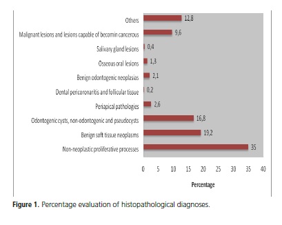
Of the 938 reports analyzed, 323 fitted into the group of non neoplastic proliferative processes of oral soft tissues, attaining a percentage of 35% (Figure 1).
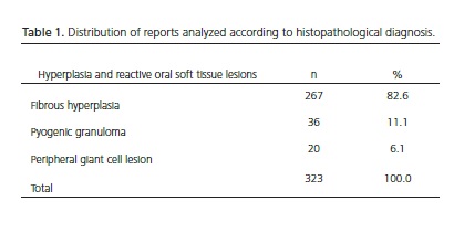
Of the 323 reports of reactive oral soft tissue lesions, the lesion most frequently found was fibrous hyperplasia (n=267, 82.6%), followed by pyogenic granuloma (n=36; 11.1%), and peripheral giant cell lesions (n=20; 6.1) (Table 1).
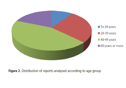
As regards sex, the female sex was most frequently affected, with 241 cases (73%) and 87 cases in the male sex (27%), with significant difference (p < 0.001). The predominant age-range was from 40-59 years, attaining a percentage of 44% (n=143), followed by the group from 20-39 years (n=89; 27%) and subjects over the age of 60 years (n=63; 19%), proving statistically significant difference between the age groups (p < 0.001). The 19 year-old age group was less affected, with 33 cases (10%) (Figure 2).
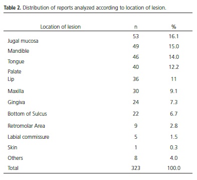
The majority of the lesions (n=310; 95%) produced no symptomatology (p < 0.001) vs. 5% (n=18) of the patients who related pain resulting from the lesion. The biopsy most frequently used was the excisional type (p < 0.001), attaining 97% of the cases (n=317). The most frequent location of non neoplastic proliferative processes was the jugal mucosa (n=53; 16.1%), followed by the mandible (n=49; 15%) and tongue (n=46; 14%) (Table 2).
DISCUSSION
In this study, the group of lesions belonging to the non neoplastic proliferative processes attained the percentage of 35% of all the lesions found, corroborating the 33% found by Simões et al.10 in their research at the Oral Pathology Laboratory of the Federal University of Pernambuco and 32% found by Prado et al.14, in their research with record charts from the semiology clinic of the University of the City of São Paulo, however the finding is in disagreement with the 18.63% found by Amadei et al.1 in their study of the surgical pathology files of the School of dentistry of São José dos Campos, of the "Universidade Estadual Paulista Júlio de Mesquita". For various authors, fibrous hyperplasia was also the most common entity9-10,12,15I. Among the non neoplastic proliferative processes, fibrous hyperplasia presented a rate of 82.6%. This high rate of prevalence of this type of lesion may be associated with the number of traumatic factors capable of triggering this type of lesion in our population. The lack of access to dental treatment for the fabrication of quality dental prostheses leads to various individuals making use of poorly fitting complete or partial dentures4,10.
Local irritation or infection acts as a coadjuvent factor in the appearance of another NNPP, such as pyogenic granuloma10 which, in this research, showed a percentage of 11.1%, a vale compatible with the value of 9.68% found by Amadei et al.1.
The findings encountered in this research showed that the peripheral giant cell lesion was the 3rd most frequent lesion, in 6.1% of the diagnoses found. According to Capelozza et al.7, the occurrence of peripheral giant cell lesions may vary from 6.5 to 12.7%.
As regards sex, Bertoja et al.9, Simões et al.10, Kniest et al.11, Xavier et al.13, Marin et al.15, Deboni et al.16, Aquino et al.17 in their findings, they showed that the female sex was the most affected, corroborating the findings of this research, in which the value found was 73%. This suggests that women take more care of their oral health, and seek specialized attendance with greater frequency than men do10,18. Another possible hypothesis for this fact would be the existence of some systemic factor, such as hormone alterations, inherent to the female sex, which may favor the appearance of oral pathologies2.
The predominant age range in this research was from 40-59 years of age, corresponding with the results found by Bertoja et al.9, Kniest et al.11, Xavier et al.13 e Aquino et al.17, in addition to corroborating the results found by Simões et al.10 who pointed out the 6th decade of life as being the most affected, and diverging from the results found by Marin et al.15, who pointed out the 2nd to 4th decades of life. Prado et al.14, Marin et al.15 and Aquino et al.17 are unanimous in stating that the biopsy most frequently performed in their studies was the excisional type, presenting 74%, 54%, 67% and 59,4% of occurrence, respectively; whereas in this study, 97% of the cases were of the excisional type.
The most frequent location of non neoplastic proliferative processes in this study was the jugal mucosa affecting 16.1% of the cases, in agreement with the study of Amadei et al.1, who related that for fibrous hyperplasia, the area most affected was the jugal mucosa, in 21.97% of the cases.
CONCLUSION
Non neoplastic proliferative processes are found in the population in general, affecting a wide age range, predominantly in individuals in the fifth and sixth decades of life, with fibrous hyperplasia being the most frequently found lesion. It is more common in the female sex, often causes no painful symptomatology, however, it may lead to serious damage to the oral soft tissues. The dental surgeon should have knowledge of these lesions, removal of the etiological factors, and indication of the correct treatment.
Collaborators
ARBLS PALMEIRA and AG FLORÊNCIO participated in the research and in writing the article. JP SILVA FILHO was responsible for the data analysis and interpretation, and participated in writing the article. UH SILVA and NS ARAÚJO guided the research and participated in writing the article.
REFERENCES
1. Amadei SU, Pereira AC, Silveira VAS, Carmo ED, Scherma AP, Rosa LEB. Pevalência de processos proliferativos nãoneoplásicos na cavidade bucal: estudo retrospectivo de quarenta anos. Clin Pesq Odontol UNITAU. 2009;1(1):38-42. [ Links ]
2. Neville BW, Damm DD, Allen CM, Bouquot JE. Tumores dos tecidos moles. In: Neville BW, Damm DD, Allen CM, Bouquot JE. Patologia oral e maxilofacial. 2ª ed. Rio de Janeiro: Guanabara Koogan; 2004.
3. Avelar RL, Antunes AA, Carvalho RWF, Santos TS, Oliveira Neto PJ, Andrade ESS. Granuloma piogênico oral: um estudo epideiomógico de 191 casos. RGO - Rev Gaúcha Odontol. 2008;56(2):131-5.
4. Durso BC, Consolaro A. Hiperplasia fibrosa inflamatória: análise da casuística do serviço de anatomia patológica da Faculdade de Odontologia de Bauru- Universidade de São Paulo. Rev Int Estomat. 2005;2(4):15-22.
5. Santos MESM, Costa WRM, Silva Neto JC. Terapêutica cirúrgica da hiperplasia fibrosa inflamatória: relato de caso. Rev Cir Traumatol Buco-Maxilo-Fac. 2004;4(4):241-5.
6. Melo TRNB, Lima LHMA, Lima MG, Godoy GP. Lesão reativa hiperproliferativa: relato de caso clínico. Odontol Clín-Científ. 2009;8(1):73-7.
7. Capelozza ALA, Taveira LAA, Pagin O. Lesão periférica de células gigantes: relato de caso. Salusvita. 2007;26(1):99-104.
8. Cavezzi Junior O, Sartori JHF, Aguiar ECG. Lesão periférica de células gigantes: relato de caso clínico. Odontol Clín.-Científ. 2008;7(3):257-60.
9. Bertoja IC, Tomazini JG, Braosi APR, Zielak JC, Reis LFG, Giovanini AF. Prevalência de lesões bucais diagnosticadas pelo laboratório de histopatologia do UnicenP. RSBO. 2007;4(2):41-6.
10. Simões CA, Lins RC, Henriques ACG, Cazal C, Castro JFL. Prevalência das lesões diagnosticadas na região maxilofacial no laboratório de patologia oral da Universidade Federal de Pernambuco. IJD Int J Dent. 2007;6(2):35-8.
11. Kniest G, Stramandinoli RT, Ávila LFC, Izidoro ACAS. Frequência das lesões bucais diagnosticadas no centro de especialidades odontológicas de Tubarão (SC). RSBO. 2011;8(1):13-8.
12. Moresco FC, Nora Filho MR, Balbinot MA. Levantamento epidemiológico dos diagnósticos histopatológicos da disciplina de estomatologia da Faculdade de Odontologia da ULBRACanoas/ RS. Stomatos. 2003;9(17):29-34.
13. Xavier JC, Andrade SC, Arcoverde CAL, Lucena KCR, Cavalcanti UDNT, Carvalho AAT. Levantamento epidemiológico das lesões bucais apresentadas por pacientes atendidos no Serviço de Estomatologia da Universidade Federal de Pernambuco durante o período de janeiro de 2006 a julho de 2008. IJD. Int J Dent. 2009;8(3):135-9.
14. Prado BN, Trevisan S, Passarelli DHC. Estudo epidemiológico das lesões bucais no período de 05 anos. Rev Odontol Univ Cid São Paulo. 2010;22(1):25-9.
15. Marin HJI, Silveira MMF, Souza CFM, Pereira JRD. Lesões bucais: concordância diagnóstica na faculdade de odontologia de Pernambuco. Odontol Clín.-Científ. 2007;6(4):315-8.
16. Deboni MCZ, Traina AA, Trindade IK, Rocha EMV, Teixeira VCB, Takahashi A. Levantamento retrospectivo dos resultados dos exames anatomopatológicos da disciplina de cirurgia da FOUSP - SP. RPG Rev Pos-Grad. 2005;2005;2(2):229-33.
17. Aquino SN, Martelli DRB, Borges SP, Bonan PRF, Martelli Júnior H. Concordância entre diagnóstico clínico e histopatológico de lesões bucais. RGO - Rev Gaúcha Odontol. 2010;58(3):345-9.
18. Cruz MCFN, Almeida KGB, Lopes FF, Bastos EG, Freitas RA. Levantamento das biópsias da cavidade oral realizadas no hospital universitário - unidade Presidente Dutra/UFMA, da cidade de São Luís-Ma, no período de 1992 a 2002. Rev Bras Patol Oral. 2005;4(3):185-8.
19. White SC, Yoon DC. Comparison of sensitometric and diagnostic performance of two films. Comip Cont in Educ Dent. 2000;21(6):530 9.
Received on: 28/5/2012
Final version resubmitted on: 12/12/2012
Approved on: 23/3/2013
