


Serviços Personalizados
Artigo
 pdf em Inglês
 Artigo em XML
 Referências do artigo
 Enviar este artigo por email
Links relacionados
Compartilhar

 Permalink
RGO.Revista Gaúcha de Odontologia (Online)
versão On-line ISSN 1981-8637
RGO, Rev. gaúch. odontol. (Online) vol.61 no.4 Porto Alegre Out./Dez. 2013
ORIGINAL / ORIGINAL
Oral lesions diagnosed in a public oral pathology laboratory
Alterações bucais registradas em um serviço público de patologia bucalI
Fábia Ferreira de Albuquerque da CUNHAII; Milena Bortolotto Felippe SILVAII; Francine Kühl PANZARELLAII; José Luiz Cintra JUNQUEIRAII; Luciana Butini OLIVEIRAII
I Artigo baseado na dissertação de FFA Cunha, intitulada "Estudo retrospectivo das lesões bucais registradas no serviço de patologia bucal do laboratório público do estado de Mato Grosso". Programa de Pós-Graduação em Odontologia, Faculdade São Leopoldo Mandic; 2010.
II Faculdade São Leopoldo Mandic, Curso de Odontologia, Programa de Pós-Graduação em Radiologia. Rua José Rocha Junqueira, 13, 13045-755, Swift, Campinas, SP, Brasil.
ABSTRACT
Objective
The aim of this study was to perform a retrospective study of 1,894 maxillofacial injuries diagnosed in a public laboratory in Mato Grosso and verify the association by considering the following variables: gender, age, anatomical locationand origin of the patient (capital, interior).
Methods
A sample was selected in the period from2005 to 2008 in order to assess the prevalence of oral lesions in a Public Laboratory (MT Laboratório).
Results
Chronic gingivitis was the most prevalent lesion, with a frequency of 11.46%, followed by inflammatory fibrous hyperplasia (7.44%), mucocele (7.23%) and fibroma (5.54%). Females were affected in50.63% of cases and males49.37%. However, there is no difference in gender (p=0.435). The second decade of lifeshowed a higher prevalence of injuries. The region of the jaw proved to be the most affected (24.45%) and most patients came from the interior. There was a statistically significant association between some pathologies and sex(p<0.001) among the most prevalent anatomical locations and gender (p<0.001) and origin of the patient (p<0.001).
Conclusion
It can be concluded that chronic gingivitis was the most prevalent lesion. There was a statistically significant association between some pathologies and sex, amongst the most prevalent anatomical locations and sex and origin of the patient.
Indexing terms: Epidemiology. Oral diagnosis. Prevalence.
RESUMO
Objetivo
Realizar um estudo retrospectivo para verificar a prevalência das alterações bucais diagnosticadas em um laboratório público do Estado de Mato Grosso e determinar a associação com as seguintes variáveis: gênero, idade, localização anatômica da lesão e procedência do paciente (interior ou capital).
Métodos
Uma amostra compreendendo 1894 laudos alterações bucais foi obtida no período de 2005 a 2008 no Laboratório Público do Mato Grosso (MT Laboratório).
Resultados
Verificou-se que a lesão mais prevalente foi a gengivite crônica com uma frequência de 11,46%, seguido da hiperplasia fibrosa inflamatória (7,44%), mucocele (7,23%) e o fibroma (5,54%). O gênero feminino foi acometido por 50,63% dos casos e o masculino por 49,37, entretanto, não houve diferença significativa entre os gêneros (p=0,435). A frequência de alterações bucais foi maior na segunda década de vida. A região da maxila mostrou-se a mais atingida (24,45%) e a maioria dos pacientes procedia do interior do Estado. Houve associação estatisticamente significante entre algumas patologias e o gênero (p<0,001), entre a localização anatômica de algumas alterações e o gênero (p<0,001) e de acordo com a procedência dos pacientes (p<0,001).
Conclusão
A lesão mais prevalente foi a gengivite crônica. Houve associação estatisticamente significante entre algumas patologias e o gênero do paciente; localização anatômica e de acordo com a procedência dos pacientes.
Termos de indexação: Epidemiologia. Diagnóstico bucal. Prevalência.
INTRODUCTION
Epidemiological studies carried out in a variety of regions are of fundamental importance, since demographic factors such as age, sex, ethnicity1-3, nutrition, habits, geography and the socioeconomic status of the population, have a considerable influence on the incidence of oral diseases4.
The study of the alterations in the oral cavity and adjacent structures constitutesa material factin Dentistry and the dental surgeon has a unique role in the early recognition of these alterations, it being possible to produce a final diagnosis based on the signals and symptoms present.
The results obtained through epidemiological studiesmake it possible to plan and carry out health programs for the population. As Brazil is a country of continental proportions, it is fundamental to carry out these studies in the different regions, as the socioeconomic, cultural and climatic differences observedcan indicate possible distinctions in the prevalence of these injuries, emphasizing that pathological alterations which afflict the oral cavity, whether or not they are associated with the teeth, are commonplace in the population as a whole5.
Given these assumptions, the aim of this study was to perform an epidemiological survey of the histopathological diagnoses of the Oral Pathology Service at the Public Laboratory of the state of Mato Grosso, in order to determine the prevalence of the most common oral alterations, taking into consideration the following patient-related variables: sex, age, anatomic location and place of origin.
METHODS
This research represents a quantitative approach by way of an epidemiological, documentary, descriptive and cross-sectional investigation of the database of the Public Laboratory of Mato Grosso (MT Laboratório).
The Oral Pathology Service at the Public Laboratory of the state of Mato Grosso (MT Laboratório) is located together with the State Center of Reference for Medium and High Complexity (CERMAC) in the city of Cuiabá, in the Brazilian state of Mato Grosso.
The federal state of Mato Grosso, through Directive 195 of November 30,2004, issued by the Secretary of State for Health (SESMT), published a public policy on the care of oral and facial diseases and launched a diagnostic service for these diseases. The service acts as a reference within the state for the performance of oral lesion diagnoses and is responsible, again, within the state, for conducting anatomical and histopathological examinations of oral and facial lesions.
MT Laboratório has at its disposal 160 publicsector employees, including pharmaceutical, biochemical and biomedical staff, doctors, nurses, biologists, laboratory assistants and other health professionals. Laboratory services are being carried out in the Office of the Public Health Laboratory (LACEN), Department of Clinical Analysis and Department of Cytopathology.
The study included all the histopathological reports issued and archived by the Mato Grosso public laboratory (MT Laboratório), for the period from January 2005 to December 2008. A total of 1,894 reports issued and archived by the Public Laboratory of Mato Grosso (MT Laboratório), were analyzed for the period from January 2005, when the laboratory was implemented, to December 2008. The checking of the report files and the use of the physical facilities at the Oral Pathology Laboratory were authorized by the director general of this laboratory. The researcher responsible for the current research study collected the data between December 2009 and February 2010. The methodology consisted of a survey of data from the case histories and from official reports belonging to the Oral Pathology Service of a public laboratory in Mato Grosso - MT Laboratório.
The variables collected related to the sex (male/ female), the patient's age at the time of the diagnosis, the patient's place of origin (state capital or interior), anatomic region affected and the histopathological diagnosis.
The information was collected and input to a database created by the researcher using the SPSS software program, version 15.0. Descriptive and inferential statistical techniques were used to analyze the data. For the descriptive piece, tables, graphs and measurements of position and variance were employed. As for the inferential part, measures of association were computed between the dependent variables and the other explanatory or independent variables, employing Chi-Squared tests, assuming a level of significance of p<0.05.
Statistical analyses were conducted with the aid of the SPSS software program, version 15.0 and versions of MINITAB 15.
The present study was submitted to the Research Ethics Committee at the São Leopoldo Mandic Faculty of Dentistry and Center for Dental Research, which approved the project under reference no. 2009/0299.
RESULTS
From an analysis of the 1,894 reports, considering the distribution of the number and percentage of patients by sex, it was found that 959 (50.63%) were female and 935 (49.37%) were male. It was found that there is little difference between the sexes, a little less than 0.5%. After applying the Z Test for the two proportions, it was ascertained that there was no statistically significant difference between males and females (p=0.435).
The distribution of the number and percentage of patients in relation to agewas roughly symmetrical, between males and females. Out of the sample total, 952 were women and 933 men, with a mean age of 36.27 and 35.88, respectively. However, it was found that, on average, there was no statistically significant difference (p=0.665, Student t test) between the age groups.
The Table below describes the most prevalent alterations by sex, recorded in the MT Laboratório between 2005 and 2008, in Cuiabá, Mato Grosso.
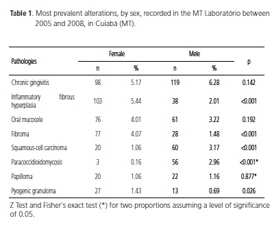
According to the data in Table 1, it was found that there was an association between the most prevalent pathologies and sex (p<0.001, Chi-squared). It should be stressed that, of these eight most prevalent alterations, a statistically significant difference was found in five of them, when compared by sex; for these the values for p are highlighted in bold type.
Table 2 describes the location of the most prevalent alterations, by sex, of patients registered with the MT Laboratório, between 2005 and 2008, in Cuiabá, MT.
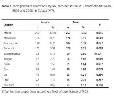
Based on Table 2, it was found that there was an association between the location of the most prevalent alterations and the patients' sex (p<0.001). It is important to highlight that, of these 11 locations of the most prevalent alterations, there is a statistically significant difference in 3, when comparing sex; in these locations the values for p are highlighted in bold type.
Table 3 shows the number and percentage of patients according to place of origin.
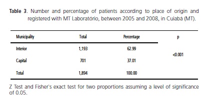
More patients were cared for in the interior (62.99%) than in the state capital(37.01), the difference being statistically significant (p <0.001).
DISCUSSION
Epidemiological studies are of fundamental importance for clinics, epidemiologists and administrators as they provide an understanding of the frequency of alterations in the oral cavity in different groups of the population, allowingtheneeds profile to be formulated for a particular region. Once this profile is sketched out, it is possible to planthe proper treatment and define the preventionstrategies by individualizing the actions in accordance with the distinct features of the studied group.
The understanding of the diverse clinical manifestations of oral alterations can contribute to the diagnosis of systemic diseases, malign and benign neoplasias, and for this reason, the performance of histopathological examinations should be routine in the day-to-day practice of dental procedures.
According to Deboni et al.6, when the documentation relating to the material sent to the Pathology laboratory is well prepared and completed, together with a detailed analysis of information contained in a set of examinations of a disease, it is able to contribute to an understanding of incidence and prevalence and furnish information on epidemiological characteristics of the population under a particular health service.
Retrospective studies on the prevalence of alterations in oral mucosa found in the literature, used samples via anatomical pathology reports issued by Oral Pathology Services in Dental Faculties, University Hospitals, histopathological diagnoses of the Stomatology services in the Dental Faculties and Public Laboratories in the state1,3,7-9. The methodology of the present study is similar to that of the other studies, since it was conducted in an Oral Pathology Public Laboratory, thus permitting a comparison of results.
According to the results obtained, it can be seen that, of the total number of 1,894 anatomical pathology reports examined, the most prevalent alterations were chronic gingivitis, inflammatory fibrous hyperplasia, oral mucocele, fibroma, squamous-cell carcinoma, paracoccidioidomycosis, papilloma and pyogenic granuloma, each of the first four exceeding 5%. Similar results were found by Henrique et al.2 in a study on the prevalence of alterations in the oral mucosa, in the city of Uberaba, which is located in the Brazilian state of Minas Gerais. Also included in this study amongst alterations in oral mucosa were the gingival inflammatory processes, and this explains why the most prevalent pathology was gingivitis, since in the diverse epidemiological studies, this morbidity is not cited, although the gums are a normal constituent of the oral mucosa, and subject to alteration. Other studies referred to in the reviewed literature also show a prevalence of alterations similar to those found in this study, probably as a result of characteristics inherent to the provision of services to the population served7-11.
In the present research, a higher number of alterations was found of infectious and irritating origin. These alterations are frequently detected in countries of low socioeconomic levels, where it is possible to find a higher number of inhabitants inadequately protected by their health services, presenting with precarious oral health conditions, poor conservation and adaptation of prostheses and lack ofinformation and education in relation to oral hygiene.
Of the total number of histological reports, we observed that the eight most frequent alterations accounted for approximately 43% of cases. We noted that these alterations correspond to proliferative non-neoplastic processes and infectious diseases. Similar frequencies are found in the study by Torrão et al.5.
Considering the distribution of the number and percentage of patients by sex, it was found that, of the 1,894 reports, 959 (50.63%) were female and 935 (49.37%) were male. It can be seen that the difference between the sexes is small, a little under 0.5%. It was found that there was no statistically significant difference between females and males (p=0.665), in agreement with the majority of studies reviewed in the literature4,6,8,11-17 although Cebeci et al.1 noted a higher prevalence of alterations in oral mucosa in males, four times higher than in women.
Of the 8 most prevalent pathologies, a statistically significant difference was found in 5,when considering the sex variable (p<0.001), which disagrees with Volkweis et al.3 who observed no statistically significant association between the sex of the patient and any group of diseases (p=0.137).
Simões et al.18 believed that women are more careful with their oral health, and these results may be a reflection of more women seeking healthcare services, although we certainly cannot discount the possibility that the female sex may in fact be more affected by oral and maxillofacial alterations.
The fact that the male population presents more cases of oral cancer is due, ostensibly, to a greater involvement by the male sex with the risk factors, remembering that multiple causes are frequently involved in the formation of cancer, such that an association of factors is often required, such as alcohol, tobacco, socioeconomic conditions, excess exposure to the sun, eating habits and deficient oral hygiene, for this pathology to occur. Rural workers in the lower classes are more affected by the disease, while in developed countries, cancer of the mouth occurs less frequently than in developing countries.
In the four years investigated, we found 59 cases of paracoccidioidomycosis (3.12%) making it the sixth most prevalent lesion. This finding differs from the studies of Bertoja et al.9 who, based on 1,963 biopsies, found just 5 cases of paracoccidioidomycosis, and Zancanaro et al.19, who found a rate of occurrence of this lesion of 42.1%, a total of 16 cases, out of a total of 4,914 histopathological results. This may be due to the fact that the Midwest region of Brazil is considered to be endemic for paracoccidioidomycosis and the aforementioned studies were conducted in different regions and different populations. Paracoccidioidomycosis mainly targets males, there being 56 cases (2.96%) in the current study. This discrepancy between the sexes can be explained by the fact that women are less susceptible to the disease due to hormonal factors. The age at which oral alterations occur mostis a topic where, in epidemiological studies demonstrating the frequency of lesions in the oral and maxillofacial complex, one finds a great diversity across the different regions. Amongst the possible factors which explain this fact, are those related to the higher life expectancy in countries with a higher socioeconomic level, when compared to the developing world, and possibly as a result of the regional and cultural differences that exist in each population where the studies are carried out.
The age distribution was roughly symmetrical between men and women, with the 25-29 age range displaying the highest prevalence of oral mucosa alterations. It was found that, in the older age groups, there is an increased prevalence of inflammatory fibrous hyperplasia, squamous-cell carcinoma and fibroma. On average, no statistically significant difference (p=0.665) was observed between the age groups, in keeping with the study by Silveira et al.19.
According to Carvalho et al.20, adults exhibit a higher prevalence of oral diseases. The authors established a need for greater care for patients aged between 35 and 44, where the majority goes to work, with the aim of changing the prevailing epidemiological picture.
There was an age variance between 0 and 87, the mean being 36 with peak occurrence in the 2nd, 3rd and 4th decade of life12,21. Some studies, however, found the 5th, 6th and 7th decades of life to be the age groups most affected3,18,22-23.
By correlating the incidence of alterations with age, we can see that up to the age of 34, the most frequent histological types were chronic gingivitis and oral mucocele. Between the ages of 35 and 54, inflammatory fibrous hyperplasia, chronic gingivitis, paracoccidioidomycosis and fibroma were the most frequent, while in the 55 to 69 age range, inflammatory fibrous hyperplasia, squamous-cell carcinoma andfibroma occurred most, and finally, between the ages of 70 and 89, only squamous-cell carcinoma. These results are probably due to the acquisition of habits and vices over the years, the continued use of poorly fitting prostheses, parafunctional habits, amongst others, promoting the emergence of specific pathologies. Pentenero et al.15, in their study on the prevalence of oral mucosa lesions, stated that the presence of lesions in the oral mucosa is directly linked to the acquisition of risk habits and to age. As for anatomic location, there was a higher prevalence of alterations in the maxilla. It was found that there was an association between the most prevalent anatomic location and sex (p<0.001), however, it should be pointed out that of the 11 most frequent anatomic locations, considering only the 3 most prevalent (maxilla, buccal mucosa and tongue) there is a statistically significant difference (maxilla p=0.015, buccal mucosa p=<0.001 and tongue p=0.049).
In the reviewed literature, the location of the alterations varied. The results obtained in the present research study corroborate the results of Deboni et al.6 in which the maxilla was the most frequently affected anatomic location, and those of Nascimento et al.8, who found the mandibular to be the most affected anatomical region, followed by the maxilla and the lower lip. Vier et al.24, however, found diverging results when considering the location of the alterations, namely the alveolar rim, followed by the tongue, lip, buccal mucosa and hard palate as the places with greatest prevalence, and similar results were recorded by Rocha et al.25 who reported that the anatomic location most affected was the buccal mucosa, followed by the palate, tongue, lip and gums. Moreira et al.26 found that the anatomic location most affected was the tongue, although the top lip was most commonly affected specifically amongst the benign neoplasias. A recent study conducted in the oral pathology laboratory at the University of Pernambuco made it clear that oral lesions in the elderly occur principally in the gingival margin and alveolar ridge27.
By analyzing the patients' origins, it can be verified that, over the four years studied, the great majority hailed from the interior of the state and that there was a statistically significant difference (p<0.001) between the prevalence of oral alterations and patients' places of origin. Such a discrepancy in patients emanating from the interior may be due to the fact that these cities do not have Oral Pathology services and the samples collected are all sent off to the state capital. Similar data were found by Borges et al.28 following a study of the epidemiology of oral cancer in the state of Mato Grosso. Spara et al.29, however, found a higher prevalence of alterations in patients living in urban areas.
Certain pathologies, such as squamous-cell carcinoma and actinic cheilitis, are found to be more prevalent in rural workers when compared to people in the urban areas, as they are constantly exposed to solar radiation; another factor relates to the different habits, where there is a greater consumption of alcoholic beverages and tobacco by rural workers than with individuals in urban areas. Paracoccidioidomycosis also occurs more frequently due to the presence of a fungus in the soil and its consequent inhalation during the tilling of land in rural activities.
By contributing to the identification of the prevalence of alterations of the oral and maxillofacial complex in the state of Mato Grosso, this study promotes an advancement in scientific understanding by highlighting the need to make the general public and professionals alike aware of the importance of early detection and treatment of oral cavity pathologies.
CONCLUSION
Based on the results obtained, it may be concluded that, of the 1,894 reports evaluated in the Mato Grosso Public Laboratory (MT Laboratório), the most prevalent alterations were of traumatic and infectious origin. The most frequently afflicted region was the maxilla. There was a statistically significant association between certain pathologies and the sex, anatomic location and place of origin of the patients.
Collaborators
FFA CUNHA performed the data collection, took part in the data analysis and interpretation and the composition of the article. MBF SILVA, FK PANZARELLA, JLC JUNQUEIRA and LB OLIVEIRA took part in the outline of the research study, the data analysis and interpretation and a critical review of the article.
REFERENCES
1. Cebeci ARI, Gülsahi A, Kamburoglu K, OrhanBK, Öztas B. Prevalence and distribution of oral mucosal lesions in an adult Turkish population. Med Oral Patol Oral Cir Bucal. 2009;14(6):E272-7. [ Links ]
2. Henrique PR, Júnior Bazaga M, Araújo VC, Junqueira JLC, Furuse C. Prevalência de alterations da mucosa bucal em indivíduos adultos da população de Uberaba, Minas Gerais. RGO - Rev Gaúcha Odontol. 2009;57(3):261-7.
3. Volkweis MR, Garcia R, Pacheco CA. Estudo retrospectivo sobre as lesões bucais na população atendida em um Centro de Especialidades Odontológicas. RGO - Rev Gaúcha Odontol. 2010;58(1):21-5.
4. Moreira ARO, Oliveira CDM, Figueiredo EP, Silva RR, Lopes FF, Bastos EG. Levantamento epidemiológico das enfermidades das glândulas salivares em São Luís - MA: casuística de vinte anos. RFO UPF. 2009;14(2):105-10.
5. Torrão ACR, Rabelo MLM, Soares PL, Nunes RB, Andrade ESS. Levantamento epidemiológico de biópsias da região bucomaxilofacial encaminhadas ao laboratório de patologia bucal da Faculdade de Odontologia de Pernambuco. ver Cons Reg Odontol Pernamb. 1999;2(2):119-25.
6. Deboni ZMC, Traina AA, Trindade IK, Rocha VEM, Teixeira CBV, Takahashi A. Levantamento retrospectivo dos resultados dos exames anatomopatológicos da disciplina de cirurgia da FOUSPSP. RPG Rev Pós Grad. 2005;12(2):229-33.
7. Moresco FC, Filho Nora MR, Balbinot MA. Levantamento epidemiológico dos Diagnosticos Histopatológicos da Disciplina de Estomatologia da Faculdade de Odontologia da ULBRA - Canoas/RS. Stomatos. 2003;9(17):29-34.
8. Nascimento JF, Paraíso DP, Goés PSA, Sobral APV. Estudo epidemiológico de 2.147 casos de lesões bucomaxilo-faciais. Rev Bras Pat Oral. 2005;4(2):82-9.
9. BertojaI C, Tomazini JG, Braosi APR, Zielak JC, Reis LFG, Giovanini AF. Prevalência de lesões bucais diagnosticadas pelo Laboratório de Histopatologia do UnicenP. Rev Sul Bras Odontol. 2007;4(2):41-6.
10. Leite Segundo AV, Silva UH, Martelli PJL. Estudo retrospectivo de exames anatomopatológicos do Laboratório de Anatomia Patológica da Faculdade de Odontologia de Caruaru/PE. Clín Científ. 2003;2(1):15-20.
11. Cruz MCFN, Almeida KGB, Lopes FF, Bastos EG, Freitas RA. Levantamento das biópsias da cavidade oral realizadas no hospital universitário - Unidade Presidente Dutra/ UFMA, da cidade de São Luis - MA, no período de 1992 a 2002. Rev Bras Pat Oral. 2005;4(3):185-8.
12. Colombo CED, Santos AL, Júnior Donzelli JC, Arisawa EAL, Silva CMOM, Canettieri ACV. Levantamento epidemiológico dos casos clínicos diagnosticados no serviço de patologia do curso de odontologia da FCS-UNIVAP. Qualivitae. 2005;23.
13. Grandi G, Maito FDM, Rados PV, Filho Sant'Ana M. Estudo epidemiológico das lesões ósseas diagnosticadas no serviço de patologia bucal da PUCRS. Rev Cir Traumatol Buco-Maxilo-Fac. 2005;5(2):67-74.
14. Lima SG, Fontes TS, Araújo ALM, Etges A, TarquinioCSB, Gomes NAP. A survey of oral and maxillofacial biopsies in children. A single-center retrospective study of 20 years in Pelotas-Brazil. J Appl Oral Sci. 2008;16(6):397-402. doi: 10.1590/S1678- 77572008000600008.
15. Pentenero M, Broccoletti R, Carbone M, Conrotto D, Gandolfo S. The prevalence of oral mucosal lesions in adults from the Turin area. Oral Diseases. 2008;14(4):356-66.doi: 10.1111/j.1601- 0825.2007.01391.x.
16. Amadei SU, Pereira AC, Silveira VAS, Carmo ED, Sherma AP, Rosa LEB. Prevalência de processos proliferativos não neoplásicos na cavidade bucal: estudo retrospectivo de quarenta anos. Clipeodonto - UNITAU. 2007;1(1):38-42.
17. Silveira EJD, Lopes MFF, Silva LMM, Ribeiro BF, Lima KC, Queiroz LMG. Lesões orais com potencial de malignização: análise clínica e morfológica de 205 casos. J Bras Patol Med Lab. 2009;45(3):233-8.
18. Simões CA, Lins RC, Henriques ACG, Cazal C, Castro JFL. Prevalência das lesões diagnosticadas na região maxilofacial no laboratório de Patologia Oral da Universidade Federal de Pernambuco. IJD. 2007;6(2):35-8.
19. Zancanaro MA, Lorandi CS, Yurgel LS, Verdi HPC. Levantamento de diagnósticos histopatológicos de um laboratório de patologia Buco-Maxilo-Facial, em um período de 5 anos. RGO - Rev Gaúcha Odontol. 1983;31(4):309-11.
20. Carvalho ES, Bastos RS, Rodrigues ADM, Mello WM, Lauris JRP, Bastos JRM, et al. Epidemiologia das doenças bucais em indivíduos na faixa etária entre 35 e 44 anos: o cenário epidemiológico do trabalhador. RGO - Rev Gaúcha Odontol. 2010;58(1):109-14.
21. Bacaltchuk M, Cumerlato ML, Zardo P, Luisi SB, Rados PV, Barbachan JJD. Avaliação da prevalência de lesões periapicais examinadas no laboratório de Patologia Bucal da FO-PUCRS nos anos de 1973, 1983, 1993 e 2003. Rev Odonto Ciên. 2005;20(50):324-9.
22. Anjos Hora IA, Pinto LP, Souza LB, Freitas RA. Estudo epidemiológico do carcinoma epidermóide de boca no Estado de Sergipe. Cienc Odontol Bras. 2003;6(2):41-8.
23. Paradella TC, Rosa LEB, Leite HF. Estudo epidemiológico descritivo do carcinoma epidermóide bucal em uma população brasileira. Cienc Odontol Bras. 2008;11(4):24-9.
24. Vier FV, Rockenbach MIB, Gabriel JG, Yurgel LS, Cherubini K, Figueiredo MAZ. Diagnósticos histopatológicos do laboratório de patologia do serviço de Estomatologia da PUCRS, nos anos de 2000 a 2002 e sua relação com o diagnóstico clínico. Rev Odonto Ciên. 2004;19(46):382-8.
25. Rocha DAP, Oliveira LMM, Souza LB. Neoplasias benignas da cavidade oral: estudo epidemiológico de 21 anos (1982 a 2002). Rev Unicid. 2006;18(1):53-60.
26. Moreira ARO, Oliveira CDM, Silva RR, Lopes FF, Bastos EG. Levantamento epidemiológico das doenças epiteliais da região bucomaxilofacilal: casuística de 20 anos. RGO - Rev Gaúcha Odontol. 2011;59(1):65-70.
27. Carvalho MV, Iglesias DPP, do Nascimento GJ, Sobral AP. Epidemiological study of 534 biopsies of oral mucosal lesions in elderly Brazilian patients. Gerodontology. 2011;28(2):111-5.doi: 10.1111/j.1741-2358.2010.00370.x.
28. Borges TF, Garbin SAC, Carvalhosa AA, Castro SHP, Hidalgo CRL. Epidemiologia do câncer de boca em laboratório público do Estado de Mato Grosso, Brasil. Cad Saúde Pública. 2008;24(9):1977- 82.doi: 10.1590/S0102-311X2008000900003.
29. Spara L, Spara P, Costa AG. Achados epidemiológicos de câncer da cavidade oral em hospital de referência avaliados no período de 1980-2003. Clín Científ. 2005;4(3):177-83.
 Endereço para correspondência:
LB OLIVEIRA
e-mail: luciana.butini@slmandic.edu.br
Received on: 6/9/2012
Final version resubmitted on: 2/3/2013
Approved on: 20/3/2013
