


Serviços Personalizados
Artigo
 pdf em Inglês
 Artigo em XML
 Referências do artigo
 Enviar este artigo por email
Links relacionados
Compartilhar

 Permalink
RGO.Revista Gaúcha de Odontologia (Online)
versão On-line ISSN 1981-8637
RGO, Rev. gaúch. odontol. (Online) vol.61 no.4 Porto Alegre Out./Dez. 2013
ORIGINAL / ORIGINAL
Influence of temporomandibular joint dysfunction on the development of the masseter muscle standard thickness
Influência da disfunção temporomandibular no padrão de desenvolvimento da espessura do músculo masseter
Adelsilene das Graças VERASI; Maria Astrid Rocha LIBERATOII; Raquel Virginia ZANETTII; Pedro Paulo FELTRINI
I Faculdade São Leopoldo Mandic, Curso de Odontologia, Programa de Pós-Graduação em Prótese Dentária. Rua José Rocha Junqueira, 13, Swift, 13045- 755, Campinas, SP, Brasil.
II Universidade do Estado do Amazonas, Curso de Ciências Biológicas. Manaus, AM, Brasil.
ABSTRACT
Objective
To evaluate the occurrence of alterations in the thickness of Masseter, through non-invasive technique of image ultrasonography, in subjects with temporomandibular joint disorder.
Methods
The thickness of Masseter muscle was measured by bilateral, transverse sections, in real time, through image ultrasonography, in thirty-five volunteers, from 19 to 45 years, who were classified in two groups: Group A - symptomatic patients (21 volunteers - 10 men and 11 women), and Group B - asymptomatic patients (14 volunteers - 7 of each gender).
Results
The results were submitted to the test t of Student for independent samples (p≤ 0.05). The masculine Masseter measured were shown to be bigger than the feminine ones in both the groups. Among the participants of the Groups A and B, the width and length of the Masseter muscles, left and right, are, on average, 23% and 12%, respectively, bigger in men. However, no differences were found in the thickness of this muscle between the subjects with or without temporomandibular disorders.
Conclusion
The measurement of the Masseter muscle thickness, by Ultrasonography images, is not conclusive for temporomandibular disorders (TMD).
Indexing terms: Bite strength. Masseter muscle. Mastication. Ultrasonography.
RESUMO
Objetivo
Avaliar a ocorrência de alterações na espessura do músculo Masseter, por técnica não invasiva de ultrassonografia de imagem, em portadores de disfunção temporomandibular.
Métodos
A espessura do músculo Masseter foi mensurada em cortes transversais, bilaterais e, em tempo real, por meio da técnica ultrassonográfica de imagem, em trinta e cinco voluntários, com idades entre dezenove e quarenta anos, que foram classificados em dois grupos: Grupo A - portadores de disfunção temporomandibular sintomáticos (21 voluntários, sendo 10 homens e 11 mulheres), e Grupo B - assintomático (14 voluntários, sendo 7 de cada sexo).
Resultados
Os resultados obtidos foram submetidos ao teste t de Student para amostras independentes (p≤ 0,05). Os Masseteres masculinos mensurados apresentaram-se maiores que os femininos em ambos os grupos. Nos participantes dos Grupos A e B a largura e o comprimento dos músculos Masseteres, esquerdo e direito, são em média 23% e 12%, respectivamente, maiores no sexo masculino. Entretanto não foram encontradas diferenças na espessura desse músculo entre os indivíduos portadores e não portadores de disfunção temporomandibular.
Conclusão
A mensuração da espessura do músculo Masseter, por imagens ultrassonográficas, é inconclusiva para o diagnóstico da disfunção temporomandibular.
Termos de indexação: Força de mordida. Músculo Masseter. Mastigação. Ultrassonografia.
INTRODUCTION
The temporomandibular joint dysfunction (TMD) is defined by McNeill et al.1 as a broad group of clinical issues that involve the masticatory musculature, the temporomandibular joint (TMJ) and its associated structures2. The analysis of the masticatory musculature is restricted to the electromyography test as well as the clinical palpitation technique3-4, once the computer tomography, which presents cumulative biological effect; and the magnetic resonance, due to its accessibility and high costs, are not typically recommended5. The technological advances, with anatomic details have been widely expanded, and among those, ultrasonography is included6. The ultrasonography of the Masseter muscle is shown to be simple, fast, non-invasive and technically reproductive, reliable in its measuring of the muscle's thickness5-6. It is a dependent operator technique, requiring, for its application, the following of the described protocol and the awareness of the transducer's necessary pressure on the skin's surface5. In many studies, this technique has been utilized in order to measure, through transverse sections, the Masseter muscle and co-relate such findings with the temporomandibular joint dysfunction, pain in muscular palpation, facial morphology, bite strength, and occlusal factors7-8. According to Heo et al.9 and Glaros & Burton10, a great number of women patients presented some necessity of treatment for TMDs, and, in this respect, Mangilli et al.11 argue that in studies utilizing the ultrasound technique it is important, in order of investigation, to evaluate differences in age, sex, facial patterns, and muscular mass.
Therefore, the objective of this work is to evaluate, though image ultrasonography technique, alterations in the thickness of the Masserter muscle in partients with TMD, in men and women.
METHODS
Research place and biological material
The research was conducted at the Centro de Ecografia Médica Diagnóstica Fetal of the Beneficência Portuguesa Hospital in Manaus (Amazon), in October, 2008. The participants were chosen among students of the Medical and Dentistry Schools of the Federal University of Amazon (UFAM), in non-randomized selection. 35 individuals participated in this research, ranging from the ages of 19 to 40 (average of 28.6 ± 5.6 years), with full dentition, not submitted to any orthodontic treatments. The participants were selected taking into consideration the presence and absence of signals and symptoms of TMD. The selective test was done through two calibrated examiners (Kappa = 0.913)13 and the signs of the TMD were calculated according to the Mandibular Cranium Index (MCI), described by Fricton & Schiffman12. The symptomatology was evaluated by standard questionnaire14 According to the results, the participants were classified in two groups: Group A: formed by 21 TMD symptomatic participants (11 women and 10 men); Group B: formed by 14 TMD asymptomatic participants (7 of each sex).
All the participants signed a Term of Agreement for this research, which was previously approved by the Ethics Committee of the Center of Graduate Studies at São Leopoldo Mandic College, protocol #: 1088, according to the resolution 196/1996 of the National Health Council.
Ultrasound test
The ultrasonography test of the Masseter Muscle thickness, in all the participants of this research, was done by the same technician.
In order to measure the Masseter muscle, the Valuson 730, GE, New York, USA was utilized and the linear multi-focal transducer for smaller structures of 6 to 12 MHz with trapezoidal function. The equipment was adjusted for a 3,5 cm of depth in three focal points, with a captured image in velocity of 31Hz per second, and with a transducer perpendicularly oriented to the branch of the mandible5-6.
The participants were accommodated with their heads kept in the same position for all the various stages of the test, and with a 90° spin to the side where the muscle would be analyzed. However, the methodology to record the measurements of the Masseter Muscle, in postural position, was modified with the purpose of preventing the compression of the tissue5-6. Besides that, it was suggested to the participant, that he or she kept a minimal distance between the teeth and avoided pressuring them15, and then remain in their lying vertical dimension.
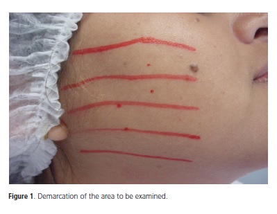
The masseter muscle was found in an objective, palpation, clinical test16, demarcating in five areas to be examined6 (Figure 1), and bilaterally scanned in five positions, from the fixed insertion to the mobile ones7,17, previously determined.
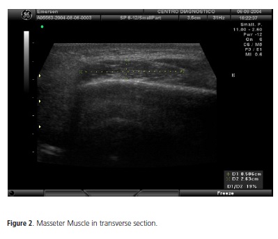
The measurement of the muscle's thickness was conducted in each transverse section, one by one separately (Figure 2). Only one measurement was evaluated, in real time, for each transverse section17. The records of the individually annotated measurements, at first, in the length of each transverse section, and then, in the width of the muscle.
Statistical analysis
The experimental delineament was in double blind and the obtained results were submitted to the test t of Student (p<0,05) for independent samples.
RESULTS
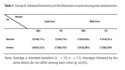
Among the Group A participants, it was observed that the width and length of the Masseter muscles, left and right, were approximately 23% bigger in men (Table 1).
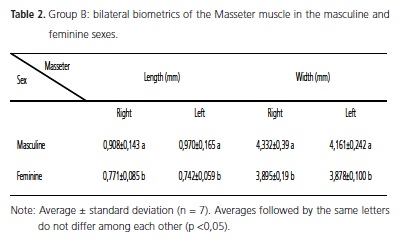
Among the Group B participants, it was also verified that the width and length of the Masseter muscles, right and left, are about 12% bigger in men (Table 2).
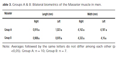
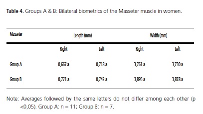
However, there is no difference in length nor width of the Masseter muscles, of both faces, among the same sex the participants between Groups A and B. (Tables 3 & 4).
DISCUSSION
The ultrasound measurement of the Masseter muscle has been utilized for the most diverse observations, for example, in order to correlate the thickness with the facial morphology and the width of the dental arch6,18-19 and the results demonstrate that this technique of image can be capable of obtaining such information. On the other hand, Emshoff & Bertram4 report difference regarding the thickness of the Masseter muscle measured by ultrasonography after the use of stabilizing plates in patients with TMD before and after the treatment, while Arijii et al.7 claim that the ultrasound images of patients with TMD demonstrate that there had been increase in the thickness of the masticatory muscle, which, according to Farella et al.20, influence positively in the slowdown of the beginning of the pain as well as prolonged resistance.
However, in the results obtained in this research, no differences were observed in the pattern of development of the Masseter Muscle thickness among TMD and Non- TMD patients, which corroborates the results of Pereira et al.13 utilizing the same technique in similar experiments.
On the other hand, works of ultrasound evaluation of the thickness of the Masseter muscle performed by Kiliaridis & Kalebo15 and by Close et al.21 demonstrate that there is a great variation in the thickness of this muscle among men and women, what also was evidenced among participants of both sexes in the groups A and B of this research. Such findings suggest that the Masseter muscle presents phenotypic variability among sexes and, therefore, it is likely that this behavior prevents the detection by ultrasound images, of alterations in their thickness, influenced by temporomandibular dysfunction.
CONCLUSION
After the results, we can conclude that the ultrasound images technique show the occurrence of differences in length and width of the Masseter muscle among men and women while there was no evidence of change in size (length and width) of the muscle of both faces among TMD and Non-TMD patients.
Collaborators
AG VERAS was responsible for the idea and execution of this research. MAR LIBERATO collaborated in the interpretation of the data. RV ZANETTI e PP FELTRIN oriented the processes of elaboration and execution of the research. All the collaborators participated in the writing process of the present article.
REFERENCES
1. McNeill C, Falace D, Attanasio R. Continuing education for TMD and orofacial pain; a Philosophical overview. J Craniomandib Disord. 1992;6(2):135-6. [ Links ]
2. Dworkin SF, LeResche L. Research diagnostic criteria for temporomandibular disorders, Review, criteria, examinations and specifications, critique. J Craniomandib Disord. 1992;6(4):301- 55.
3. Koyano K, Kin YJ, Clark GT. Eletromiographic signal changes during exercise in human chronic jaw-muscle pain. Arch Oral Biol. 1993;40(3):21-7. doi: 10.1016/0003-9969(95)98811-C.
4. Emshoff R, Bertram S. The ultrasonic value of muscle hypertrophy in patients with temporomandibular joint disorders. J Prosthet Dent. 1995;73(4):373-6. doi: 10.1016/S0022-3913(05)80334- 8.
5. Emshoff R, Emshoff I, Rudisch A, Bertram S. Reliability and temporal variation of masseter muscle thickness measurements utilizing ultrasonography. J Oral Rehabil. 2003;30(12):1168-72. doi: 10.1111/j.1365-2842.2003.01186.x.
6. Bertram S, Bodner G, Rudish A, Brandlmaier I, Emshoff R. Effect of scaning level and muscle condition on ultrasonographic cross-sectional mensurements of the anterior masseter muscle. J Oral Rehabil. 2003;30(4):403-5. doi: 10.1046/j.1365- 2842.2003.01052.x.
7. Ariji Y, Sakuma S, Izumi M, Sasaki J, Kurita K, Ogi N, et al. Ultrasonographic features of the masseter muscle in female patients with temporomandibular disorder associated with myofascial pain. Oral Surg Oral Med Oral Pathol Oral Radiol Endod. 2004;98(3):337-41. doi: 10.1016/j.tripleo.2004.06.068.
8. Bakke M, Tuxen A, Vilmann P, Jensen BR, Vilmann A, Toft M. Ultrasound image of human masseter muscle related to bite force, electromyography, facial morphology, and occlusal factors. Scand J Dent Res. 1992;100(3):164-71. doi: 10.1111/ j.1600-0722.1992.tb01734.x.
9. Heo MS, An BM, Lee SS et al. Use of advanced imaging modalities for the differential diagnosis of pathoses mimicking temporomandibular disorders. Oral Surg Oral med oral Pathol Oral radiol Endod. 2003;96(5):630-8. doi: 10.1016/S1079- 2104(03)00373-1.
10. Glaros AG, Burton E. Parafunctional clenching, pain, and effort in temporomandibular disorders. J Behav Med. 2004;27(1):91- 100. doi: 10.1023/B:JOBM.0000013646.04624.8f.
11. Mangilli LD, Sassi FC, Sernik RA, Tanaka C, de Andrade CR. Electromyographic and ultrasonographic assessment of the masseter muscle in normal individuals: a pilot study. Pro Fono R Atual. 2009;21(3):261-4. doi: 10.1590/S0104- 56872009000300014.
12. Fricton JR, Schiffman EL. Reliability of a craniomandibular index: vadity. J Prosthet Dent. 1987;58(2):222-8.
13. Pereira LJ, Gavião MBD, Bonjardim LR, Castelo PM, Andrade AS. Ultrasonography and electromyography of mastigatory muscle in a group of adolescents with signs and symptoms of TMD. J Clin Pediatr Dent. 2006;30(4):314-9.
14. Palla P. Mioartropatias do sistema mastigatório e dores orofaciais. São Paulo: Artes Médicas; 2004.
15. Killiaridis S, Kalebo P. Masseter muscle thickness measured by ultrassonography and its relatio to facial morphology. J Dent Res. 1991;70(9):1262-5. doi: 10.1177/00220345910700090601.
16. Oliveira W. Disfunção temporomandibular. São Paulo: Artes Médicas; 2002.
17. Emshoff R, Bertram S, Strobl H. Ultrasonographic cross-sectional characteristics of muscles of the head and neck. Oral Surg Oral Med Oral Pathol Oral Radiol Endod. 1999;87(1):93-106. doi: 10.1016/S1079-2104(99)70302-1.
18. McAlister RW, Harkness EM, Nicoll JJ. An ultrasound investigation of the lip levator musculature. Eur J Orthod. 1998;20(6):713-20. doi: 10.1093/ejo/20.6.713.
19. Raadsheer MC, van Eijden TM, van Spronsen PH, van Ginkel FC, Kiliaridis S, Prahl-Andersen B. A comparison of human masseter muscle thickness measured by ultrasonography and magnetic resonance imaging. Arch Oral Biol. 1994;39(12):1079-84.
20. Farella M, Bakke M, Michelotti A, Rapuano A, Martina R. Masseter thickness, endurance and exercise-induced pain in subjects with different vertical craniofacial morphology. Eur J Oral Sci. 2003;111(3):183-8. doi: 10.1034/j.1600-0722.2003.00035.x.
21. Close PJ, Stokes MJ, L'Estrange PR, Rowell J. Ultrasonography of masseter muscle size in normal young adults. J Oral Rehabil. 1995;22(2):129-35. doi: 10.1111/j.1365-2842.1995.tb00246.x.
 Endereço para correspondência:
RV ZANETTI
e-mail: rvzanett@uol.com.br
Received on: 11/8/2010
Final version resubmitted on: 28/9/2010
Approved on: 17/1/2011
