


Serviços Personalizados
Artigo
 pdf em Inglês
 Artigo em XML
 Referências do artigo
 Enviar este artigo por email
Links relacionados
Compartilhar

 Permalink
RGO.Revista Gaúcha de Odontologia (Online)
versão On-line ISSN 1981-8637
RGO, Rev. gaúch. odontol. (Online) vol.63 no.3 Porto Alegre Jul./Set. 2015
ORIGINAL / ORIGINAL
Botryoid odontogenic cyst: case report with etiopathogenic, diagnostic and therapeutic considerations
Cisto odontogênico botrióide: relato de caso com considerações etiopatogênicas, diagnósticas e terapêuticas
Rhoner GONÇALVESI; Ophir RIBEIRO JÚNIORI; Alexandre Meireles BORBAI; Annelise Nazareth Cunha RIBEIROII; Norberto Nobuo SUGAYAI; Jayro GUIMARÃES JÚNIORI
I Universidade de São Paulo, Faculdade de Odontologia, Departamento de Estomatologia. Av. Professor Lineu Prestes, 2227, Cidade Universitária, 05508-000, São Paulo, SP, Brasil
II Universidade de São Paulo, Faculdade de Odontologia, Departamento de Ortodontia. São Paulo, SP, Brasil
ABSTRACT
The botryoid odontogenic cyst is a rare asymptomatic lesion characterized by its typical multilocular aspect similar to a bunch of grapes commonly affecting the mandible bicuspids and canine region. The possibility of this lesion representing a clinical variation of the lateral periodontal cyst is source of discussion and doubt among many authors. Our article reports a rare case of the polemic odontogenic cyst and presents considerations related to its etiopathogenic, diagnostic and therapeutic aspects.
Indexing terms: Odontogenic cysts. Periodontal cyst. Pathology oral. Surgery oral.
RESUMO
O cisto odontogênico botrióide é uma lesão rara caracterizada pelo aspecto multilocular típico semelhante a um cacho de uvas. A possibilidade de essa lesão representar uma variante clínica do cisto periodontal lateral é fonte de discussão e indefinição entre diversos autores. O presente trabalho relata um caso desse polêmico cisto odontogênico e apresenta considerações relacionadas aos seus aspectos etiopatogênicos, diagnósticos e terapêuticos.
Termos de indexação: Cistos odontogênicos. Cisto periodontal. Patologia bucal. Cirurgia bucal.
INTRODUCTION
The botryoid odontogenic cyst (BOC) is a rare lesion, described by Weathers, Waldron in 19731. Its main characteristics are the typical multilocular aspect, similar to a bunch of grapes, justifying its terminology (botryoid: from the greek botrys= bunch of grapes; oeides= in a shape of), commonly affecting the mandible bicuspid and canine region without symptoms. Other evidences suggest the possibility of this lesion represent a variation of the lateral periodontal cyst (LPC), promoting discussion and doubt among authors.
This paper presents a case report of this rare and polemic odontogenic cyst, contributing to the small number of publications of this pathology in the literature. Moreover a discussion with focus on its etiopathogenic, diagnostic and therapeutic considerations is presented by the authors.
CASE REPORT
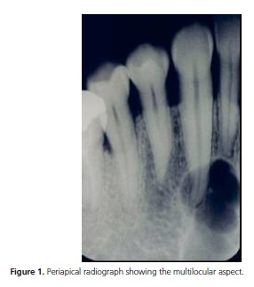
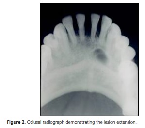
A male, 44 years old, melanoderma, came up complaining of a swelling in the anterior left region of the mandible for already two months. The intra-oral examination showed a discreet but consistent and hard swelling adjacent to the canine without alterations in the local mucosa. Periapical and oclusal radiographies demonstrated a radiolucent multilocular image of approximately 20 mm over the canine root with extension to the adjacent incisive teeth (Figures 1 and 2). Dental vitality test showed positive result of the mentioned teeth, discarding inflammatory origin. Considering the possibility of a BOC, an excisional biopsy using enucleation followed by peripheral ostectomy technique was performed.
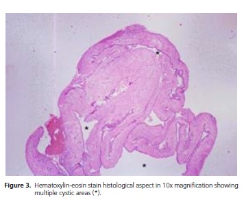
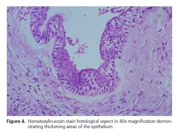
During surgery, strong adhesion of the cystic capsule to the canine root was observed. The surgical specimen was sent to histopathologic examination in which was observed a multilocular lesion 15 x 17 mm, characterized by multiple cysts and separated by discreet connective tissue septa associated to the capsule (Figure 3). The epithelial boundary of these small cysts showed regions of nodular enlargement composed by glycogenrich clear cells (Figure 4). In face of this, the final diagnosis was of a BOC.
After 15 days, a new dental vitality test was accomplished and the canine vitality was maintained. Postoperative periapical radiography taken after 60 days demonstrated normal bone healing process. The patient continues in follow-up in case of any recurrence is observed.
DISCUSSION
There are many doubts related to the etiopathogenesis of the BOC and its condition as an individual entity. The hypothesis of such lesion representing a multilocular variation of the LPC is defined by some authors based in clinical and histopathologic similarities1-2. Both present predilection to the inferior canine and bicuspid region, as presented in the present case, and demonstrate epithelium with clear cells nodule3-5.
It is believed that the BOC and the LPC have the same origin, although the type of odontogenic epithelium is debated. The participation of the reduced enamel epithelium or the epithelium rests of Malassez was suggested in the etiopathogenesis of these lesions3. However, recent publications defend the cystic degeneration of dental lamina rests, found in the alveolar ridge, in function of the observed histopathologic characteristics4. The determining point that supports this theory is the presence of clear cells in the epithelial boundary of these cysts, similar as in the dental lamina. On the other hand, those cells are not identified in dentigerous and radicular cysts, which present etiopathogenesis related to the reduced epithelium and the epithelium rests of Malassez, respectively.
The histopathologic characteristics of the BOC are quite similar to the LPC, differing only in the multilocular aspect. Multilocular lesions are characterized by the presence of cavities, cystic or not, separated by connective or osseous septa. In the case of the BOC, cystic cavities of different sizes and quantities are limited by fine connective tissue septa contiguous to the capsule. Such aspect is explained by epithelium nodule growth towards the capsule, with subsequent cystic degeneration1, or by new cysts formation from the proliferative stimulation of adjacent epithelium remnants4-5. Although this characteristic is essential to the histopathologic diagnosis of the BOC, the eventual damage of this aspect during surgery or by laboratorial manipulation may difficult its differentiation from the LPC6.
The BOC generally occurs in older patients and shows greater proportions that the LPC, being sometimes observer in edentulous patients2,5. Although these differences indicate a distinction between these two entities, a critical analysis is necessary. As the development of a multilocular lesion presents slow growth, a LPC not diagnosed in the youth may present later, between the fifth and seventh decades, larger and with a botryoid aspect, as described in the literature4,7. The possibility of identification of a BOC in edentulous areas also does not mean much since a residual LPC may continue its development after the extraction of the adjacent teeth.
For all of above mentioned reasons, we consider that the BOC and the LPC are a unique nosologic entity in different periods of evolution. However, we believe that is necessary to classify the variant botryoid due to its apparently higher recurrence rate and for its necessary treatment peculiarities. Although the recurrence rate is presented between 15 and 30%, the presented incidence number is low to determine confident data4-5,8. In these studies, recurrence is observed around ten years after enucleation.
Even tough some reports point out the proliferative behavior of the BOC, it is believed that the recurrences are related to the difficulty to the complete removal of the lesion due to its multilocular aspect and its thin capsule5,8. This idea is supported in the histopathologic finds of Phelan et al.9, that showed microcysts in the capsule of recurred LPCs (which perhaps could classified as BOC due to its multilocular aspect).
To accomplish the therapeutic of the BOC, the complementary treatment of the surgical bed may be used after enucleation. As alternatives, the peripheral ostectomy or Carnoy solution application fit perfectly in order to eliminate possible remnants of this lesion10-11. Although the diagnosis of the BOC is only confirmed after histopathologic examination, clinical, radiographic or macroscopic characteristics suggestible of this lesion may help in the indication of complementary treatment modalities during surgery. Even with the use of the techniques, we suggest follow-up for a long period due to the scarce scientific knowledge related to this odontogenic cyst.
CONCLUSION
The etiopathogenesis of the BOC seems to be related to the degeneration of the dental lamina rests, as nowadays accepted for the LPC. Both lesions have similar characteristics and may represent the same nosologic entity in distinct evolution periods. The tendency for recurrence presented by the botryoid variant might be controlled by surgical complementary approaches, like peripherical ostectomy and application of Carnoy Solution.
Collaborators
All authors provided the conception of the manuscript, acquisition of data, drafting the manuscript and final approval of the submitted version.
REFERENCES
1. Weathers D, Waldron C. Unusual multilocular cysts of the jaw (botryoid odontogenic cysts). Oral Surg Oral Med Oral Pathol. 1973;36(2):235-41. [ Links ]
2. Mendes RA, van der Wall I. An unusual clinicoradiographic presentation of a lateral periodontal cyst--report of two cases. Med Oral Patol Oral Cir Bucal. 2006;11(2):185-7.
3. Shear M, Pindborg JJ. Microscopic features of the lateral periodontal cyst. Scand J Dent Res. 1975;83(2):103-10.
4. Carter LC, Carney YL, Perez-Pudlewski D. Lateral periodontal cyst: multifactorial analysis of a previously unreported series. Oral Surg Oral Med Oral Pathol Oral Radiol Endod. 1996;81(2):210- 6.
5. Üçok Ö, Yaman Z, Günhan Ö, Üçok C, Dogan N, Baykul T. Botryoid odontogenic cyst: report of a case with extensive epithelial proliferation. Int J Oral Maxillofac Surg. 2005;34(6):693-5. doi:10.1016/j.ijom.2005.01.005
6. Lindh C, Larson A. Unusual jaw-bone cysts. J Oral Maxillofac Surg. 1990;48(3):258-63. doi:10.1016/0278-2391(90)90390-N
7. Gurol M, Burkes EJ, Jacoway J. Botryoid odontogenic cyst: analysis of 33 cases. J Periodontol. 1995;66(12):1069-73. doi:10.1902/jop.1995.66.12.1069
8. Greer RO, Johnson M. Botryoid odontogenic cyst: clinicopathologic analysis of ten cases with three recurrences. J Oral Maxillofac Surg. 1988;46(7):574-9. doi:10.1016/0278- 2391(88)90147-4
9. Phelan JA, Kritchman D, Fusco-Ramer M, Freedman PD, Lumerman H. Recurrent botryoid odontogenic cyst (lateral periodontal cyst). Oral Surg. 1988;66(3):345-8. doi: 10.4103/0976-237X.103629
10. Lee PK, Samman N, Ng IO. Unicystic ameloblastoma: use of Carnoy's solution after enucleation. Int J Oral Maxillofac Surg. 2004;33(3):263-7. doi:10.1006/ijom.2003.0496
11. Ribeiro Júnior O, Borba AM, Alves CAF, Guimarães Júnior J. Complicações da solução de Carnoy no tratamento de tumores odontogênicos. RGO, Rev Gaúch Odontol. 2007;55(3):263-6.
 Correspondence to:
R GONÇALVES
e-mail: rhoner@usp.br
Received on: 3/7/2013
Final version resubmitted on: 19/11/2013
Approved on: 14/2/2014
