


Serviços Personalizados
Artigo
 pdf em Inglês
 Artigo em XML
 Referências do artigo
 Enviar este artigo por email
Links relacionados
Compartilhar

 Permalink
RSBO (Online)
versão On-line ISSN 1984-5685
RSBO (Online) vol.7 no.1 Joinville Mar. 2010
SHORT COMMUNICATION
Identification of root canals in maxillary molars using cone beam computed tomography
Identificação de canais radiculares nos molares superiores utilizando tomografia computadorizada
Allan AbuabaraI; Giuseppe Valduga CruzII; Claudia Gastaldi Lopes CorreaII; Liliane GuerinoIV; Flares Baratto FilhoIII
IDDS, Specialit in Dental and Maxillofacial Radiology, City Hall of Joinville, SC, Brazil
IIMSc, DDS, Professor of the University of the Region of Joinville (Univille), Joinville, SC, Brazil
IIIPhD, MSc, DDS, Professor of the University of the Region of Joinville (Univille), Joinville, SC and Positivo University, PR, Brazil
IVDDS, Specialist in Dental and Maxillofacial Radiology. Private clinic
ABSTRACT
INTRODUCTION: Knowledge on variations in the root canal system is essential for a successful endodontic treatment.
OBJECTIVE: To present a case of identification of root canals in the maxillary left first molar, including the fourth root canal in the mesiobuccal root, using cone beam computed tomography (CBCT).
CONCLUSION: The CBCT system proved to be relevant in providing important information on the root canal system for the planning of endodontic treatment in cases of persistent infection or access difficulties.
Keywords: maxillary first molar; root canal; root canal ramifications; Endodontics; cone beam computed tomography.
RESUMO
INTRODUÇÃO: O conhecimento sobre as variações dos sistemas de canais radiculares é essencial para o sucesso do tratamento endodôntico.
OBJETIVO: Apresentar um caso de identificação dos canais radiculares do primeiro molar superior esquerdo, incluindo o quarto canal na raiz mesiobucal, por meio da utilização da tomografia computadorizada do feixe cônico.
CONCLUSÃO: O sistema de tomografia do feixe cônico mostrou-se relevante no fornecimento de informações importantes sobre o sistema de canais radiculares para o planejamento do tratamento endodôntico em casos de infecções persistentes ou dificuldades de acesso.
Palavras-chave: primeiro molar superior; canal radicular; ramificações do canal radicular; Endodontia; tomografia computadorizada do feixe cônico.
Introduction
Endodontic problems are difficult to diagnose using two-dimensional radiography. A cone beam computed tomography (CBCT) system specifically designed for hard tissue imaging of the maxillofacial region has recently become commercially available, and it provides an accurate, non-invasive, practical method for the management of endodontic problems [3, 6]. The literature describes wide variations in root canal morphology of maxillary first molars, such as first molars with two palatal roots [4].
The root anatomy of the maxillary first molar predominantly comprises three roots (over 95%); two-rooted teeth are rarely reported and may be a result of fusion of the distobuccal root with the palatal root or fusion of the distobuccal root with the mesiobuccal root [2]. Single root or conical shape of the maxillary first molar are rarely reported. The internal root canal system morphology reflects the external root anatomy. However, the prevalence of two canals in the mesiobuccal root is nearly 57% according to the literature [2]. Less variation is found in the distobuccal and palatal roots. Using CBCT, this case identified the root canals of the maxillary left first molar, including the fourth root canal in the mesiobuccal root.
Case report
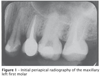
A 39-year-old Caucasian woman was referred to the Maxillofacial Radiology Center for evaluation and was submitted to CBCT (i-CAT, Imaging Sciences International, Hatfield, PA) due to suspected root fracture of the maxillary left first molar because of a history of occlusal trauma, coronal fissure and persistent sensitivity to percussion. Her medical history was noncontributory. Clinically, the tooth presented occlusal restoration. No alterations at the periapical region could be observed on the periapical radiography (figure 1). Endodontic coronal access, initial instrumentation and irrigation with 2.5% sodium hypochlorite of three root canals had already been performed after clinical diagnosis of irreversible pulpitis.
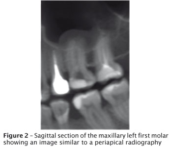
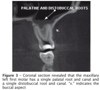
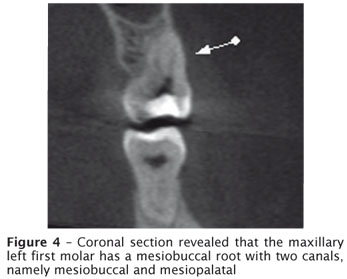
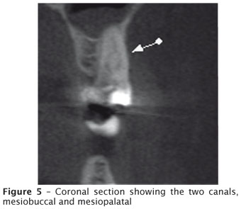
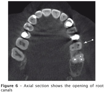
A CBCT of the maxilla was obtained in order to investigate the presence of root fracture. The involved tooth was focused, and the morphology was obtained in coronal, axial, and sagittal sections with 1-mm thickness. The sagittal section showed an image similar to a periapical radiography (figure 2). Coronal sections revealed that the maxillary left first molar has a single palatal root and canal (figure 3), a single distobuccal root and canal (figure 3 – "v." indicates the buccal aspect) and a mesiobuccal root with two canals, namely mesiobuccal and mesiopalatal (figures 4 and 5). The axial section, displayed in figure 6, shows the opening of root canals. The hypothesis of root fracture was ruled out according to the CBCT images. A detailed analysis of the root canal system might be accomplished using CBCT. Mucosal hyperplasia of the maxillary sinus was also observed, which was not associated with dental infection.
Discussion and conclusion
A key objective of successful nonsurgical endodontic treatment is the cleaning and shaping of the root canal system. Knowledge on possible root canal morphologies is a factor in achieving this goal [7]. Although it has been reported that non-microbial factors might be implicated in the failure of root canal treatment, literature suggests that persistent interradicular or secondary infections are the major causes of unsuccessful root canal treatment [8]. Knowledge on variations in the root canal system is essential for a successful endodontic treatment. The advantages of CBCT include increased accuracy, higher resolution and scan-time reduction. The patient receives an absorbed dose similar to a periapical survey of the dentition [5]. Baratto Filho et al. (2009) [1] demonstrated that operating microscope and CBCT have been important for locating and identifying root canals, and CBCT can be used as a good method for initial identification of maxillary first molar internal morphology.
Although artifacts from metallic objects can be disturbing, CBCT system proved to be relevant in providing information on the root canal system for the planning of endodontic treatment.
References
1. Baratto Filho F, Zaitter S, Haragushiku GA, Campos EA, Abuabara A, Correr GM. Analysis of the internal anatomy of maxillary first molars by using different methods. J Endod. 2009;35(3): 337-42. [ Links ]
2. Cleghorn BM, Christie WH, Dong CC. Root and root canal morphology of the human permanent maxillary first molar: a literature review. J Endod. 2006;32(9): 813-21. [ Links ]
3. Cotton TP, Geisler TM, Holden DT, Schwartz SA, Schindler WG. Endodontic applications of cone-beam volumetric tomography. J Endod. 2007;33(9): 1.121-32. [ Links ]
4. Gopikrishna V, Reuben J, Kandaswamy D. Endodontic management of a maxillary first molar with two palatal roots and a single fused buccal root diagnosed with spiral computed tomography: a case report. Oral Surg Oral Med Oral Pathol Oral Radiol Endod. 2008;105(4): e74-8. [ Links ]
5. Hatcher DC, Aboudara CL. Diagnosis goes digital. Am J Orthod Dentofacial Orthop. 2004;125(4): 512-5. [ Links ]
6. Patel S, Dawood A, Ford TP, Whaites E. The potential applications of cone beam computed tomography in the management of endodontic problems. Int Endod J. 2007;40(10): 818-30. [ Links ]
7. Sert S, Bayirli GS. Evaluation of the root canal configurations of the mandibular and maxillary permanent teeth by gender in the Turkish population. J Endod. 2004: 30(6): 391-8. [ Links ]
8. Siqueira Jr JF. Aetiology of root canal treatment failure: why well-treated teeth can fail. Int Endod J. 2001;34(1): 1-10. [ Links ]
 Address for correspondence:
Allan Abuabara
Rua Fernando Machado, n.º 400
CEP 89204-400 – Joinville – SC
E-mail: allan.abuabara@gmail.com
Received on July 30, 2009.
Accepted on August 28, 2009.
Recebido em 30/7/2009.
Aceito em 28/8/2009.
