


Serviços Personalizados
Artigo
 pdf em Inglês
 Artigo em XML
 Referências do artigo
 Enviar este artigo por email
Links relacionados
Compartilhar

 Permalink
RSBO (Online)
versão On-line ISSN 1984-5685
RSBO (Online) vol.8 no.2 Joinville Jun. 2011
ORIGINAL RESEARCH ARTICLE
Evaluation of the dentin remaining after flaring using Gates Glidden drills and Protaper rotary files
Bruno Carvalho-SousaI; José Ribamar Costa-FilhoII; Fábio de Almeida-GomesII; Cláudio Maníglia-FerreiraII; Eduardo Diogo Gurgel-FilhoII; Diana Santana de AlbuquerqueIII
ISchool of Dentistry, Federal University of Ceara – Sobral – CE– Brazil
IISchool of Dentistry, University of Fortaleza – Fortaleza – CE – Brazil
IIISchool of Dentistry, University of Pernambuco – Recife – PE – Brazil
ABSTRACT
INTRODUCTION: The application of rotary instruments for root canal preparation requires a safe, not harming procedure to the root structure remaining.
OBJECTIVES: The purpose of this study was to analyze the root thickness in 28 mesial canals of lower permanent first molars before and after flaring using two rotary instruments: Gates-Glidden drills and ProTaper rotary files.
MATERIAL AND METHODS: Teeth were embedded into a muffle system. Samples were obtained by cutting 2mm below the furcation. The images were captured by a digital video system (8X and 12X magnification). For image analysis and processing, Pro-Image Plus 4.1 software was used. Each image captured by the computer was gauged, eliminating any possible distortion. Gates-Glidden drills were used in decreasing order of size (GG#4, GG#3, GG#2). ProTaper was used according to the manufacturer's recommendations, with hand-piece powered by an electric motor with low torque. 5.25% sodium hypochlorite was utilized as irrigant.
RESULTS: The average thickness between the canal and furcation before and after use of rotary instruments were: 0.857 mm and 0.561 mm for Gates-Glidden drills, and 0.858 mm and 0.486 mm for ProTaper, respectively. No statistical differences were found in the root thickness of specimens shaped with ProTaper rotary files and Gates Glidden drills.
CONCLUSION: The use of Gates-Glidden drills is as safe as ProTaper rotary files with respect to danger of perforation on the distal side of the mesial roots of lower molars.
Keywords: root canals; instrumentation; dental instruments.
Introduction
Flaring the coronal portion of the root canal during endodontic therapy has several advantages and functions. It allows the removal of interferences in the coronal and middle thirds and gives the operator better control of the instruments in the apical third [17, 8]. Greater coronal diameter allows greater penetration of the irrigation needle that improves the efficiency of the irrigant [1]. Better shaping with enlargement of cervical and middle thirds facilitates three dimensional filling [16].
Many instrumentation techniques have been proposed to optimize the process of cleaning and shaping the root canal system. The preparation of both cervical and middle thirds is unanimously, a constant concern for every proposed technique to accomplish cleaning and shaping [2, 5].
Gates-Glidden drills are one of the most common instruments used to achieve this goal [16]. The search for solutions for ideal endodontic preparation has resulted in the introduction of rotary instruments that are meant to facilitate the radicular preparation and to provide a uniform final shape to the canals [6].
The ability to enlarge a canal without deviation from the original curvature is a primary objective [17, 3]. Unfortunately, procedural accidents can occur, such as ledge formation or transportation of the apical foramen in degrees ranging from zipping to apical perforation or stripping [18, 9, 15]. The incorrect use of instruments during root canal preparation may cause a root perforation in the danger zone of lower molars [1].
The objective of this study was to evaluate ex vivo the dentin remaining of mesiobuccal canals of lower molars after flaring using Gates Glidden drills and ProTaper rotary system.
Material and methods
Twenty-eight mandibular molars with slightly curved mesial roots (Schneider Method) were selected from the tooth bank of the University of Fortaleza, Brazil. The study was approved by the Ethics Committee in Research of the University of Fortaleza – under protocol number #310/05.
Standard access cavities were made and the mesial canals were located and negotiated with size #10 and #15 K-files (Dentsply-Maillefer, Ballaigues, Switzerland), until the tip was visible at the apex. Sodium hypochlorite (5.25%) was used to irrigate the canal during all steps.
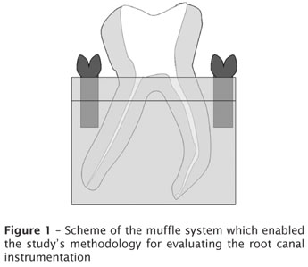
The teeth were then embedded into epoxy clear resin (Arazyn, São Paulo, Brazil) blocks up to furcation. These blocks were encased in a mold specially developed for this study using PVC tubing and two parallel metal threads on its lid. Thus, it was possible to remove the resin blocks out of the mold, cut them with a low-speed saw (Isomet, Buhler, Ltd. Lake Bluff, NY, USA), using a diamond disc (Æ 125 mm x 0.35 mm x 12.7 mm – 330C), under constant irrigation with water to prevent overheating. The cuts were made 2 mm below the root bifurcation [4] and the teeth were analyzed. Finally the blocks were reassembled into the mold to enable root canals instrumentation (figure 1).
The image of the resin blocks with the cervical third of the roots was examined under a microscope (Axioscoppe, Carl Zeiss Vision Gmbh, Hallbergmoos, Germany) and the images captured by a digital video system using 8X to 12X magnification. The images were submitted to software evaluation. For image analysis and processing Pro-Image Plus 4.1 software (Media Cybernetics, Silver Spring, MD, USA) were used. The software, through numerical integration, precisely measured the smallest radicular thickness between the furcation and the mesial root. Each image captured by the computer was gauged, eliminating any possible distortion.
After the obtainment and initial analysis of the mesial canal, the canals were shaped by using Gates-Glidden drills (Dentsply-Maillefer, Ballaigues, Switzerland), in decreasing order of size: GG#4, GG#3, GG#2. A conventional motor at low rotation speed of 2500 rpm was used. Rotary instruments were applied with slight apical pressure, up and down movements, with only one penetration with each drill. The drill's depth was determined by its adaptation within root canal [2]. At every drill change, canal was negotiated through apical file (#10) and irrigated with 2 ml of 5.25% sodium hypochlorite.
ProTaper rotary files (Dentsply-Maillefer, Ballaigues, Switzerland) were used by a hand-piece powered by an electric motor (Endo Plus, Driller, Brazil), at 16:1 reduction, and torque of 0.2 N. A digital display indicating the velocity allowed the maintenance of a constant speed of 300 rpm and torque of 0.2N Each instrument was used five times before being replaced.
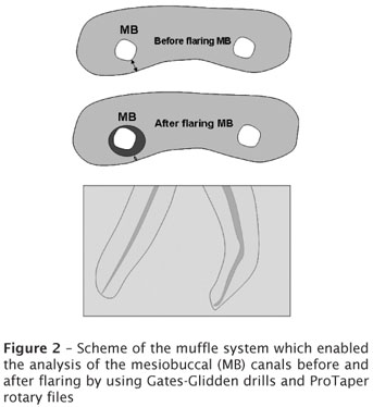
ProTaper rotary files were used according to the manufacturer's recommendations: SX, S1, and S2 (which are the files used for enlarging the cervical and middle thirds of the canal); F1 and F2 (which are the files used for enlarging the apical third of the canal) (Dentsply-Maillefer) (figure 2).
At every drill change, canal was negotiated through apical file (#10) and irrigated with 2 ml of 5.25% sodium hypochlorite.
After instrumentation, canals had their image digitized and evaluated according to the previously selected features.
An Excel spreadsheet was created recording the smallest distance to the root bifurcation, before and after the instrumentation with GG and ProTaper rotary system. The results were statistically analyzed (SPSS 12.0) by Kruskal-Wallis test.
Results
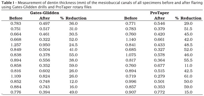
Table I shows all the measurements of dentin thickness (mm) of the mesiobuccal canals of all specimens, before and after flaring using Gates-Glidden drills and ProTaper rotary files.
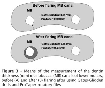
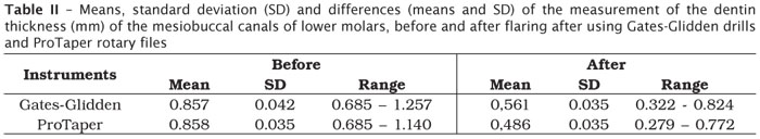
The thickness means between the mesial root canal and the furcation, before and after the preparation were: 0.857 mm and 0.561 mm by GG drills, and 0.858 mm and 0.486 mm by ProTaper, respectively (table II).
Figure 3 shows the means of the measurement of the dentin thickness (mm) of the mesiobuccal canals of lower molars before and after flaring after using Gates-Glidden drills and ProTaper.
The radicular thickness remaining between the mesial root canal and the furcation prepared by GG drills and ProTaper rotary system did not show a significant different (p = 0.1611). No statistical significant differences between groups before (p = 0,919) and after (p = 0.2565) canals preparation by GG and ProTaper were found.
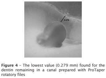
Strip perforation that could provoke any damage to the root was not observed in the selected molars. The lowest value found for the dentin remaining was 0.279 mm, in a canal prepared with ProTaper rotary files (figure 4).
Discussion
Several methods were already used with the objective of analyzing the use of rotary instruments in the cleaning and shaping of the root canal system [4, 14]. This study employed a novel method constituted by a precise model ("muffle system"), which can be used several times and enables root canal evaluation before and after its preparation [4].
The teeth were horizontally sectioned at 2mm below the furcation because this is the area that presents the greatest decrease of dentin remaining during flaring [8]. Measurement of the minimum remaining structure after preparation of cervical and middle thirds of mesiobuccal canals of lower molars after using Gates-Glidden drills and ProTaper rotary files revealed no significant differences between any technique. The means of the remaining root thickness were 0.561 mm and 0.486 mm for Gates-Glidden drills and ProTaper, respectively.
The ability of the operator seems to be an important clinical factor of instrument failure [10]. In this study all specimens were instrumented by the same operator. One ProTaper rotary file (F2) instrument fractures after using 5 times, despite of all instruments had been constantly checked for defects that would alert the operator prior to fracture [12], and low torque [7]. All the instruments were used only 5 times [11].
All canals were negotiated with #10 K-file, and #15, #20 Flexofile, at working length. This provides a safer use of the Gates-Glidden drills and rotary instruments [18]. Between the uses of each instrument the canal was irrigated with 2ml of 5.25% sodium hypochlorite.
It should be noted that no perforations occurred during the study, but there was little remaining tooth structure in some cases (0.279 mm, 0.322 mm and 0.327 mm). Other study showed no perforations when Protaper rotary system was used [13]. Perforation or fracture may occur during the process of root canal obturation as a result of the small amount of tooth structure remaining after flaring and instrumentation [8, 11].
Conclusion
This study suggests that the use of Gates Glidden drills is as safe as ProTaper rotary files with respect to danger of perforation on the distal side of the mesial roots of lower molars. Also, the Gates-Glidden drills should be use in decreasing size order, without risk of perforation.
References
1. Abou-Rass M, Frank AL, Glick DH. The anticurvature filing method to prepare the curved root canal. J Am Dent Assoc. 1982;101:792-4. [ Links ]
2. Abou-Rass M, Jastrab RJ. The use of rotary instruments as auxiliary aids to root canal preparation of molars. J Endod. 1982;8:78-82. [ Links ]
3. Bertrand MF, Lupi-Pégurier L, Médioni E, Muller M, Bolla M. Curved molar root canal preparations using Hero 642 rotary nickel-titanium instruments. Int Endod J. 2001;34:631-6. [ Links ]
4. Bramante CM, Berbet A, Borges RP. A methodology for evaluation of root canal instrumentation. J Endod. 1987;13:243-5. [ Links ]
5. Coutinho-Filho T, Yamasake S, De Deus GA, Gurgel-Filho ED. Análise da estrutura remanescente após o uso decrescente das brocas de Gates-Glidden na sua penetração máxima em raízes curvas. JBE. 2001;2:68-75. [ Links ]
6. Coutinho-Filho T, De Deus G, Pinto TG, Gurgel-Filho ED, Maniglia-Ferreira C. A computer evaluation of the dentin remaining after cervical preparation in curved canals: gates-glidden drills vs orifice shaper. Braz J Oral Science. 2002;1:116-20. [ Links ]
7. Garambini G. Rationale for the use of low-torque endodontic motors in root canal instrumentation. Endod Dent Traumatol. 2000;16:95-100. [ Links ]
8. Isom TL, Marshall JG, Baumgartner JC. Evaluation of root thickness in curved canals after flaring. J Endod. 1995;21:368-71. [ Links ]
9. Kosa DA, Marshall G, Baumgartner C. An analysis of canals centering using mechanical instrumentation techniques. J Endod. 1999;25:441-5. [ Links ]
10. Mandel E, Adib-Yazdi M, Benhamou LM, Lachkar T, Mesgouez C, Sobel M. Rotary Ni-Ti profile systems for preparing curved canals in resin blocks: influence of operator on instrument breakage. Int Endod J. 1999;36:436-43. [ Links ]
11. Martin LR, Gilbert B, Dickerson AW. Management of endodontic perforations. Oral Surg Oral Med Oral Pathol Radiol Endod. 1982;54:668-77. [ Links ]
12. Martin B, Zelada G, Varela P, Bahillo JG, Magán F, Ahn S et al. Factors influencing the fracture of nickel-titanium rotary instruments. Int Endod J. 2003;36:262-6. [ Links ]
13. Matsubara FMB, Pereira JGB, Baratto-Filho F, Haragushiku GA, Fagundes FS. Avaliação do desgaste da zona de perigo de molares inferiores utilizando o sistema rotatório Protaper. RSBO. 2005 Nov;2:12-6. [ Links ]
14. Peters OA, Rüegsegger P, Barbakow F. Three dimensional analysis of root canal geometry using high resolution computed tomography. J Dent Res. 2000;79:1405-9. [ Links ]
15. Peters OA, Peters CI, Schönenberger K, Barbakow F. ProTaper rotary root canal preparation effects of canal anatomy on final shape analyzed by micro CT. Int Endod J. 2003;36:86-92. [ Links ]
16. Schilder H. Filling root canal in three dimensions. Dent Clin North Am. 1967;11:723-44. [ Links ]
17. Schilder H. Cleaning and shaping the root canal. Dent Clin North Am. 1974;18:269-96. [ Links ]
18. Weine FS, Kelly RF, Lio PJ. The effect of preparation procedures on original canal shape and on apical foramen shape. J Endod. 1975;1:255-62. [ Links ]
 Corresponding author:
Bruno Carvalho-Sousa
95, Paulo Faustino St., apt. #101 – Luciano Cavalcante
Zip code 60813-530 – Fortaleza – Ceara
E-mail: brunendo@hotmail.com
Received for publication: November 9, 2010
Accepted for publication: November 22, 2010.
