


Serviços Personalizados
Artigo
 pdf em Inglês
 Artigo em XML
 Referências do artigo
 Enviar este artigo por email
Links relacionados
Compartilhar

 Permalink
RSBO (Online)
versão On-line ISSN 1984-5685
RSBO (Online) vol.8 no.2 Joinville Jun. 2011
SHORT COMMUNICATION
Atypical appearance of epidermoid cyst in tongue's ventral surface
Claudio Maranhão PereiraI; Patrícia Freire GasparettoII; Tessa de Lucena BotelhoII
IDepartment Oral of Diagnosis, School of Dentistry, Paulista University – Goiania – GO – Brasília – DF – Brazil
IIDepartment Oral of Diagnosis, School of Dentistry, Paulista University – Goiania – GO – Brazil
ABSTRACT
INTRODUCTION: Epidermoids cysts are benign and rare lesions in oral cavity. It presents as a nodular lesion of sessile base, soft to palpation. In the oral cavity, it most happens on the floor of the mouth's midline. Without treatment, these lesions can lead to dysphagia, dysphonia and dyspnea due to reach a large size.
OBJECTIVE: To report a case of a 12-year-old boy presenting a lesion on the tongue's ventral surface causing difficult to swallow.
CASE REPORT: The patient was referred to the School of Dentistry of Paulista University, complaining about an asymptomatic nodule on the tongue's ventral surface, lasting for 10 months. Clinical examination showed the extensive fibrous mass on the tongue's medium anterior ventral surface.
CONCLUSION: With a clinical diagnosis of dermoid, epidermoid cyst, or lipoma, an excisional biopsy was performed. Histological examination was consistent to the diagnosis of epidermoid cyst. The patient was followed up and 2 years after surgery there was no sign of recurrence.
Keywords: epidermoid cysts; congenital abnormalities; mouth.
Introduction
Epidermoid cyst is a rare group of benign lesions that occur in the body, although they are found in larger frequency in ovaries and testicles with 7% of the cases occurring in the head and neck area and 1.6% within the oral cavity [4]. In oral cavity, they represent less than 0.01% of all cysts [3]. The floor of the mouth is frequently involved and the swelling can make difficult to swallow and speak [1, 5]. This lesion has a slow asymptomatic growth, and if congenital, the diagnosis usually occurs during the second or third decade of life [1, 2].
This lesion is classified according to the anatomical area into three different types: lateral cyst, located at the submaxillary region; median genioglossal cyst, located above the geniohyoid muscles; and median geniohyoid cyst, at the submentual region between the geniohyoid and the mylohyoid muscles [2].The treatment of the epidermoid cyst is surgical enucleation. The extra oral approach is generally preferred in case of either median geniohyoid or very large sublingual cysts, whereas the intraoral approach is typically used for smaller sublingual cysts. The prognosis is excellent and the recurrence is rare [1, 2].
Case report
A 12-year-old white male was referred to the School of Dentistry of Paulista University, Goiania, Goias, Brazil, in June 2008, complaining about the asymptomatic nodule on the tongue's ventral surface lasting for 10 months. Clinical examination showed the extensive fibrous mass on the tongue's medium anterior ventral surface (figures 1, 2 and 3). The patient reported that the lesions caused considerable discomfort and affected his normal oral function. Personal and family histories and laboratory tests were uneventful.
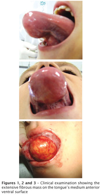
During clinical examination, the patient had a 6.0 X 5.0 cm nodule on the tongue's ventral surface. The patient was submitted to a computed tomography for evaluation of the lesion's limits and relations to the adjacent anatomic structures. CT showed a well-circumscribed lesion suggestive of a cystic lesion. Clinical diagnosis comprised dermoid or epidermoid cyst, and lipoma.The excisional biopsy was performed and the sample was sent to histological analysis. Microscopic examination revealed the presence of a fibrous capsule covered by epithelium, without any skin adnexa. This was consistent to the diagnosis of epidermoid cyst. The patient was followed up, and 2 years after surgery there is no sign of recurrence.
Discussion
Epidermoid cysts are benign and rare lesions in oral cavity. The complementary tests by imaging technique include ultrasonography, computed tomography and magnetic resonance, which help their diagnosis. Ultrasonography is the most used technique because is a low-cost, fast, reliable imaging method, without x-ray exposure. Consequently, it is the first choice, also being easily suitable to young patients. However, the others techniques present better quality when compared to ultrasonography, because the image is more precise, showing the exact relation of the lesion to the muscles of the floor of the mouth [2]. Our patient was submitted to computed tomography that confirmed the lesion's limits.
When the lesion is on the floor of the mouth, a differential diagnosis must be performed regarding to other lesions that resembling clinically an epidermoid cyst, such as: ranula, infectious process, lymphatic malformation, and heterotypic gastrointestinal cyst. On the other hand, when the lesion is on the tongue, differential diagnosis must be performed regarding to lesions, such as: tumor of granular cells, schwanoma, lipoma and neurofibroma, because they occur in a lager frequency in this site [5]. Thus, besides the clinical examination, other complementary tests are necessary to achieve a differential diagnosis and eliminate other diseases.
References
1. Koca H, Seckin T, Sipahi A, Kazanc A. Epidermoid cyst in the floor of the mouth: report of a case. Quintessence Int. 2007;38:473-7. [ Links ]
2. Longo F, Maremonti P, Mangone GM, De Maria G, Califano L. Midline (dermoid) cysts of the floor of the mouth: report of 16 cases and review of surgical techniques. Plast Reconstr Surg. 2003;112:1560-5. [ Links ]
3. Rajayogeswaran V, Eveson JW. Epidermoid cyst of the buccal mucosa. Oral Surg Oral Med Oral Pathol. 1989;67:181-4. [ Links ]
4. Turetschek K, Hospodka H, Steiner E. Case report: epidermoid cyst of the floor of the mouth: diagnostic imaging by sonography, computed tomography and magnetic resonance imaging. Br J Radiol. 1995;68:205-7. [ Links ]
5. Walstad WR, Solomon JM, Schow SR, Ochs MW. Midline cystic lesion of the floor of the mouth. J Oral Maxillofac Surg. 1998;56:70-4. [ Links ]
 Corresponding author:
Claudio Maranhão Pereira
School of Dentistry of Paulista University, Brasília campus
Dentistry coordination
SGAS Quadra 913, s/n.º – Conjunto B – Asa Sul
Zip code 70390-130 – Brasília – DF
E-mails: odontologiabrasilia@unip.br / claudiomaranhao@hotmail.com
Received for publication: August 25, 2010.
Accepted for publication: October 13, 2010.
