


Serviços Personalizados
Artigo
 pdf em Inglês
 Artigo em XML
 Referências do artigo
 Enviar este artigo por email
Links relacionados
Compartilhar

 Permalink
RSBO (Online)
versão On-line ISSN 1984-5685
RSBO (Online) vol.8 no.3 Joinville Jul./Set. 2011
CASE REPORT ARTICLE
Two root canals in maxillary central incisor
Fábio de Almeida-GomesI; Nadine Luísa Soares de Lima GuimarãesI; Claudio Maniglia-FerreiraI; Roberto Alves dos SantosII; Marcelo de Morais VitorianoI; Bruno Carvalho de SousaIII
I Department of Endodontics, University of Fortaleza – Fortaleza – CE – Brazil
II Department of Endodontics, University of Pernambuco – Recife – PE – Brazil
III Department of Endodontics, University Federal of Ceara – Fortaleza – CE – Brazil
ABSTRACT
Introduction and objective: The success of endodontic treatment requires the knowledge of tooth morphology and its variations. Case report: This clinical article reports an unusual root canal configuration that was detected in a maxillary central incisor with two root canals, demonstrated by radiographic and computerized tomography exams. Conclusion: Knowledge of endodontic anatomy as well as the obtainment of both preoperative radiographs and tomography is important to detect abnormal tooth morphology.
Keywords: dental anatomy; root canal; maxillary central incisor.
Introduction
One of the main objectives from nonsurgical endodontic treatment is the elimination of infections from root canal system and the prevention of its reinfection. A clear understanding of root canal morphology of the human dentition is a pre-requisite for conventional endodontic procedures. The anatomical complexities of root canal anatomy have been highlighted in literature and the clinician's necessity of understanding probable aberrations was emphasized 6.
Consistent high levels of success in endodontic treatment require an understanding of root canal anatomy and morphology and that the entire root canal system must be shaped, disinfected and filled. Thus, it is necessary for the clinician to have knowledge of not only understand dental anatomy but also its variations 4.
According to Ingle 9, one of the most important causes of endodontic treatment failure is the incomplete obturation of the root canal system. Therefore, the correct location, clean, shape and obturation of all canals are indispensable procedures. Similarly, Vertuci 17 and De Grood, Cunninghan 7 reported that a considerable number of failures could be assigned to anatomical variations, such as the presence of unusually root canals.
The presence of an additional root canal in the maxillary central incisor is extremely rare 14. A number of studies of root canal anatomy have described that the maxillary central incisor has only one root and one canal in 100% of examined cases 10,17, with variations only in the number of lateral canals and the position of the apical foramen.
Many studies have described root canal variations 1,2,3,12 and some of these studies described a maxillary central incisor with two canals or two roots 8,14,16.
The purpose of the present article is to present and describe a clinical case of endodontic treatment of a maxillary central incisor with two root canals, demonstrated by radiograph and computed tomography (CT) examinations.
Case report
A 42-year-old Caucasian female was referred to the clinic of Endodontics of the University of Fortaleza for endodontic treatment of the right maxillary central incisor. In the referral letter, it was reported that the tooth presented a root canal calcified and internal root resorption.
Clinical examination revealed that there was no swelling or history of trauma and there was no tenderness to palpation and percussion. After the pulp vitality tests, the tooth was diagnosed with pulp necrosis.
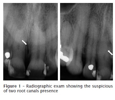
Radiographic examination did not allow a perfect view of the root canal, which led us to suspect the presence of two root canals (figure 1).
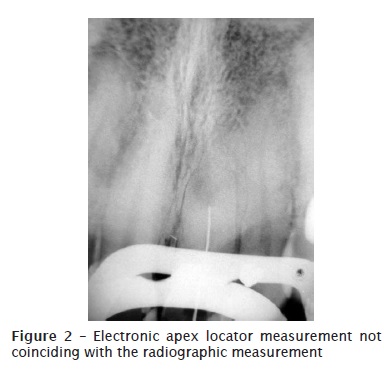
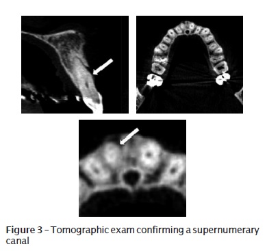
The tooth was isolated with rubber dam and access was gained to the pulp chamber with high-speed round diamond number #1012 (KG Sorensen, Barueri, SP, Brazil). During canal negotiation, the file was moved to the buccal surface. Working length was attempted to be determined by using Root ZX electronic apex locator (J Morita Corp., Kyoto, Japan), but the locator measurement did not coincide with the radiographic measurement (figure 2). Therefore, these conditions led to suspect the presence of a supernumerary canal at the buccal surface. Computed tomography examination confirmed the supernumerary canal (figure 3).
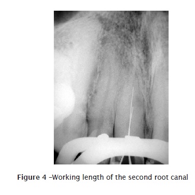
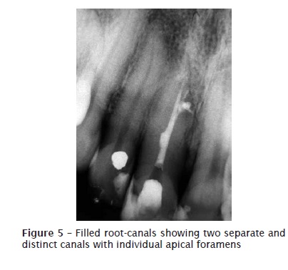
The other canal was found and working length was determined (figure 4). After that, the chemomechanical preparation by Crown-Down technique with K-Flexofile (Maillefer-Dentsply, Switzerland) was carried out. Sodium hypochlorite (2.5%) and EDTA (17%) solutions were used as irrigants. The canals were filled by Lateral Condensation technique using gutta-percha points and root canal sealer (Endofill, Denstply, Petropolis, Rio de Janeiro, Brazil). Treatment was executed in a single visit. After the filling, the final radiographic exam showed two distinct canals with separate apical foramens (figure 5).
Discussion
The case reported here presents an unusual case of a maxillary central incisor with two root canals. Reports of cases with unusual morphology have an important didactic value. Their documentation in case reports may facilitate the recognition and successful management of similar cases that should require endodontic therapy.
Most endodontic and anatomy texts describe the human maxillary central incisors with single root and single canal 17. There were few case reports describing an additional canal in maxillary central incisors 13,16 and most of them present morphological alterations, such as macrodontia, fused and geminated tooth 5,11.
Although the maxillary central incisor usually has one canal, clinicians need to be aware of unexpected root canal morphology when performing root canal therapy. Accurate preoperative radiographs, straight and angled, using a parallel technique are essentials in providing clues to the number of roots that exist 15.
In the present clinical report, it was possible to suspect the presence of the supernumerary root canal through the radiographic exam. Computed tomography examination confirmed the unusual anatomy.
Endodontic success in teeth with a number of canals above that normally found requires a correct diagnosis and careful clinical radiographic inspection. Morphological variations in pulpal anatomy must be considered before treatment onset. The case presented a maxillary central incisor with 2 root canals. Determining the developmental origin of this anatomical anomaly appeared to have clinical significance.
Conclusion
Knowledge of dental anatomy is fundamental for proper endodontic practice. When root canal treatment is performed, the clinician should be aware that both external and internal anatomy may be abnormal. Radiograph and computer tomography can help identifying abnormal tooth anatomy.
References
1. Almeida-Gomes F, Sousa BC, Santos RA. Unusual anatomy of mandibular premolars. Aust Endod J. 2006;32(1):43-5. [ Links ]
2. Almeida-Gomes F, Maniglia-Ferreira C, Santos RA. Two palatal root canals in a maxillary second molar. Aust Endod J. 2007;33(2):82-3.
3. Almeida-Gomes F, Maniglia-Ferreira C, Carvalho-Sousa B, Santos RA. Six root canals in maxillary root canals. Oral Surg Oral Med Oral Pathol Oral Radiol Endod. 2009;108(3):e157-9.
4. Baratto-Filho F, Fariniuk LF, Ferreira EL, Pecora JD, Cruz-Filho AM, Sousa-Neto MD. Clinical and macroscopic study of maxillary molars with two palatal roots. Int Endod J. 2002;35(9):796-801.
5. Cimili H, Kartal N. Endodontic treatment of unusual central incisors. J Endod. 2002;28(6):480-1.
6. De Deus QD. Frequency, location and direction of the lateral, secondary and accessory canals. J Endod. 1975;1(11):361-6.
7. De Grood ME, Cunningham CJ. Mandibular molar with five canals: report of case. J Endod. 1997;23(1):60-2.
8. Ghoddusi J, Javidi M, Vatanpour M. Treatment of a two-canal maxillary lateral incisor. NY State Dent J. 2010 Apr;76(3):40-1.
9. Ingle JI. Endodontic. 3.ª ed. Philadelphia: Saunders; 1985.
10. Kasahara E, Yasuda E, Yamamoto A, Anzai M. Root canal system of the maxillary central incisor. J Endod. 1990;16(4):158-61.
11. Libfeld H, Stabholz A, Friedman S. Endodontic therapy of bilateral geminated permanent maxillary central incisor. J Endod. 1986;12(5):214-6.
12. Maniglia-Ferreira C, Almeida-Gomes F, Sousa BC, Lins CCSA, Santos RA. A case of unusual anatomy in second mandibular molar with four canals. Eur J Dent. 2008;2(3):217-9.
13. Rodrigues EA, Silva SJA. A case of unusual anatomy: maxillary central incisor with two root canals. Int J Morphol. 2009;27(3):827-30.
14. Sheikh-Nezami M, Mokhber N. Endodontic treatment of a maxillary central incisor with three root canals. J Oral Sci. 2007;49(3):245-7.
15. Silha RE. Paralleling long cone technique. Dental Rad Photo. 1968;41:3-19.
16. Sponchiado Jr. EC, Ismail HA, Braga MR, Carvalho FK, Simões CA. Maxillary central incisor with two root canals: a case report. J. Endod. 2006;32(10):1002-4.
17. Vertucci FJ. Root canal anatomy of the human permanent teeth. Oral Surg Oral Med Oral Pathol. 1984;58(5):589-99.
 Correspondence:
Fábio de Almeida-Gomes
Rua Arquiteto Reginaldo Rangel, n. 155, apto. 1403 – Parque do Cocó
CEP 60191-250 – Fortaleza – CE – Brasil
E-mail: fabiogomesce@yahoo.com.br
Received for publication: November 16, 2010
Accepted for publication: December 1, 2010
