


Serviços Personalizados
Artigo
 pdf em Inglês
 Artigo em XML
 Referências do artigo
 Enviar este artigo por email
Links relacionados
Compartilhar

 Permalink
RSBO (Online)
versão On-line ISSN 1984-5685
RSBO (Online) vol.8 no.4 Joinville Out./Dez. 2011
ORIGINAL RESEARCH ARTICLE
Influence of root curvature's initial position on apical deviation occurrence after oscillatory preparation in simulated root canals
Tiago André Fontoura de MeloI; Luciana Dellinghausen SilveiraI; Vanessa Santos Simões PiresI; Simone Viegas da SilvaI; Luis Eduardo Duarte IralaI
I Department of Endodontics, São Leopoldo Mandic, Sobracursos School – Porto Alegre – RS – Brazil
ABSTRACT
Introduction and objective: This study aimed to analyze the influence of root curvature's initial position on apical deviation occurrence after oscillatory system preparation. Material and methods: For this purpose, we used twenty simulated root canals with 21 mm length and 30 degree angle, which were divided into two experimental groups according to curvature's initial position: 8 mm (group A) and 12 mm (group B) short of the canal orifice. The canals were prepared using crown-down technique, and memory instrument was size #30. For apical deviation analysis, before and after preparation, canals were filled with Indian ink and standardly photographed with the aid of a platform. After that, the images were manipulated by Adobe Photoshop® software, through superimposing pre- and post-operative images. Deviation occurrence was measured 1 mm short of working length and at the middle of the curvature by using the ruler tool. Data were subjected to analysis of variance (ANOVA) with significance level set at 5%. Results: Although group B showed a significantly greater deviation mean than group A, no significant interaction was verified between the analysis site and the experimental group. Conclusion: According to the present data, it could be observed that the smaller the curvature radius, the greater the deviation. Concerning to the analysis site, it could be noted that the area 1 mm short of working length presented a higher deviation than the point at the middle of the curvature.
Keywords: stainless steel; endodontics; root canal preparation.
Introduction
Despite of the technological advancement, it is difficult to maintain the foramen position and not to alter root canal's original conformation when performing root preparation 21.
Therefore, teeth requiring endodontic treatment and displaying an increased degree of curvature, root dilaceration, or the presence of alterations (e.g. pulp calcification or atresia) show a higher probability of occurring operative accidents. Apical deviation is one of the most frequent accidents in endodontic therapy and is of great relevance because it could compromise treatment success.
Curve root canal preparation has a higher tendency toward promoting deviations, zips, transportations, and perforations 1.
After the use of oscillatory system with Hedströem instruments, it has been verified an increased number of zips, apical deviations, irregularities, and excessive weariness of root walls 14.
Campos and Pastora 5 used K type instruments connected to oscillatory system for endodontic preparation and observed root canal deviation toward the inner wall of the curvature, at the cervical and middle thirds as well as toward the outer wall of the apical third.
Kosa et al. 12 also analysed the deviation occurrence after the preparation of mesial canals of lower molars. For this purpose, the authors compared different systems: NiTi Profile Series 29 and Quanted 2000 instruments used with continuous rotation as well as stainless steel Flex R and Hedströem instruments coupled to oscillatory system. The results demonstrated deviation occurrence in all systems.
Considering the aforementioned discussions, this study aimed to analyze the influence of root curvature's initial position variation on deviation occurrence, through using the oscillatory system during endodontic preparation.
Material and methods
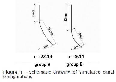
Twenty epoxy resin blocks (Odontofix®, Ribeirão Preto, Brazil) were used, containing artificial root canals with 21 mm length and 0.20 mm of apical diameter. All canals had 30º of curvature; 10 canals presented the curvature initial position at 8 mm short of apical foramen (group A), the following 10 canals showed the curvature initial position at 12 mm short of apical foramen (group B) (figure 1 and table I).
Concerning to endodontic instruments, stainless steel Flexo-File (size #15 to #40) and K type (size #45 to #50) (Dentsply/Maillefer Instruments S.A., Ballaigues, Switzerland) were used in the two experimental groups. Each instrument set was used for the preparation of five simulated canals.
During preparation execution, the simulated canals were fixed to a counter mini clamp (Worker®) to facilitate their instrumentation. Additionally, canals were laterally surrounded by foil so that the operator could not know to which experimental group the canals belong. The operator only was aware of the direction of root canal's curvature degree, which was standardly positioned toward the operator's right hand.
Prior to and during the preparation of the two experimental groups, at every endodontic instrument change, canals were copiously irrigated with distilled water (Iodontosul – Industrial Odontológica do Sul Ltda., Porto Alegre, Brazil) to remove resin debris, followed by anionic detergent irrigation with Tergensol (Inodon, Porto Alegre, Brazil) to lubricate the canal. Both irrigants were stored in a 10-ml disposable syringe (Plastipakkkkú Indústria Cirúrgica Ltda., Curitiba, Paraná, Brazil ), coupled to 25 x 04 hypodermic needle (Becton-Dickinson Indústria Cirúrgica Ltda., Curitiba, Paraná, Brazil). Aspiration was performed with a point coupled to a cannula size 40-20 (Ibrás CBO Indústria Cirúrgica e Óptica S.A., Campinas, São Paulo, Brazil) adapted to the suction unit of the dental chair (Gnatus Equipamentos Médico-Odontológicos Ltda., São Paulo, São Paulo, Brazil), and placed into pulp chamber entrance during irrigation.
Firstly, Flexo-File instruments were used at the pre-established real working length (RWL), in filing movements, sequentially from size #15 to #20. RWL was standardized in all simulated canals at 20 mm of length.
Following, root canal preparations by using a contra-angle handpiece with an automatized system of oscillatory movement (NSK Adiel Super Endo, Ribeirão Preto, Brazil), at 10:1 speed reduction and 45º oscillation, coupled to a pneumatic micromotor (Dabi Atlante, Ribeirão Preto, Brazil). A single operator executed root canal preparation in both groups.
Chemical-mechanical preparation was executed by crown-down technique with oscillation movements, gentle pressure, and brushing movements toward root canal's walls, with sequential endodontic instruments size #50, #45, #40, #35, #30, #25, and #20, up to reach RWL. Larger instruments were pre-curved prior to their insertion.
At every instrument change, a size #15 endodontic instrument was manually inserted up to RWL to promote resin debris removal from canal's apical area.
Following, apical preparation was executed by using the sequence of instruments size #20, # 25, and #30, at RWL. Size #30 was standardized as the memory instrument for all simulated canals.
Root canal preparation was finished by step-back technique with a reduction of 1 mm for each file from size #35 to #50; size #30 instrument was inserted at RWL, at every instrument change.
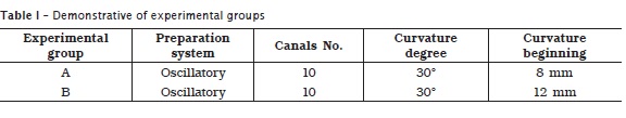
For deviation analysis, before and after preparation, canals were positioned onto a platform and photographed with a digital camera, by using always the same object-to-focus distance (figure 2). To improve the photograph visualization contrast, Indian ink (Corfix, Porto Alegre, Brasil) was injected into root canals.
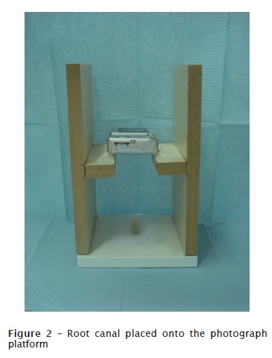
Next, the obtained images were manipulated by Adobe Photoshop® version 6.0. To transform the image in millimeters, a rule of three was used by associating the original length of the canal with the image length on the computer screen. Consequently, the image pixels were not reduced, maintaining the image sharpness.
By using this same software, the images were submitted to contrast adjustment. Each post-operative image was transformed into 50% transparence layer, and, each one was superimposed to the pre-operative image. Therefore, we observed through transparence, the both images superimposed.
Following the ruler tool was employed to localize exactly the image's sites where the deviations would be measured. This analysis was pre-determined at 1 mm short of RWL (figure 3) and at the middle of the curvature area (figure 4).
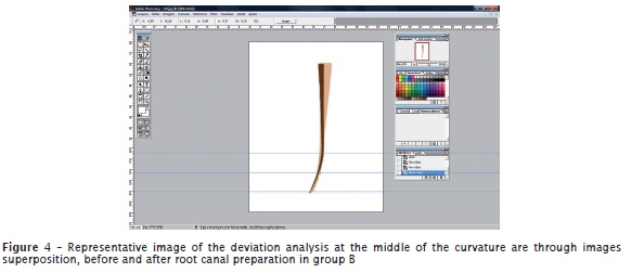
Deviation measurement was performed with aid of the ruler tool onto the both points to be analyzed. The distance was measured from the lateral wall opposite to canal's curvature area prior to and after the preparation.
Data were submitted to ANOVA with level of significance set at 5%.
Results
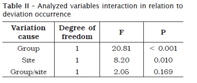
ANOVA showed that, through randomized block design with 5% level of significance, no significant interaction between the analysis site and the experimental group (table II).
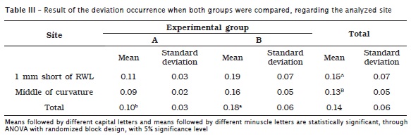
Concerning to the main effects, both variables were significant, that is, regardless of the site, group B mean deviation was significantly higher than group A; and regardless of the experimental group, deviation mean at 1 mm short of RWL was significantly higher than at the middle of the curvature area (table III).
Discussion
Anatomical complexity often presents in a curved root canal may cause accidents or undesirable complications during chemical-mechanical preparation. Apical deviation is one of the most frequent accidents and is of great relevance in endodontic therapy because it can compromise treatment success. Therefore, this study aimed to analyze the influence of root curvature's initial position on deviation occurrence by using oscillatory system.
For this purpose, we used simulated canals. These allowed a better length standardization, anatomic diameter, and root canal's curvature degree when compared with the use of human teeth 3. Studies conducted in simulated canals are ideal for assessing preparation techniques 8. Other researches employing these experimental methods were those from Troian et al. 25, Faria et al. 10, Otoboni Filho et al. 18 and Silva et al. 23.
We decided to standardize the curvature degree at 30º based on the study of Schneider and Austin 22, in which root canals were classified into: straight (less than 5º of curvature), moderate (10º to 20º), and severe (25º to 70º). Several studies utilized simulated canals with curvatures ranging from 30º to 40º 10,16,18,20. On the other hand, the definition of the curvature's initial position is in agreement with the studies of Thompson and Dummer 24, Calberson et al. 3,4 and Troian et al. 25, who also determined the initial point at 8 and 12 mm short of apex.
Concerning to the instrumentation method, we employed the oscillatory system advocated by Batista et al. 2, because it better reproduces the movement manually performed in comparison to continuous rotation system. This system allowed the adaptation of conventional hand files for root canal preparation, and it has been very used due to its easy technique and low cost when compared to NiTi rotary instruments 26,28.
We use an Indian ink within root canals in order to facilitate the deviation visualization, similarly to the studies of Melo et al. 15 and Otoboni Filho et al. 18. To standardize the photographic images, we employed a platform so that no distortion would occur when pre- and post-operative images were superimposed. In the studies of Faria et al. 10, Javaheri and Javaheri 11, Pires et al. 19 and Limongi et al. 13 a device for this type of analysis was also used.
We defined the apical third analysis at 1 mm short of RWL because this is the site where deviations normally take place. On the other hand, the middle area of the curvature was chosen because this is the site where the instrument undergoes the most tensile and compression loading within curved root canal.
Our results (deviation observed in both groups) are in agreement with the findings of Nagy et al. 17 and Cruz Filho et al. 7, who affirmed that curved root canals' preparation by only oscillatory system allowed a higher tendency toward deviations.
The verification of a higher deviation rate in canals with curvature's initial site at 12 mm short of apex (curvature radius = 9.14 mm) in relation to the position at 8 mm short of apex (curvature radius = 22.13 mm) is in agreement with the studies of Thompson and Dummer 24 and Claberson et al. 4.
The influence of curvature radius can be also observed in the study of Troian et al. 25, in which six of the endodontic instruments fractures occurred in the group presenting the smallest curvature radius.
The knowledge of curvature radius enables a better preparation planning, decreasing the impact showed by the difficulties existing in root canal morphology and by the limitations concerning to the physical properties of endodontic instruments 9.
We found a higher deviation rate at 1mm short of RWL compared to the point at the middle of the curvature area. This could be justified by the composition of the stainless steel alloy, which does not exhibit a good capacity of elastic memory. Also, this alloy presents a smaller flexibility when larger endodontic instruments are employed, tending to alter the anatomical configuration of curved root canals 6. This result was also found by the study of Melo et al. 15.
Root canal curvature presence jeopardizes endodontic instrument controlling by the clinician because of the leverage developed by the curvature's inner wall together with the material's loss of flexibility due to the increasing instrument diameter, tending to deviation occurrence at apical third 27.
Therefore, further studies are necessary to relate our results to human tooth clinical use, aiming to promote favorable conditions to chemical-mechanical preparation of curved root canals without the occurrence of any type of accident.
Conclusion
According to the methodology employed and the present data, it can be concluded that:
• oscillatory preparation of group B canals (curvature initial position at 12 mm short of apex) presented a higher deviation than group B canals (curvature initial position at 8 mm short of apex);
• concerning to the site of analysis, there was higher deviation at 1 mm short of RWL than at the middle of the curvature, after root canal preparation through oscillatory technique.
References
1. Ayar LR, Love RM. Shaping ability of ProFile and K3 rotary Ni-Ti instruments when used in a variable tip sequence in simulated curved root canals. Int Endod J. 2004 Sep;37(9):593-601. [ Links ]
2. Batista A, Costa ALC, Sydney GB, Melo LL, Mattos NHR. Análise do preparo de canais simulados realizado manualmente e com sistema de rotação alternada com instrumentos de níquel-titânio. JBE. 2003 Jan-Mar;4(12):51-8.
3. Calberson FL, Deroose CA, Hommez GM, Raes H, De Moor RJ. Shaping ability of GTTM Rotary Files in simulated resin root canals. Int Endod J. 2002 Jul;35(7):607-14.
4. Calberson FL, Deroose CA, Hommez GM, De Moor RJ. Shaping ability of ProTaper nickel-titanium files in simulated resin root canals. Int Endod J. 2004 Sep;37(9):613-23.
5. Campos HF, Pastora JV. Digitalizacion, análisis y procesamiento de imágenes dentales de la preparación de los condutos, efectuada com el sistema mecánico M4 (Kerr). Endodoncia. 1999 Jan;17(1):25-34.
6. Camps JJ, Pertot WJ. Torsional and stiffness properties of Canal Master U stainless steel and nitinol instruments. J Endod. 1994 Aug;20(8):395-8.
7. Cruz Filho AM, Alencar CSM, Carvalho Júnior JR, Borges AH, Baratto-Filho F. Análise ex vivo do desvio apical em canais radiculares curvos por meio de tomografia computadorizada cone beam 3D. RSBO. 2009 Dec;6(4):353-9.
8. Dummer PM, Alodeh MH, Al-Omari MA. A method for the construction of simulated root canals in clear resin blocks. Int Endod J. 1991 Mar;24(2):63-6.
9. Estrela C, Bueno MR, Sousa-Neto MD, Pécora JD. Method for determination of root curvature radius using cone-bean computed tomography images. Braz Dent J. 2008 Apr-Jun;19(2):114-8.
10. Faria AGM, Rocha RG, Perez FEG. Análise do índice e ângulo do desvio apical através de técnica de instrumentação manual e automatizada realizada por alunos de graduação em odontologia da Universidade Federal do Pará e do Centro Universitário do Pará. Rev Odontol Univ São Paulo. 2006 Sep-Dec;18(3)211-7.
11. Javaheri HH, Javaheri GH. A comparison of three Ni-Ti rotary instruments in apical transportation. J Endod. 2007 Mar;33(3):284-6.
12. Kosa DA, Marshall G, Baumgartner JC. An analysis of canal centering using mechanical instrumentation techniques. J Endod. 1999 Jun;25(6):441-5.
13. Limongi O, Klymus AO, Baratto-Filho F, Vanni JR, Travassos R. Avaliação, in vitro, da presença de desvio apical quando do uso de peças automatizadas de giro contínuo e alternado no preparo do canal radicular. J Appl Oral Sci. 2004 Jul-Sep;12(3):195-9.
14. Lloyd A, Jauberzins A, Dhopatkar A, Bryant S, Dummer PmH. Shaping of simulated root canals by the M4 handpiece and safety hedstrom files when oriented incorrectly. Braz Endod J. 1997 Jan;2(1):7-15.
15. Melo TAF, Weber A, Menon D, Soares RG, Salles AA. Análise da influência do grau de curvatura na ocorrência de desvios apicais após o preparo oscilatório em canais simulados. RSBO. 2010 Jul-Sep;7(3):312-9.
16. Miranzi BAS, Silva MM, Silva CVM, Miranzi AJS, Miranzi MAS, Vansan LP et al. Avaliação in vitro das alterações promovidas em canais radiculares artificiais curvos após instrumentação com as técnicas ápico-cervical e cérvico-apical. J Bras Endod. 2005 Mar-Jun;5(20):349-53.
17. Nagy CD, Bartha K, Bernáth M. The effect of root canal morphology on canal shape following instrumentation using different techniques. Int Endod J. 1997 Mar;30(2):133-40.
18. Otoboni Filho JA, Holland R, Souza V, Bernabé PFE, Nery MJ, Dezem-Júnior ED et al. Avaliação da preparação do canal com sistema de rotação alternada e diferentes limas de aço inoxidável. Rev Odontol Univ São Paulo. 2006 Sep-Dec;18(3):251-6.
19. Pires LB, Albergaria SJ, Fagundes Tomazinho FS, Tomazinho LF. Avaliação radiográfica do desvio apical de canais radiculares curvos após emprego da instrumentação manual e rotatória. RSBO. 2009 Sep;6(3):279-85.
20. Santos MD, Marceliano MF, Silva e Souza PR. Evaluation of apical deviation in root canals instrumented with K3 and ProTaper Systems. J Appl Oral Sci. 2006 Dec;14(6):460-4.
21. Schilder H. Cleaning and shaping the root canal. Dent Clin North Am. 1974 Apr;18(2):269-96.
22. Schneider SW, Austin DDS. A comparison of canal preparations in straight and curved root canals. Oral Surg Oral Med Oral Pathol Oral Radiol Endod. 1971 Aug;32(2):271-5.
23. Silva KT, Soares RG, Limongi O, Irala LED, Salles AA. Wear analysis promoted in simulated canals apical third after endodontic preparation with ProTaper Universal Sistem. J Appl Oral Sci. 2009 Sep-Oct;17(5):145-51.
24. Thompson SA, Dummer PM. Shaping ability of ProFile.04 Taper Series 29 rotary nickel-titanium instruments in simulated root canals. Part 1. Int Endod J. 1997 Jan;30(1):1-7.
25. Troian CH, Só MV, Figueiredo JA, Oliveira EP. Deformation and fracture of RaCe and K3 endodontic instruments according to the number of uses. Int Endod J. 2006 Aug;39(8):616-25.
26. Vanzin ACM, Barletta FB, Fontanella V. Comparative assessment of root canal preparation by undergraduate students using manual and automated devices. Rev Odonto Ciênc. 2010;25(1):69-73.
27. Volpato WM, Prokopowitsch I, Yamasaki AK, Cardoso LN, Carlos Filho CU, Moura Netto C. Análise comparativa do preparo químico-cirúrgico através das técnicas automatizada híbrida e escalonada em canais curvos. Rev Odontol Univ São Paulo. 2006 May-Aug;18(2):123-8.
28. Wagner MH, Barletta FB, Reis MS, Mello LL, Ferreira R, Fernandes ALR. NSK reciprocating handpiece: in vitro comparative analysis of dentinal removal during root canal preparation by different operators. Braz Dent J. 2006 Jan-Mar;17(1):10-4.
 Correspondence:
Tiago André Fontoura de Melo
Rua Coronel Lucas de Oliveira, n.º 336 – apto. 206 – Mont Serrat
CEP 90440-010 – Porto Alegre – RS – Brasil
E-mail: ana_vidotto@hotmail.com
Received for publication: February 28, 2011
Accepted for publication: April 26, 2011
