


Serviços Personalizados
Artigo
 pdf em Inglês
 Artigo em XML
 Referências do artigo
 Enviar este artigo por email
Links relacionados
Compartilhar

 Permalink
RSBO (Online)
versão On-line ISSN 1984-5685
RSBO (Online) vol.8 no.4 Joinville Out./Dez. 2011
ORIGINAL RESEARCH ARTICLE
Influence of chlorhexidine digluconate on bond strength durability of a self-etching adhesive system
Synara Santos HerênioI; Natália Maria Porto de CarvalhoI; Darlon Martins LimaII
I Undergraduate, Federal University of Maranhao – Sao Luís – MA – Brazil
II Department of Dentistry I, Federal University of Maranhao – Sao Luís – MA – Brazil
ABSTRACT
Introduction and objective: The aim of this study was to evaluate in vitro the effect of 2% chlorhexidine on bond strength durability of a self-etching adhesive system (ClearFill SE Bond). Material and methods: Forty bovine incisors' crowns had their labial surfaces abraded to dentin exposure, in order that the standard adhesion area reached 4 mm in diameter. Subsequently, they were divided into four groups, according to the treatments performed on the surfaces and storage time: G1 – adhesive system without chlorhexidine for 24 hours (control group); G2 – adhesive system without chlorhexidine for 6 months (control group); G3 – adhesive system with chlorhexidine for 24 hours (experimental group); G4 – adhesive system with chlorhexidine for 6 months (experimental group). After dentin surface treatments, cylinders of composite resin (Z350) were constructed. Then, the specimens were stored in distilled water according to each group design and storage time. Following, the four groups were subjected to shear bond strength test, at a crosshead speed of 0.5 mm / min. The obtained values were subjected to statistical analysis. Results: The results indicated a significant decrease of bond strength in the group treated with chlorhexidine followed by 24-hour storage when compared to control group. However, there was no significant difference in 6-month storage between the experimental and control groups (p>0.05). Conclusion: The application of 2% chlorhexidine was deleterious for bond strength after 24-hour storage.
Keywords: dentin; chlorhexidine; bond strength.
Introduction
The search for a material that promotes the ideal sealing of restoration / tooth structure interface has determined the constant evolution in researches aiming to adhesive restorations' clinical success. Accordingly, it is also notable the development of studies aiming to assure not only the establishment, but also the durability of the adhesion to dental tissues, mainly in dentin, because enamel etching 5 enables a stable adhesion.
Basically, adhesion to dentin mechanism occurs by the use of acid substances on it, resulting in: mineral content demineralization; organic content exposure, particularly of the collagen fibril net; and posterior impregnation with an adhesive resin, which propagates among the collagen net, forming the hybrid layer 25. Among the several adhesive systems capable of hybridizing dentin, self-etching adhesive systems are highlighted by their practicability. Widely researched, these adhesive system groups do not require the washing step 14, decreasing wet technique sensibility. Also, their mechanism of action is based on smear layer incorporation during the hybridization process, that is, smear layer dissolution and/or modification is achieved instead of its complete removal when phosphoric acid is applied 21,28,36.
The operative technique of self-etching adhesives can be executed in one or two steps 15. In two-step technique, acidic monomers are incorporated to the primer solution (acid primer) and the adhesive is applied separately. On the other hand, in one-step technique, the primer (acid primer) and the adhesive are inside one single flask or in two flasks (liquid A + liquid B), also so-called all-in-one adhesives. When all-in-one adhesives are presented in two flasks, the liquids must be mixed in the moment of their application and the resulting mixture should be applied onto the tooth 37. A better sealing could occur with such adhesives, since there would not be a discrepancy between the etching deepness and the leakage extension of resin monomer infiltration in the substrate, resulting in smaller or lack of post-operative sensibility 11,34.
Aiming to promote the cavity preparation for adhesive techniques, substances have been applied, e.g. chlorhexidine, which is commonly used as antimicrobial agent 12. Chlorhexidine utilization is attractive from a clinical point of view, because chlorhexidine has frequently been employed for the cleaning of cavity preparations, either in caries-affected enamel or dentin, prior to restorations 1,10,16,18,30. Due to its composition of 98% of water content, chlorhexidine may also act in the expansion maintenance of the demineralized collagen fibril net, which is a necessary requisite for resin monomer infiltration, consequently forming the hybrid layer 19. However, controversial results have been found on how chlorhexidine affects the adhesion 17.
Long-term analysis has been considered the ideal method for validating the effectiveness of adhesive restorative materials. The search for the ideal adhesion comprises the effective establishment of an adhesion that be resistant not only immediately, but also as time goes by. In this context, alternatives have been researched aiming to promote an effective long-term adhesion to tooth substrates. One of the possibilities studied in the so-called adhesive systems that require washing due to phosphoric acid application was chlorhexidine utilization. Studies have reported 8,18 that 2% chlorhexidine prevent hybrid layer long-term degradation, more precisely in 6-month storage group; in 6-month storage group without chlorhexidine application, bond strength values were significantly smaller and hybrid layer was degraded.
Considering the large use of adhesive systems to dentin as well as their known performance in immediate bond strength tests, the aim of this study was to investigate the effect of different storage periods (immediate and 6 months) on shear bond strength of a two-step self-etching adhesive system applied onto dentin previously treated by 2% chlorhexidine.
Material and methods
For this in vitro experimental study, 40 bovine incisors were used and cleaned with periodontal curettes (Trinity®, São Paulo/SP, Brazil). Biosecurity protocol comprises the teeth sterilization with moist heat at 121ºC, for 30 minutes, because autoclave is the most reliable method for tooth disinfection, not exerting any influence on adhesive strength values 31. The teeth had their incisal edges regularized through 80-grit sandpaper adapted into a horizontal polishing device (DP– 10 model, Panambra Industrial e Técnica S.A., São Paulo/SP, Brazil), to standardize the incisal edge surfaces. Crown and root was separated by a cut performed onto the enamel-cementum junction of all teeth, through carborundum discs (Pontas Schelble Ltda., Petrópolis/RJ, Brazil) mounted on a straight handpiece (Dabi Atlante®, Ribeirão Preto/SP, Brazil) under copious water irrigation. Following, tooth remanent (crown portion) underwent tooth prophylaxis with pumice and water to remove debris.
Next, the crowns were immersed into chemically-activated acrylic resin (JET®, São Paulo/SP, Brazil) through using pre-fabricated PVC-reducer bushing (Tigre®, Joinville/SC, Brazil), with 2.5 cm height and 2.0 cm inner diameter. Accordingly, enough amount of chemically-activated acrylic resin was handled and poured into PVC tubes; following, the bovine crown was embedded in a way that the crown's labial surface was externally exposed.
After, all set (tooth crown, PVC tube and chemically-activated acrylic resin) was inserted onto a horizontal polishing device (DP-10 model, Panambra Industrial e Técnica S.A., São Paulo/SP, Brazil) for abrasion of labial surface up to obtain a homogenous dentin surface. For this purpose, 80-grit water sandpaper was used for abrasion beginning. Following, to standardize the smear layer 33, 120-, 360-, 600-, and 800-grit sandpaper (3M, Campinas/SP, Brazil) were respectively used for 60 seconds each, under copious water irrigation.
After surface preparation, the samples (n = 40) were divided into four groups of 10 samples each, as following:
• G1 (control) – application of Clearfil SE/Bond adhesive system on dentin, without chlorhexidine and shear bond strength test after 24-hour storage;
• G2 (control) – application of Clearfil SE/Bond adhesive system on dentin, without chlorhexidine and shear bond strength test after 6-month storage;
• G3 (experimental) – application of Clearfil SE/Bond adhesive system self-etching primer, application of 2% chlorhexidine for 20 seconds, application of Clearfil SE/Bond system adhesive and shear bond strength test after 24-hour storage;
• G4 (experimental) – application of Clearfil SE/Bond adhesive system self-etching primer, application of 2% chlorhexidine for 20 seconds, application of Clearfil SE/Bond system adhesive and shear bond strength test after 6-month storage.
The adhesion area standardization was executed with aid of an adhesive tape (3M, São Paulo/SP, Brazil), with an inner perforation of 4 mm, to delimitate the dentin area to be studied. Clearfil SE Bond® adhesive system (Kuraray Co. Ltda., Osaka, Japan) was properly applied following manufacturer's instructions. Firstly, self-etching primer was actively applied onto the delimited surface, for 20 s through a microbrush; then, an air jet was used to evaporate the solvent. Following, the adhesive was applied and again a gentle air jet was used for 2 seconds. Then, the adhesive was light-cured for 20 seconds through halogen light (Schuster Comércio de Equipe Odontológica Ltda., Santa Maria/RS, Brazil) with intensity previously calibrated by radiometer at 450 mW/cm².
Concerning to experimental groups, 2% chlorhexidine (FGM®, Joinville/SC, Brazil) was actively applied through microbrush after Primer E and prior to adhesive application. The solution excess was removed through gentle air jet, leaving the dentin surface saturated with moisture.
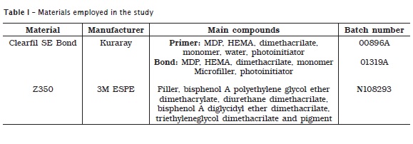
Composite resin samples were constructed with aid of a bipartite stainless steel matrix (4 mm height, central perforation of 4 mm diameter). Therefore, two halves of the matrixes were placed onto the delimited dentin surface and composite resin was inserted. The matrix was inserted in two increments of nanoparticle composite resin (Filtek™ Z350, 3M Espe, Saint Paul, MN, EUA) shade A3. Each increment had 2 mm width and each one was light-cured for 20 seconds. Following, the bipartite stainless matrix was removed and the specimens stored in distilled water at 37ºC until their utilization. All materials used in this study are listed in table I. After 24 hours and 6 months of distilled water storage, the specimens were coupled into a device and mounted into universal mechanical testing machine (Tira Maschinenbau Gmbh, Schalkau, Germany). We used a 20 kN load and 0.5 mm/min velocity, under shear bond strength through orthodontic wire No. 07. The movement was stopped when a rupture or failure of the specimens occurred. Data were collected through specific software connected to the machine. The final values of bond strength (MPa) were calculated by dividing the maximum load values in Newton (N) by the specimens' bonding area in mm2.
To complement the study, after shear bond strength tests, dentin and resin surfaces corresponding to the adhesion area were observed with aid of a stereoscopic magnifying glass (Quimis Aparelhos Científicos Ltda. Q7355-TZ, Diadema/SP, Brazil), at x 15 magnification, to verify the type of failures, which were classified into:
• Adhesive: when the adhesive was present in composite resin, dentin, or both;
• Cohesive in composite resin: when the fracture occurred in composite resin, and both sides of the specimen were covered with composite resin;
• Cohesive in dentin: when the fracture occurred in dentin, and both sides of the specimen had dentin remanent;
• Mixed: when there were two or more types of failures, as previously described.
In this study, the groups were statistically analyzed by one-way ANOVA, with level of significance set at 5%. Since there was significant variability, paired Turkey test was applied with level of significance set at 5%. We used the software Statistical Package for the Social Sciences (SPSS) for data tabulation and analysis. Microsoft Excel software was used for graphic presentation.
Results
With the result means, we could observe that one-way ANOVA showed different results. However, ANOVA comparisons do not allow us to conclude which means would lead to significant different results. To reach such situation, paired Tukey's test was applied (table II).
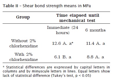
There was no statistical significant difference in the group treated with chlorhexidine after 6-month storage compared with the group without treatment surface. This same fact was also observed by comparing the groups with surface treatment to the groups without surface treatment, after 6-month storage and the strength values of the immediate groups. On the other hand, a significant decrease of bond strength was evidenced in the group treated by chlorhexidine, after 24-hour storage.
The analysis of the fracture result occurring in each experimental condition is represented in graph 1. We adopted four categories to classify the fractures: adhesive fracture, cohesive in dentin (CD), cohesive in resin (CR), and mixed.
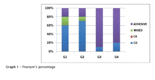
The results indicated that the fracture pattern distribution was variable in each group. However, a tendency toward cohesive failure in dentin was predominantly repeated in the groups which chlorhexidine treatment of dentin surface was not applied (G1 and G2), regardless of storage time. On the other hand, in the groups treated with chlorhexidine (G3 and G4), the most frequent failure was adhesive, both for immediate and 6-month storage.
Discussion
Premature loss of adhesion is one of the problems affecting adhesive restorations 24 and it has been primarily attributed to hybrid layer degradation at tooth/restoration interface 32.
Literature has reported that adhesion to enamel and dentin superficial layer does not demonstrate statistically significant differences between human and bovine teeth 26. According to Bouillaguet et al. 4, bovine dentin could never be considered a perfect substitute in adhesion studies. However, studies 29 have revealed that bovine dentin is one of tooth substrates that are closest to human tooth structures, both in tubule amount and diameter, without presenting significant differences.
In vivo 18 and in vitro 9 studies have proposed that 2% chlorhexidine digluconate solution applied onto human dentin etched with 37% phosphoric acid could maintain the adhesion for a longer time, at the same time that they indicated a decrease in bond strength of teeth without chlorhexidine application as well as a progressive fibril portion disintegration.
This present study aimed to investigate how it would be the immediate and long-term effect of 2% chlorhexidine use on a two-step self-etching adhesive. These adhesive systems' hybrid layer is obtained by the use of a primer comprising acid monomers, which is directly applied onto the smear layer, consequently eliminating the necessity of the initial controlling of surface moisture, mandatory for conventional adhesive systems 38. It is believed that self-etching adhesive systems demineralize the dentin and infiltrate their monomers simultaneously, avoiding collagen fibrils' collapse by air drying and also the occurrence of unprotected fibrils by the applied resin 35.
In this present research, the vertical analysis of the means revealed statistically significant differences for bond strength values between the groups with and without chlorhexidine, 24 hours prior to rehearsal, revealing a decrease in bond strength values of the group receiving surface treatment. Such findings corroborate the study of Campos et al. 7, in which the adhesion of the employed self-etching adhesive system was extremely affected by 2% chlorhexidine. This is probably explained by the interactions that may occur between chlorhexidine and the adhesive components, maybe decreasing the adhesive wettability. In this present study, this could have occurred because the adhesive was applied after chlorhexidine and the latter could have diluted it, determining the low values of bond strength 7. Additionally, the fractured specimens in the groups treated with the antimicrobial agent presented mostly adhesive failures (graph I). Notwithstanding, Castro et al. 10 by evaluating the effect of 2% chlorhexidine on human dentinal substrate, during a period of 24 hours, in different adhesive systems (including self-etching adhesives) reported no significant differences for the values obtained for all evaluated groups. Such findings are in agreement with the results of other studies 6,22.
Other studies employing conventional 8,33 and/or all-in-one self-etching adhesive system 6 demonstrated that after a period of 6 months, the groups treated with chlorhexidine presented a significant better improvement in bond strength values than the groups where the antimicrobial agent was not used. The authors attributed this fact to a better preservation of collagen fibrils, consequently increasing its longevity. Our results do not agree with such findings, because after 6-month storage, the groups treated with chlorhexidine did not show significant difference when compared to the groups without surface treatment. Also, 6-month storage groups either with or without chlorhexidine did not reveal statistically significant difference when compared to groups which were submitted to shear bond strength test after 24-hour storage.
Concerning to the horizontal analysis of both groups' means, we observed that they did not exhibit significant differences at 24-hour and 6-month storages, contrasting to previous studies 6,19 which reported a bond strength decrease as time goes by for the groups with and without surface treatment. In another recent in vivo study 27 using caries-affected occlusal surfaces and conventional adhesive system, the use of chlorhexidine as dentin adjuvant did not produce any detrimental effect on immediate bond strength; chlorhexidine treatment surface was also capable of degrading the hybrid layer in the first months after the restorations. This is in contrast to our study, in which the adhesion was impaired by chlorhexidine application at the immediate period. Therefore, we hypothesize that chlorhexidine interference on bond strength to dentin may be related to the adhesive system used; also, self-etching adhesive systems have their adhesiveness altered by this substance application.
Additionally, literature has diverged regarding both to the moment when chlorhexidine should be applied and to the way its excess should be removed. In our study, we opted by the active utilization after the application of self-etching primer. Our intention was to prevent hybrid layer degradation by metalloproteinases, as observed by conventional adhesive systems, favoring the establishment of a more stable bond strength. Since we removed the solution only by gentle air jet, this could have also influenced our results due to impede an adequate interaction among bond, primer and tooth substrate. Currently, more than a powerful microbial agent for cavity application 23, chlorhexidine is a potential adjuvant in the establishment of a better bonding to dentin. However, this application together with self-etching adhesive systems requires further studies to be well understood since there is not a consensus. Some studies used chlorhexidine prior to 2,3,7,9,33 and after 6,8,18,19,20,27 acid etching with conventional adhesive systems; others used it prior to self-etching primer 6,7 with self-etching adhesive systems.
Concerning to chlorhexidine excess removal from dentin surface, literature have reported several ways to perform this action: absorbent paper points 6,8,19,27, air jet 9,18, water/air spray 2,3, and water washing followed by gentle air jet 2. This fact should be taken into consideration because the amount of chlorhexidine remnant on tooth substrate could influence, positively or negatively, bond strength results due to more or less moisture of dentin surface.
We also have to consider the evaluation mechanism of the adhesive interface. Most of the aforementioned studies 2,6,7,8,9,19,20,27,33 used microtensile test. The shear bond strength test used in our study, in spite of presenting the advantage of being a simpler method, it is frequently criticized due to lack of standard tensile patterns with uniform distribution, which would lead to bond failure at a plane determined by the test and not by the adhesive characteristic itself 13.
Conclusion
Within the limitations of this study, the use of 2% chlorhexidine digluconate solution did not influence the bond strength of the tested self-etching adhesive system, from 24-hour (immediate) to 6-month (long-term) storage. However, 2% chlorhexidine application was deleterious at the moment of shear bond strength tests were executed, in 24-hour storage group. Further in vivo and in vitro studies are necessary to improve the understanding of chlorhexidine interaction with self-etching adhesive system components.
References
1. Basrani B, Ghanem A, Tjaderhane L. Physical and chemical properties of chlorhexidine and calcium hydroxide-containing medications. J Endod. 2004;30(6):413-7. [ Links ]
2. Bengtson CRG, Bengtson AL, Bengtson NG, Turbino ML. Efeito da clorexidina 2% na resistência de união de dois sistemas adesivos à dentina humana. Pesq Bras Odontoped Clín Integr. 2008;8(1):51-6.
3. Bocangel JS, Kraul AOE, Vargas AG, Demarco FF, Matson E. Influence of disinfectant solutions on the tensile bond strength of a fourth generation dentin bonding agent. Pesq Odontol Bras. 2000;14(2):107-11.
4. Bouillaguet S, Gysi P, Wataha JC, Ciucchi B, Cattani M, Godin C et al. Bond strength of composite to dentin using conventional, one-step, and self-etching adhesive systems. J Dent. 2001;29(1):55-61.
5. Buonocore MG. A simple method of increasing the adhesion of acrylic filling materials to enamel surfaces. J Dent Res. 1955;34(6):849-53.
6. Campos EA, Correr GM, Leonardi DP, Barato-Filho F, Gonzaga CC, Zielak JC. Chlorhexidine diminishes the loss of bond strength over time under simulated pulpal pressure and thermo-mechanical stressing. J Dent. 2009;37(2):108-14.
7. Campos EA, Correr GM, Leonardi DP, Pizzatto E, Morais EC. Influence of chlorhexidine concentration on microtensile bond strength of contemporary adhesive systems. Braz Oral Res. 2009;23(3):340-5.
8. Carrilho MR, Carvalho RM, Goes MF, Hipolito V, Geraldeli S, Tay FR et al. Chlorhexidine preserves dentin bond in vitro. J Dent Res. 2007;86(1):90-4.
9. Carrilho MR, Geraldeli S, Tay F, Goes MF, Carvalho RM, Tjäderhane L et al. In vivo preservation of the hybrid layer by chlorhexidine. J Dent Res. 2007;86(6)529-33.
10. Castro FL, Andrade MF, Duarte Júnior SL, Vaz LG, Ahid FJ. Effect of 2% chlorhexidine on microtensile bond strength of composite to dentin. J Adhes Dent. 2003;5(2):129-38.
11. Cunha LA, Ribeiro CF, Dutra-Corrêa M, Rocha PI, Miranda CB, Pagani C. Análise de fatores etiológicos relacionados à sensibilidade pós-operatória na odontologia estética adesiva. Revista de Odontologia da Universidade Cidade de São Paulo. 2007;19(1):68-76.
12. Fardal O, Turnbull RS. A review of the literature on use of chlorhexidine in dentistry. J Am Dent Assoc. 1986;112(6):863-9.
13. Garcia FCP, D'Alpino PHP, Terada RSS, Carvalho RM. Testes mecânicos para avaliação laboratorial da união resina/dentina. Rev Fac Odontol Bauru. 2002;10(3):118-27.
14. Garcia RN, Goes MF, Giannini M. Effect of water storage on bond strength of self-etching adhesives to dentin. J Contemp Dent Pract. 2007;8(7):46-53.
15. Garcia RN, Schaible BR, Lohbauer U, Petschelt A, Frankenberger R. Resistência de união de sistemas adesivos autocondicionantes em dentina profunda. RSBO. 2008;5:39-47.
16. Gendron R, Grenier D, Sorsa T, Mayrand D. Inhibition of the activities of matrix metalloproteinases 2, 8, and 9 by chlorhexidine. Clin Diagn Lab Immunol. 1999;6(3):437-9.
17. Geraldo-Martins VR, Robles FR, Matos AB. Chlorhexidine's effect on sealing ability of composite restorations following Er:YAG laser cavity preparation. J Contemp Dent Pract. 2007;8(5):26-33.
18. Hebling J, Pashley DH, Tjaderhane L, Tay F. Chlorhexidine arrests subclinical degradation of dentin hybrid layers in vivo. J Dent Res. 2005;84(8):741-6.
19. Komori PC, Pashley DH, Tjäderhane L, Breschi L, Mazzoni A, Goes MF et al. Effect of 2% chlorhexidine digluconate on the bond strength to normal versus caries-affected dentin. Oper Dent. 2009;34(2):157-65.
20. Loguercio AD, Stanislawczuk R, Polli LG, Costa JA, Michel MD, Reis A. Influence of chlorhexidine digluconate concentration and application time on resin-dentin bond strength durability. Eur J Oral Sci. 2009;117(5):587-96.
21. Lohbauer U, Nikolaenko SA, Petschelt A, Frankenberger R. Resin tags do not contribute to dentin adhesion in self-etching adhesives. J Adhes Dent. 2008;10(2):97-103.
22. Machado SO, Pretto SM. Avaliação, in vitro, do uso da solução anti-séptica de clorexidina na resistência de união à dentina de um sistema adesivo universal de frasco único. Rev Odonto Ciênc. 2002;17(35):103-10.
23. Meiers JC, Shook LW. Effect of disinfectants on the bond strength of composite to dentin. Am J Dent. 1996;9(1):11-4.
24. Mjör IA, Moorhead JE, Dahl JE. Reasons for replacement of restoration in permanent teeth in general dental practice. Int Dent J. 2000;50(6):361-6.
25. Nakabayashi N, Kojima K, Masuhara E. The promotion of adhesion by infiltration of monomers into tooth substrates. J Biomed Mater Res. 1982;16(3):265-73.
26. Nakamichi I, Iwaku M, Fusayama T. Bovine teeth as possible substitutes in the adhesion test. J Dent Res. 1983;62(10):1076-81.
27. Ricci HA, Sanabe ME, Souza Costa CA, Pashley DH, Hebling J. Chlorhexidine increases the longevity of in vivo resin-dentin bonds. Eur J Oral Sci. 2010;118(4):411-6.
28. Sano H, Takatsu T, Ciucchi B, Horner JA, Matthews WG, Pashley DH. Nanoleakage: leakage within the hybrid layer. Oper Dent. 1995;20(1):18-25.
29. Schilke R, Lisson JA, Baub O, Geurtsen W. Comparison of the number and diameter of dentinal tubules in human and bovine dentine by scanning electron microscopic investigation. Archs Oral Biol. 2000;45(5):355-61.
30. Schmalz G, Ergucu Z, Hiller KA. Effect of dentin on the antibacterial activity of dentin bonding agents. J Endod. 2004;30(5):352-8.
31. Silva MF, Mandarino F, Sassi JF, Menezes M, Centola ALB, Nonaka T. Influência do tipo de armazenamento e do método de desinfecção de dentes extraídos sobre a adesão à estrutura dental. Rev Odontol Univ São Paulo. 2006;18(2):175-80.
32. Spencer P, Wang Y. Adhesive phase separation at the dentin interface under wet bonding conditions. J Biomed Mater Res. 2002;62(3):447-56.
33. Stanislawczuk R, Amaral RC, Grande CZ, Gagler D, Reis A, Loguercio AD. Chlorhexidine-containing acid conditioner preserves the longevity of resin-dentin bonds. Oper Dent. 2009;34(4):481-90.
34. Tay FR, King NM, Chan KM, Pashley DH. How can nanoleakage occur in self-etching adhesive systems that demineralize and infiltrate simultaneously? J Adhes Dent. 2002;4(4):255-69.
35. Tay FR, Pashley DH. Aggressiveness of contemporary self-etching systems. I: Depth of penetration beyond dentin smear layers. Dent Mater. 2001;17(4):296-308.
36. Tay FR, Pashley DH. Have dentin adhesives become too hydrophilic? J Can Dent Assoc. 2003;69(11):726-31.
37. Van Landuyt KL, Peumans M, Munck J, Lambrechts P, Van Meerbeek B. Extension of a one-step self-etch adhesive into a multi-step adhesive. Dent Mater. 2006;22(6):533-44.
38. Watanabe I, Nakabayashi N, Pashley DH. Bonding to ground dentin by a phenyl-P self-etching primer. J Dent Res. 1994;73(6):1212-20.
 Correspondence:
Darlon Martins Lima
Rua 19, quadra O, n.º 10 – Cohaserma
CEP 65072-330 – São Luís – MA – Brasil
E-mail: darlonmartins@yahoo.com.br
Received for publication: January 27, 2011
Accepted for publication: May 9, 2011
