


Serviços Personalizados
Artigo
 pdf em Inglês
 Artigo em XML
 Referências do artigo
 Enviar este artigo por email
Links relacionados
Compartilhar

 Permalink
RSBO (Online)
versão On-line ISSN 1984-5685
RSBO (Online) vol.8 no.4 Joinville Out./Dez. 2011
CASE REPORT ARTICLE
Hereditary gingival fibromatosis – a case report and management using a novel surgical technique
K. Butchi BabuI; Kalwa PavankumarII; B. R. AnuradhaII; Nupur AroraI
I Department of Periodontics, Sri Sai College of Dental Surgery – Andhra Pradesh – India
II Department of Periodontics, M.N.R. Dental College & Hospital – Andhra Pradesh – India
ABSTRACT
Introduction: Hereditary gingival fibromatosis (HGF) is a rare condition presenting varied degrees of gingival enlargement. HGF can present as an isolated entity or as part of a syndrome. Current literatures report a defect in the Son of sevenless-1 gene (SOS-1) on chromosome 2p21-p22 (HGF1) as a possible cause of this condition. Case report: A case of a 16-year-old female is reported who presented generalized extensive gingival overgrowth, involving the maxillary and mandibular arches covering almost two thirds to three quarters of all teeth. Diagnosis of HGF was substantiated by the patient's clinical features, family history and histopathological examination. Treatment was excision of the gingival tissue by a modified gingivectomy technique with both manual instrumentation and electrosurgery. The postoperative course was uneventful and the patient's aesthetic concerns were addressed. Post-surgical follow-up after 18 months demonstrated no recurrence. Conclusion: Hereditary gingival fibromatosis stands apart from other gingival enlargements in the varied treatment options available and the nature of recurrence post treatment. There is no consensus among authors related to the mode of treatment. Here, in this present case report we highlight a novel surgical technique to deal with the extensive nature of enlargement seen in HGF cases.
Keywords: hereditary gingival fibromatosis (HGF); gingival enlargement; electrosurgery; gingivectomy.
Introduction
Healthy gingival tissue completely fills the interproximal spaces between the teeth and surrounds the neck of the teeth in a collar like fashion with a knife or feather edged margin. The gingival tissues are constantly subjected to various mechanical, chemical, and bacteria aggressions. In return these tissues respond in a number of enigmatic ways, one such response is gingival overgrowth or enlargement. One such form of gingival overgrowth is known as hereditary gingival fibromatosis (HGF). It has also been designated with other terms such as idiopathic fibromatosis, congenital familial fibromatosis, gingivomatosis, and elephantiasis gingivae 11.
Hereditary gingival fibromatosis is a rare disease (1 in 750,000) and belongs to a group of benign disorders characterized by firm, enlarged gingival tissues that cover most of the anatomic crowns. It was recognized probably more than a century ago, the first case was reported by Gross in 185663 3.
It is reported to have a phenotype frequency of 1:175,000 and a gene frequency of 1:350,000 5. HGF is more commonly associated with an autosomal dominant gene 4. Pedigree analyses of HGF families were consistent with simple mendelian transmission pattern, although autosomal recessive cases have been reported in the literature. Recently, Son-of- sevenless (SOS-1) has been identified as the prime etiology for non-syndromic HGF. SOS-1 is a guanine nucleotide-exchange factor that functions in the transduction of signals that control cell growth and differentiation 9,10.
The gingiva is normal in colour, stippled (often exaggerated), firm in consistency. The tissue is fibrotic (feels like bone on palpation) and displays a nodular or minutely pebbled surface. Both attached and free gingiva is involved but does not extend beyond mucogingival junction 1,11.
Histological features of hereditary gingival fibromatosis are non specific. Fibrotic tissues shows increased amount of connective tissue. It is characterized by densely arranged collagen bundles, numerous fibroblasts, and connective tissue which is avascular along with well structured epithelium with elongated and thin papillae inserted in fibrous connective tissueeeee 2.
The present case report depicts one of the unusual presentations of hereditary gingival fibromatosis which was associated with massive destruction of periodontal tissues and deals with the management employing a novel surgical procedure.
Case report
A 16-year-old female patient accompanied by her mother and sister reported to the Department of Periodontics, Rajah Muthiah Dental College & Hospital, Chidambaram, Tamilnadu, India with a chief complaint of gingival swelling in her mouth. On further questioning, the patient revealed that she had first noticed the swelling post eruption of her permanent teeth. The swelling slowly progressed involving the gingiva in both the arches and attained the current size. Masticatory problems and esthetic concerns made the patient report for treatment.
The patient was thoroughly questioned regarding her physical and mental status to rule out any syndromes associated with the enlargement. Her medical history was non contributory. Her family history yielded significant information, as both her mother and sister presented with a similar clinical presentation of the gingiva.
Clinical examination
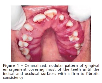
Intra oral examination revealed generalized, nodular pattern of gingival enlargement covering most of the teeth to the incisal and occlusal surfaces with a firm to fibrotic consistency (figure 1). Periodontal assessment revealed extensive mobility, especially of the maxillary left and right posterior teeth, presence of deep pockets and moderate calculus deposits with bleeding on probing in certain areas. The patient's medical history did not reveal any pathological condition. Therefore, surgical excision of the lesion was proposed to the patient.
Treatment
Considering the severity of the enlargement and anticipating the resultant bleeding during surgery, a blended surgical approach comprising of both electrosurgery and manual instrumentation was chosen. A quadrant-by-quadrant gingivectomy Goldman technique (1950) was chosen as the preferred surgical technique. Oral prophylaxis has been performed and the patient was recalled after two weeks for surgery.
Surgical procedure
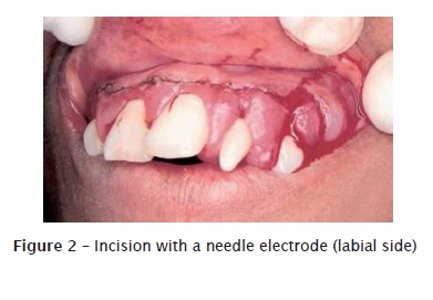
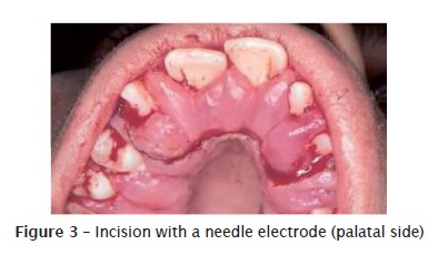
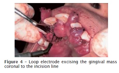
The surgical intervention was carried out under local anesthesia. Following administration of local anesthesia and intraoral disinfection with 0.12% chlorhexidine mouth rinse, the pocket is reviewed to mentally picture the 3-dimensional topography. Next, a periodontal probe is used to outline the base of the pockets with series of small bleeding points on both sides of the gingival enlargement. The bleeding points outline the incision. The orientation of the pocket marker should be parallel to the tooth or the incision will be too deep or too shallow. The incision line was then delineated with the needle electrode (figures 2 and 3) all the way down to the base of pocket, at the bevel of 45 degree, and end on the root surface at the bleeding points. A light and gentle ‘paintbrush-like' stroke was used to guide the electrode while performing the incision. Owing to the bulk of the enlargement, initial shallow cuts were made prior to refining the incision deep in to the gingival enlargement. A loop electrode was then employed to excise the gingival mass coronal to the incision line utilizing gentle sweeping motion (figure 4). Taking into consideration the amount of heat generated while using the needle or the loop electrode, an 8 to 15 seconds cooling period was advocated between successive incisions or recontouring of the gingiva.
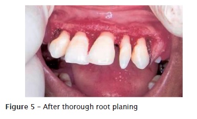
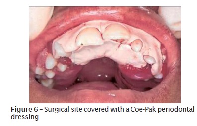
Internal bevel incision was then given with a Bard Parker blade No. 15 to further thin the flap margin. A full thickness mucoperiosteal flap was elevated and thorough degranulation was done. The exposed roots were thoroughly root planed with curettes and ultrasonic instrumentation (figure 5). Mobile teeth were extracted during the course of the surgery. Mucoperiosteal flaps were then sutured in their original position using interdental interrupted 4-0 non-absorbable black silk suture. The surgical site was given a dressing with Coe-Pak periodontal dressing (figure 6). The patient was given a prescription for an antibiotic and an analgesic. Post surgical care was followed by a regular 0.2% chlorhexidine rinse twice a day for 2 weeks. Sutures and periodontal dressing were removed after one week. The interval between each surgical procedure was one week.
Postoperative healing was uneventful. The patient was recalled 6 weeks post surgery, gingival swelling was still evident in relation to maxillary left posterior region where the teeth had been extracted. Surgical excision was repeated and the tissue was sent for histopathological examination.
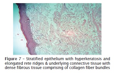
Histopathological examination revealed stratified epithelium with hyperkeratosis and elongated rete ridges. The underlying connective tissue was abundant with dense fibrous tissue comprising of collagen fiber bundles. Occasional inflammatory cell infiltrate was noticed (figure 7).
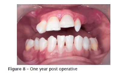
The clinical and histopathological findings were consistent with hereditary gingival fibromatosis. The postoperative course was uncomplicated and there was no lesion recurrence up to one year of follow-up (figure 8).
Discussion
Gingival enlargement, either localized or generalized might be attributed to a number of reasons, ranging from inflammation, leukemic infiltration, and association with use of medicines like phenytoin, cyclosporine, and nifedipine etc. 16.
Here, we report a case of hereditary gingival fibromatosis. HGF is transmitted as either autosomal dominant or recessive. We justify our diagnosis of the gingival fibromatosis as hereditary and as an autosomal dominant, solely relying on the occurrence of the enlargement in both her mother and sister and further corroborated by histopathological findings.
In our present case report, HGF occurred as an isolated entity. But a thorough look into the literature reveal that it can be a part and parcel of multi-system syndromes, such as Zimmermann-Laband syndrome (Ear, nose, bone and nail defects with hepatosplenomegaly), Murray-Peretic-Drescher syndrome (juvenile hyaline fibromas), Rutherfurd syndrome (corneal dystrophy, mental retardation, impairment of dental eruption by radicular resorption), Jones syndrome (progressive deafness) and Cross syndrome (microphthalmia, mental retardation, athetosis and hypopigmentation) 6,7,8,13,14. In this case, a thorough evaluation of the patient, revealed no association with any of the clinical features associated with the above syndromes.
Numerous treatment modalities have been employed for the excision of the enlarged gingival tissues, including of conventional surgery, electrosurgery, an apically positioned flap and lasers 15. Surgical intervention using conventional means like scalpel may sometimes be technically difficult and impractical for example in children or mentally handicapped, or in patients suffering from impaired haemostasis. Hence use of electrosurgery in these situations would be beneficial.
Due to the presence of extensive enlargement coupled with poor tissue tone in certain areas of the enlargement, a blended approach utilizing a quadrant- by- quadrant modified gingivectomy technique, primarily a modification of the ledge and wedge technique (modified partial-thickness palatal flap) comprising both manual instrumentation and electrosurgery was favored upon as the preferred surgical technique 12. Unlike the ledge and wedge technique which consists of a primary gingivectomy followed by two incisions, our surgical technique comprised of a primary gingivectomy followed by a single internal bevel incision.
Electrosurgery was utilized to carry on the primary horizontal incision, as it had the added advantage to minimize bleeding and simultaneously reduce the bulk of the tissue. Following which, internal bevel incision was carried out to thin down the flap for proper adaptation and permit uneventful healing.
Gingivectomy has been chosen in spite of advanced periodontitis around posterior teeth 1. A periodontal flap procedure may be preferred for the treatment of gingival enlargement if there are large areas of gingival overgrowth or attachment loss and osseous defects. Gingivectomy plus periodontal flap technique was used in the present case due to the presence of an extreme bulk of gingival tissue along with alveolar bone loss.
Subgingival calculus may be present on deep root surfaces. In the present case when the flaps were reflected subgingival calculus was revealed. The reflected flaps allowed improved access for its removal. Following surgery, the patient had less postoperative discomfort as a result of the minimal cut tissue surface using flaps compared to a gingivectomy.
Our case report presented similar histopathological findings as of the gingival lesions in hereditary gingival fibromatosis which include hyperplastic epithelium with elongated rete ridges and a connective tissue interspersed with abundant collagen-fiber bundles, numerous fibroblasts and mild presence of inflammatory cells.
Conclusion
Hereditary gingival fibromatosis stands apart from other gingival enlargements in the varied treatment options available and the nature of recurrence post treatment. There is no consensus among authors related to the mode of treatment. Here, in this present case report we highlight a novel surgical technique to deal with the extensive nature of enlargement seen in HGF cases.
Acknowledgements
Dr. Rajanikanth, M.D.S. (Oral Pathology).
Conflict of interest statement
There is no conflict of interest in the present manuscript.
Informed consent statement
The patient signed an informed consent, kept in the records, in the archives of the Rajah Muthiah Dental College & Hospital.
References
1. Baptista IP. Hereditary gingival fibromatosis: a case report. J Clin Periodontol. 2002;29(9):871-4. [ Links ]
2. Collan Y, Ranta H, Vartio T, Perheentupa J, Raeste AM. Histochemical and biochemical study of hereditary fibrous hyperplasia of the gingiva. Scand J Dent Res. 1982;90:20-8.
3 . Fletcher J. Gingival abnormalities of genetic origin: a preliminary communication with special reference to hereditary generalized gingival fibromatosis. J Dent Res. 1966;45:597-612.
4. Gorlin R, Cohen M, Levi L. Syndromes of the head and the neck. 3. ed. Nova York: Oxford Press; 1990. p. 847-52.
5. Gross SD. Case of hypertrophy of the gums. Louis Ville Rev. 1856;1:232-7.
6. Hartsfeild Jr JK, Bixler D, Hazen RH. Gingival fibromatosis with sensorineural hearing loss: an autosomal dominant trait. Am J Med Genet. 1985;22:623-7.
7. Holzhausen M, Gonçalves D, Corrêa Fde O, Spolidorio LC, Rodrigues VC, Orrico SR. A case of Zimmermann-Laband syndrome with supernumerary teeth. J Periodontol. 2003;74:1225-30.
8. Houston IB, Shotts N. Ruthurfurd's syndrome: a familial oculo-dental disorder. A clinical and electrophysiologic study. Acta Paediatr Scand. 1966;55:23333-8.
9. Lee EJ, Jang SI, Pallos D, Kather J, Hart TC. Characterization of fibroblasts with Son of Sevenless-1 mutation. J Dent Res. 2006;85:1050-5.
10. Hakkinen L, Csiszar A. Hereditary gingival fibromatosis: characteristics and novel putative pathogenic mechanisms. J Dent Res. 2007;86:25-34.
11. Newman MG, Takei HH, Carranza FA. Carranza's clinical periodontology. 9. ed. Philadelphia: WB Saunders Company; 2002. p. 285.
12. Ochsenbein C, Bohannan HM. The palatal approach to osseous surgery. I. Rationale. J Periodontol. 1963;34:60-4.
13. Piatelli A, Scarano A, Di Belluci A, Matarasso S. Juvenile hyaline fibromatosis of the gingiva: a case report. J Periodontol. 1996;67:451-3.
14. Ramon Y, Berman W, Bubis JJ. Gingival fibromatosis combined with cherubism. Oral Surg Oral Med Oral Pathol. 1967;24:435-48.
15. Coletta RD, Graner E. Hereditary gingival fibromatosis: a systematic review. J Periodontol. 2006;77:753-64.
16. Seymour RA, Heasman PA. Drugs and the periodontium. J Clin Periodontol. 1988:15:1-16.
 Correspondence:
Kalwa Pavankumar
S/o K.Vijay Kumar,
Murali Cloth Stores
Main Road, (Post) Devarakadra
(Dist) Mahabubnagar – 509204 – Andhra Pradesh – India
E-mail: drpavankumarmds@gmail.com
Received for publication: February 8, 2011
Accepted for publication: March 29, 2011
