


Serviços Personalizados
Artigo
 pdf em Inglês
 Artigo em XML
 Referências do artigo
 Enviar este artigo por email
Links relacionados
Compartilhar

 Permalink
RSBO (Online)
versão On-line ISSN 1984-5685
RSBO (Online) vol.9 no.1 Joinville Jan./Mar. 2012
ORIGINAL RESEARCH ARTICLE
Comparison between electronic and radiographic method for the determination of root canal length in primary teeth
Sérgio Luiz Pinheiro I; Iris Nogueira Bincelli II; Talita Faria I;Carlos Eduardo da Silveira Bueno II;Rodrigo Sanches Cunha III
I Pontifical Catholic University of Campinas – Campinas – SP – Brazil.
II Center of Dentistry Researches – São Leopoldo Mandic – Campinas – SP – Brazil.
III University of Manitoba – MB – Canada.
ABSTRACT
Introduction: There are few researches in literature that mention the use of the apex locator in deciduous teeth and working length is obtained through radiographies. Objective: The purpose of this research was to compare the radiographic and the electronic method to obtain the working length in deciduous molars. Material and methods: Twelve molar teeth were used. The specimens in the visual method had their root length measured through the passive insertion of a 10 K-file with a silicone stop within root canal until its tip was seen at the apical foramen. The working length was measured through radiographs or using the apex locator Root ZX II. The mean between the examiners was submitted to the variance analysis (ANOVA). Results: Statistically significant differences were found between the visual method and the radiographic method (p < 0.001). There was no significant difference between the working length measurements in visual method and those obtained with the apex locator (p = 0.1319). Conclusion: The apex locator is indicated as a clinical implementation for endodontic treatment in primary teeth.
Keywords: tooth Apex; Endodontics; primary teeth; radiographic magnification; apical foramen.
Introduction
Endodontic treatment in deciduous teeth is a therapeutic option for teeth with infected pulps9. Treatment of the root canals of deciduous teeth with pulpal necrosis helps to keep the integrity of primary teeth until their physiological exfoliation 7,23,29.
Radiographs are traditionally used to obtain information on the anatomy of the root canal, its working length and the tissues surrounding the apex. In order to achieve successful endodontic treatment in deciduous teeth, it is mandatory to know the exact working length of the root canals 4 since this reduces the risks of insufficient cleaning or damage to the periapical tissues due to over-instrumentation 3,6,25.
The radiographic method is the most commonly used method to determinate the working length in deciduous teeth; however, it is often difficult to obtain, in pediatric dentistry, due to the children's behavior, as they may move their heads, legs and arms during the radiographic scan. Anatomical variations and the superposition of images, such as the permanent tooth germs overlapping the roots of deciduous teeth, can make the location of the radicular apex difficult, leading to misleading results, particularly in cases where resorption is present 1,23. Inappropriate radiographic techniques due to the oral cavity size of the children, the positioners' size and the radiographic films can also make accurate location of the radicular apex difficult 11,27. Therefore, endodontic treatment in children can be challenging for the professional, due to the fact that it depends on radiographic images to analyze the relationship of deciduous and permanent teeth, the support tissues and the determination of the working length 8. Technique errors may result in an increased risk of over-instrumentation and/or overfilling, which can damage the permanent tooth germ 23.
Currently, the electronic method is used to determine the working length with accuracy and also to facilitate and reduce the operation time in deciduous teeth 4,21,23. The accuracy of the electronic apex locators is influenced by two factors: moisture content in root canals and diameter of the apical foramen 12,13,26.
The first generation of electronic apex locators was based on the oral mucosa and the periodontal ligament 21. The second and third generations were based on the principle of impedance 2,21,28. Root ZX II is a third-generation locator, which uses the impedance ratio instead of the impedance difference. This method simultaneously measures impedance values in two frequencies (8 and 400 Hz) and calculates the quotient of the impedances 4,25,31. The main advantage is the direct determination of the apical constriction (AC), instead of calculating the distance from AC to the radiographic apex 17.
The purpose of this research was to compare the radiographic and the electronic method to obtain the working length in deciduous molars.
Material and methods
Tooth selection
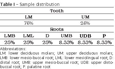
The study was approved by the Institutional Review Board (Committee on human experimentation, Helsinki Declaration). Twelve deciduous molars were used from the Human Teeth Bank at the Pontifical Catholic University of Campinas (PUC-Campinas) (table I).
Before the test, the teeth were stored in formalin solution (10%), removal of calculus and stains was performed with periodontal curettes and prophylaxis was done with pumice (SS White, Rio de Janeiro, RJ, Brazil). The selected teeth were observed with a magnifier (x10) (Guangdong Baijia Baiter Industry, China), and the inclusion criteria were as follows:
– Deciduous molars with physiological radicular resorption in the apical and medium third were select (Teeth with radicular resorption in the cervical third were excluded);
– Absence of root splits and cracks;
– Absence of furcation perforations and/or root perforations.
Teeth preparation
The endodontic access cavity was performed with a sterile carbide round bur #2 (KG Sorensen, São Paulo, Brazil); and after locating the canals, their entrances were enlarged with a diamond tip #3082 (KG Sorensen, Barueri, São Paulo).
Root canal measurement
Group I: measurement by visual method
The visual method used was obtained through the passive insertion of a 10 K-file (Dentsply Maillefer, Ballaigues, Switzerland) with silicone stop within the root canal until its tip was seen at the apical foramen considering the physiological radicular resorption. This was performed with the aid of an optical light microscope (DF Vasconcelos, São Paulo, Brazil) at x 8 magnification. When the file tip was observed at the apical foramen, the stop was stabilized at the incisal edge of the tooth, the file was removed, and the distance between the stop and the file tip was measured with a millimeter ruler (Dentsply-Maillefer Ballaigues, Switzerland) 3.
Group II: measurement by radiographic method
The teeth were bonded with utility wax (Wilson, Cotia, São Paulo, Brazil) to the base of the positioner (Indusbello, Londrina, Paraná, Brazil) and a radiograph was taken in the buccolingual direction using the standardized radiographic technique by using the extension cone paralleling system. A Kodak periapical film was used (São José dos Campos, SP, Brazil) in the RX device (ProDental, Lisbon, Portugal) with an exposure time of 0.8 seconds to obtain the working length. Radiographic developing and fixing time was about 40 seconds and 2 minutes, respectively, in a handheld dark chamber (VH, Softline, São Paulo, Brazil). The working length was obtained through the measurement of the roots in crown-apex direction, on the radiograph, with the aid of a millimeter ruler (Sunward, Taiwan, China). The reference point corresponded to the cusp tip of the analyzed root and considered the physiological radicular resorption.
Group III: measurement by Root ZX II
The determination of the root canal length through the electronic method was performed by using Root ZX II (J Morita Corp., Tokyo, Japan). The teeth were fixed in alginate (Jeltrate Plus, Dentsply, Rio de Janeiro, Brazil) mixed according to the manufacturer's instructions and put in 50 ml disposable plastic 4.5 cm diameter x 4 cm high vessels (Copaza, Içara, Santa Catarina, Brazil). The alginate was continuously moistened by saline irrigation. The root canals were also filled with saline. Cotton pellets were used to remove excess of saline from the pulp chamber. The electronic device Root ZX II was used according to the manufacturer's specifications: the electrode that should be fixed at the lips of the subject was fixed in the alginate base; a 10 K-file with silicone stop was introduced within root canal until the audible sign on the device's display would signal the radicular apex and the last green bar was seen. Then, the silicone stop was positioned in the cusp tip corresponding to the analyzed root and the file was removed for the working length to be measured in millimeters, with the ruler (Dentsply-Maillefer, Ballaigues, Switzerland).
Statistical analysis
The measurements for group 1 and 2 were performed in triplicate by two calibrated examiners under controlled blind conditions. The arithmetical mean was calculated and tabulated. The means between the examiners were submitted to descriptive analysis and variance analysis (ANOVA) for the paired samples. The mean difference between the values obtained with the radiographic method / visual method; electronic apex locator / visual method; electronic apex locator / radiographic method (mm) was submitted to t test for the paired samples
Results
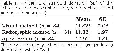
Statistically significant differences between the length measurements of the roots obtained through the passive insertion of the 10 K file (visual method) and the radiographic method were found (p < 0.01). There was no significant difference between the working length measured by passive insertion of the file (visual method) and using the apex locator (p > 0.05) (table II).
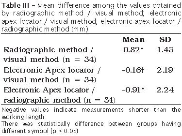
A statistical difference was found among the mean values obtained with radiographic method / visual method; electronic apex locator / visual method; electronic apex locator / radiographic method (p < 0.05) (table III).
Discussion
The determination and retention of the apical limit for instrumentation is undoubtedly an important step for the outcome of root canal treatment 4,20,21. An accurate determination of the working length contributes to safe and effective instrumentation 21,22,24.
The use of radiographs in pediatric endodontic treatment should be carefully considered 4,21,23. Radiographic measurements have also some limitations; the most important one is that they do not account for the location of the apical foramen 5,11,23.
The diagnostic value is often limited and children's exposure to X-rays should be limited to a minimum 21,23. Furthermore, radiographic assessment is difficult, particularly in cases where the physiological resorption in primary teeth occurs on buccal or lingual aspects of the root 4,21. This will not often be visible radiographically, resulting in an increased risk of over-instrumentation and/or overfilling 23 causing damage to the permanent tooth germ 21.
The results of this research agree that there is a radiographic limitation for the measurement of the radicular apex, since all measures obtained through radiographs presented significant differences in relation to the measures obtained by passively inserting a file within the root canal until visualization of its tip in the apex (visual method). It is important to emphasize that, according to this methodology, radiographs of human deciduous teeth were used to obtain the working length, since the second radiographic scan with the file within the root canal is not conducted in pediatric dentistry to confirm the apex limit of instrumentation due to cooperation issues related to children. The anatomical limitations of the oral cavity in children and their behavior were not factors that could influence our results, because the samples used in this work were donated by a Human Teeth Bank. Those factors could adversely affect the accuracy of the radiographic method for the measurement of the radicular apex 8,11,12,21,28.
The usage of electronic devices to determine the working length has gained increasing popularity among dentists in recent years to avoid the hazards of radiation 17,26,30. Modern apex locators are able to determine an area between the minor and major apical foramina by measuring the impedance between the file tip and the canal with different frequencies enabling tooth length measurements in the presence of electrical conductive media in the root canals 18,25,27,31. At the dentin wall of the radicular canal there is electric impedance, and as it gets closer to the apical third, the dentin tissue layer becomes less thick, which decreases the capacity of electric isolation. This gradual decreasing is electrically interpreted as reduction of dentinal impedance. The Root ZX II used in this study follows this principle and has been analyzed in many studies, both in vitro and in vivo. It is a third-generation electronic device, in which no calibration is required and a microprocessor calculates the impedance quotient 19. The third-generation devices use multiple frequencies in order to determine the distance from the end of the canal 6,12,15,25.
In endodontics, studies are presently performed with permanent teeth comparing the accuracy of the apex locator and the radiographic methods 8,10,11,12,15,17,20. Shanmugaraj et al. 28 showed that the radiographic method presents many limitations, but the results are similar to the apex locator. Hassanien et al. 11 added that the accuracy of the apex locator is an excellent aid to the radiographic method. Kim et al. [18] observed that the apex locator concurrent to the radiographic method is recommended for the determination of the working length, although the results of the apex locator are more accurate than those of radiographic method. The apex locator is more accurate to determinate the root canal length when compared to the radiographic method 10,11,20.
This work observed the absence of significant differences between the measurements of the apex locator and the measurement of the radicular canals of deciduous molars through the passive insertion of the file until its visualization at the radicular apex (visual method). In order to control the visualization errors, each measurement was repeated three times and the mean value was calculated. Those results are in accordance with literature 1,4,14,16,21,23,30 which observed that the electronic method can be used safely in deciduous teeth with or without resorption, and in both cases, the measurement can be performed accurately. Angwaravong & Panitvisai 1 added that, when compared to direct canal measurements, the Root ZX measurement at meter reading "Apex" had a smaller error (0.01 mm) than the meter reading "0.5 bar" (-0.33 mm), which is consistent with this work in the fact that it has presented a correlation between the measurements of Root ZX measurement at meter reading "Apex" and the visual method.
Root length determination is one of the most important factors in root canal treatment in both permanent and primary teeth 4,16,21,23. Electronic root length determination may be helpful in overcoming the shortcomings of the radiographic method, especially in teeth with root resorption 4,21,23. The measurements of Root ZX II in this work were similar to the measurement of the root canals of deciduous molars through the passive insertion of the file until its visualization in the radicular apex (visual method), unlike the radiographic method, that presented higher measurements compared to the electronic method.
Conclusion
Radiography is an auxiliary resource for diagnosis in deciduous teeth to be endodontically treated, which favors the evaluation of whether there is injury in the furcation and/or periapical area and resorption areas additionally to obtaining an estimate of the radicular behavior. The electronic methods must be used in order to complement the evaluation of the radicular length obtained by the radiographic method in order to avoid the over-instrumentation and/or overfilling that would be hazardous for both the support tissues and the permanent successor.
References
1. Angwaravong O, Panitvisai P. Accuracy of an electronic apex locator in primary teeth with root resorption. Int Endod J. 2009;42(2):115-21. [ Links ]
2. Baldi JV, Victorino FR, Bernardes RA, Moraes IG, Bramante CM, Garcia RB et al. Influence of embedding media on the assessment of electronic apex locators. J Endod. 2007;33(4):476-9.
3. Bernardes RA, Duarte MAH, Vasconcelos BC, Moraes IG, Bernardineli N, Garcia RB et al. Evaluation of precision of length determination with 3 electronic apex locators: Root ZX, elements diagnostic unit and Apex Locator, and Romi Apex D-30. Oral Surg Oral Med Oral Pathol Oral Radiol Endod. 2007;104(4):e91-4.
4. Bodur H, Odabas M, Tulunoglu Ö, Tinaz AC. Accuracy of two different apex locators in primary teeth with and without root resorption. Clin Oral Invest. 2008;12(2):137-41.
5. Brunton PA, Abdeen D, MacFarlane TV. The effect of an apex locator on exposure to radiation during endodontic therapy. J Endod. 2002;28(7):524-6.
6. D'Assunção FLC, Albuquerque DS, Salazar-Silva JR, Ferreira LCQ, Bezerra PM. The accuracy of root canal measurements using the Mini Apex Locator and Root ZX II: an evaluation in vitro. Oral Surg Oral Med Oral Pathol Oral Radiol Endod. 2007;104(3):e50-3.
7. Dandashi MB, Nazif MM, Zullo T, Elliot MA, Schneider LG, Czonstkowsky M. An in vitro comparison of three endodontic techniques for primary incisors. Pediatr Dent. 1993;15(4):254-6.
8. El Ayouti A, Lõst C. The ability of Root ZX apex locator to reduce the frequency of overestimated radiographic working length. J Endod. 2002;28(2):116-9.
9. Fuks AB, Eidelman E. Pulp therapy in the primary dentition. Curr Opin Dent. 1991;1(5):556-63.
10. Haffner C, Folwaczny M, Galler K, Hickel R. Accuracy of electronic apex locators in comparison to actual length – an in vivo study. J Dent. 2005;33(8):619-25.
11. Hassanien EE, Hasbem A, Chalfin H. Histomorphometric study of the root apex of mandibular premolar teeth: an attempt to correlate working length measured with electronic and radiography methods to various anatomic positions in the apical portion of the canal. J Endod. 2008;34(4):408-12.
12. Herrera M, Ábalos C, Planas AJ, Llamas R. Influence of apical constriction diameter on root ZX apex locator precision. J Endod. 2007;33(8):995-8.
13. Huang L. An experimental study of the principle of electronic root canal measurement. J Endod. 1987;13(2):60-4.
14. Katz A, Mass E, Kaufman AY. Electronic apex locator: a useful tool for root canal treatment in the primary dentition. J Dent Child. 1996;63(6):414-7.
15. Kaufman AY, Keila S, Yoshpe M. Accuracy of a new apex locator: an in vitro study. Int Endod J. 2002;35(2):186-92.
16. Kielbassa AM, Muller U, Munz I, Monting JS. Clinical evaluation of the measuring accuracy of root ZX in primary teeth. Int Endod J. 2003;95(1):94-100.
17. Kim E, Lee SJ. Electronic apex locator. Dent Clin N Am. 2004;48(1):35-54.
18. Kim E, Marmo M, Lee CY, Oh NS, Kim IK. An in vivo comparison of working length determination by only root-ZX apex locator versus combining root-ZX apex locator with radiographs using a new impression technique. Oral Surg Oral Med Oral Pathol Oral Radiol Endod. 2008;105(4):e79-83.
19. Kobayashi C, Suda H. New electronic canal measuring device based on the ratio method. J Endod. 1994;20(3):111-4.
20. Krajczár K, Marada G, Gyulai G, Tóth V. Comparison of radiographic and electronical working length determination on palatal and mesio-buccal root canals of extracted upper molars. Oral Surg Oral Med Oral Pathol Oral Radiol Endod. 2008;106(2):e90-3.
21. Leonardo MR, Silva LAB, Nelson-Filho P, Silva RAB, Raffaini MSGG. Ex vivo evaluation of the accuracy of two electronic apex locators during root canal length determination in primary teeth. Int Endod J. 2008;41(4):317-21.
22. Martin CL, Gijon R, Luque F, De Mondelo NR. In vitro evaluation of the accuracy of three electronic apex locators. J Endod. 2004;30(4):231-3.
23. Mente J, Seidel J, Buchalla W, Koch MJ. Electronic determination of root canal length in primary teeth with and without root resorption. Int Endod J. 2002;35(5):447-52.
24. Miguita KB, Cunha RS, Davini F, Fontana CE, Bueno CES. Análise comparativa de dois localizadores apicais eletrônicos na definição do comprimento de trabalho na terapia endodôntica: estudo in vitro. RSBO. 2011;8(1):27-32.
25. Plotino G, Grande NM, Brigante L, Lesti B, Somma F. Ex vivo accuracy of three electronic apex locators: root ZX, elements diagnostic unit and apex locator and ProPex. Int Endod J. 2006;39(5):408-14.
26. Satio T, Yamashita Y. Electronic determination of root canal length by a newly developed measuring device: the influences of the diameter of the apical foramen, the size of K-file and root canal irrigants. Dent Jpn. 1990;27(1):65-72.
27. Shabahang S, Goon WY, Gluskin AH. An in vivo evaluation of root ZX electronic apex locator. J Endod. 1996;22(11):616-8.
28. Shanmugaraj M, Nivedha R, Mathan R, Balagopal S. Evaluation of working length determination methods: an in vivo / ex vivo study. Indian J Dent Res. 2007;18(2):60-2.
29. Takushige T, Cruz EV, Asgor MA, Hoshino E. Endodontic treatment of primary teeth using a combination of antibacterial drugs. Int Endod J. 2004;37(2):132-8.
30. Tosun G, Erdemir A, Eldeniz AU, Sermet U, Sener Y. Accuracy of two electronic apex locators in primary teeth with and without apical resorption: a laboratory study. Int Endod J. 2008;41(5):436-41.
31. Venturi M, Breschi L. A comparison between two electronic apex locators: an ex vivo investigation.21
 Correspondence:
Rodrigo Sanches Cunha
Department of Restorative Dentistry
D226C – 780, Bannatyne Avenue
Winnipeg – Manitoba – Canada
E-mail: cunhars@cc.umanitoba.ca
Received for publication: August 17, 2010
Accepted for publication: October 29, 2010
