


Serviços Personalizados
Artigo
 pdf em Inglês
 Artigo em XML
 Referências do artigo
 Enviar este artigo por email
Links relacionados
Compartilhar

 Permalink
RSBO (Online)
versão On-line ISSN 1984-5685
RSBO (Online) vol.9 no.1 Joinville Jan./Mar. 2012
ORIGINAL RESEARCH ARTICLE
Analysis of two different endodontic desobturation techniques through clearing teeth technique
Mário Luiz Pinto de QueirózI; Elias Pandonor Motcy de Oliveira I; Tiago André Fontoura de MeloI; Érica Pozo Mautone I; Aline Colpo I
I Department of Endodontics, Lutheran University of Brazil, campus Canoas – Canoas – RS – Brazil.
ABSTRACT
Introduction and Objective: This study aimed to analyze in vitro the efficacy of two endodontic techniques employed for the removal of the filling material inside root canals. Material and methods: Twenty single-rooted lower bicuspids were selected, prepared by crown-down technique and filled with gutta-percha and Endofill® sealer, through lateral condensation technique. The provisional restoration was executed with Cimpat® and the teeth remained 15 days at 37°C and 100% humidity. Next, the teeth were divided into two groups according to the technique of desobturation and re-instrumentation. In group A, it was used ProTaper Universal® instrument sizes D1, D2 and D3 for desobturation procedure associated with the same system instrument sizes F4 and F5 for re-instrumentation. In group B, it was employed K type hand files for desobturation and re-instrumentation. After clearing teeth technique, the teeth examination was performed by two examiners previously trained and calibrated. Results and Conclusion: Data were subjected to Fisher's exact test, with significance level of 5%, which showed no statistical difference between the two techniques.
Keywords: endodontics; retreatment; dental instruments.
Introduction
One of the greatest causes of endodontic therapy failures is the infection persistency within root canals, which damages the treatment and consequently leads to a new endodontic reintervention 25.
Endodontic retreatment aims to the complete removal of the filling in order to access the dentinal tubules and the apical foramen, facilitating root canal system cleaning and shaping 22.
According to Peciuliene et al. 20, endodontic treatment failure is not related to patient's race, age, gender and tooth type, but to both the clinician's lack of ability and to the microorganisms' presence.
Currently, due to the difficulty in performing the complete removal of the endodontic filling, several techniques and materials have been tested. With the appearance of nickel-titanium instruments, several studies have reported their efficacy, cleaning ability, and safety during endodontic filling removal2,6,12,15,19.
In 2003, Basso et al. 7 evaluated the efficacy of hand preparation with K type/Hedströem instruments and rotary preparation with Profile .04 and ProTaper on endodontic retreatment. The authors verified that the rotary systems were more effective. At the cervical and medium thirds, they did not find statistically significant differences among groups, but at the apical third, Profile .04 showed the best results.
Carvalho Maciel & Zaccaro Scelza 9 compared different techniques of endodontic filling removal. For this purpose, 100 human filled teeth were retreated through the following techniques: group I – Gates-Glidden burs associated with K type instruments; group II – Profile system; group III – ProTaper system; group IV – K3 system; and group V – Micro Hero 642 system. The retreated teeth were radiographically analysed to verify any material presence. The authors found no statistically significant difference among the tested techniques for the endodontic filling removal. Hand instruments showed the lowest efficacy compared to K3 and ProTaper instruments.
Schirrmeister et al. 24 evaluated the efficacy of gutta-percha removal in curved root canals by employing hand (FlexMaster) and the rotary technique (ProTaper and Race). The authors verified that hand technique with the use of Flexmaster instruments resulted in larger areas of filling remnants than Race and ProTaper systems.
Taşdemir et al. 29 also evaluated the efficacy of three different rotary instruments on the removal of the endodontic filling material and concluded that ProTaper system exhibited the smallest means of material remnant on root canal's walls compared to R-Endo, Mtwo and Hedströem instruments.
Therefore, the aim of this study was to analyze, in vitro, two different techniques of endodontic filling material removal through clearing teeth.
Material and methods
This study was approved by the Ethical Committee in Research of the Lutheran University of Brazil, under protocol number #2007/354H. Twenty single-rooted lower bicuspids with straight canals were used.
Exclusion criteria comprised teeth with incomplete rizogenesis, presence of intraradicular posts, previous endodontic treatment, presence of tooth resorption, calcified root canals, dilacerations, and root fractures.
After the selection, the tooth crowns were cut at the enamel-cementum junction with the aid of a carborudum disk to standardize the roots' length from 14 to 16 mm.
Following pulp chamber access surgery, the working length was set at 1 mm short of the foramen.
Root canal preparation and obturation
Root canal preparation was executed with K type instruments (Dentsply/Maillefer, Ballaigues, Switzerland), employing crown-down technique through oscillatory and milling movements with pressure towards root canal's walls.
Firstly, cervical enlargement was performed with the instrument which better adjusted in the cervical third up to size #35 file reached the medium third. At that moment, we executed the root canal's cervical area preparation with the aid of Peeso-Largo burs size #1 and #2 (Microdont, São Paulo, Brazil), reaching 3 mm and 2 mm within root canals, respectively.
Next, according to the crown-down technique, the instruments were used in descending order up to reach working length. Canal length instrument was standardized at size #55.
At every instrument change, canals were copious irrigated with 1% sodium hypochlorite (Farmácia Escola da Ulbra, campus Canoas/RS, Brazil). At the preparation ending, canals were irrigated with 17% EDTA (Iodontosul – Industrial Odontológica do Sul Ltda., Porto Alegre, Brazil), for 3 minutes, followed by the irrigation with 1% sodium hypochlorite. Teeth were prepared by a single endodontist.
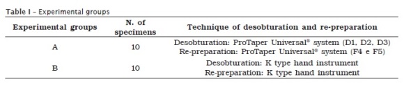
Next, the teeth were randomly divided into two groups (n = 10) (table I).
Prior to obturation, root canals were dried and the main gutta-percha point (Tanari®, Manaus, Amazonas, Brazil) was selected to tug fit in each root canal.
The obturation comprised Endofill® sealer (Dentsply/Maillefer, Ballaigues, Switzerland) and accessory gutta-percha points, always according to the manufacturer's instructions. Gutta-percha accessory points were embedded in the endodontic sealer and taken to the open spaces in the obturation mass. This procedure was repeated until the operator was not capable to insert the bidigital spacer (Dentsply/Maillefer, Ballaigues, Switzerland). All root canals obturation was executed by the same operator through lateral condensation technique and vertical adaptation with Paiva's condenser size #3 (SSWhite, Rio de Janeiro, Brazil).
After that, teeth were sealed with provisional restorative material (Cimpat®, Septodont Brasil Ltda., São Paulo, Brazil), with about 2 mm width below the root cervical surface.
The teeth were stored in flasks containing distilled water and incubated at 37ºC, 100% humidity for 30 days.
Root canal desobturation and re-preparation
After the aforementioned period, desobturation and re-preparation of groups A and B was performed as follows:
• Group A: root canal desobturation was executed through the retreatment instruments of the ProTaper Universal® system mounted in low-speed handpiece (Dabi Atlante, Ribeirão Preto, São Paulo, Brazil) powered by Endo Pro Torque® electric motor (VK Driller Equipamentos Elétricos Ltda., São Paulo, Brazil), at a constant speed of 300 rpm and 2 N/cm² torque 9. The enlargement movements with continuous rotation were executed in the following order: instruments size D1, D2, and D3 acting on the cervical, medium, and apical third, respectively. Root canal re-preparation was performed with ProTaper Universal® Finishing File size #4 (F4) and size #5 (F5) at working length.
• Group B: root canal desobturation was executed through K type hand instruments up to K type size #40 reached the working length and the operator did not see any filling material leaving root canal. Then, root canal preparation was performed with endodontic instruments size #45 and #50, by using filing movements, at the working length.
During the desobturation and re-preparation procedures, in both groups, root canal was copiously irrigated with 2 ml of 1% sodium hypochlorite at every instrument change followed by its aspiration. During root canal desobturation, irrigation was followed by the application of a solvent (Eucalyptol, Iodontosul – Industrial Odontológica do Sul Ltda., Porto Alegre, Brazil). In both groups, root canal final cleaning was executed with 17% EDTA for 3 minutes within root canals. During the last minute, the solution was activated by and endodontic instrument. EDTA was removed under irrigation/aspiration of 1% sodium hypochlorite.
The maximum use of each instrument was 5 usages, to diminish the fracture risk and maintain the cutting capacity. After 5 usages, the instruments were discarded and they were not used in the following steps.
Clearing teeth technique
After desobturation and re-preparation procedures, the teeth underwent clearing teeth technique.
Prior to this, pulp chamber cavity was sealed with glass ionomer cement (Vidrion R, SSWhite, Rio de Janeiro, Brazil) to avoid that the solutions employed during the clearing teeth technique did not interfere in the results.
Firstly, the teeth were decalcified in 5% chloridric acid (Farmácia Escola da Ulbra, campus Canoas/RS, Brazil) for 3 days, washed in running water for 24 hours and dehydrated in ascending alcohol solutions (70%, 80%, 90% e 100%) (Farmácia Escola da Ulbra, campus Canoas/RS, Brazil), for 4 hours each.
Teeth were cleared in methylsalicylate (Farmácia Escola da Ulbra, campus Canoas/RS, Brasil), where they were kept until the analysis.
Tooth examination
Tooth examination was performed with the aid of a stereomicroscope at x4 magnification, by two examiners specialized in Endodontics, who were previously trained and calibrated. Concerning to the examiners' calibration, Kappa test was used. The obtained results were submitted to statistical analysis through Fisher's exact test, with level of significance set at 5%.
Results
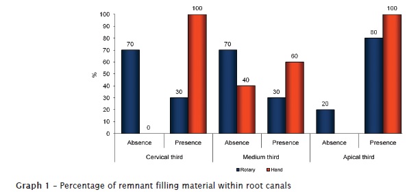
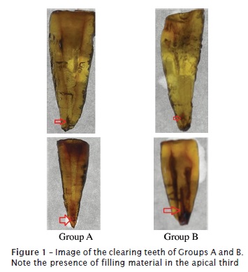
The results found in Kappa test between the two examiners were: 0.68 (cervical third analysis), 0.70 (medium third analysis), and 1 (apical third analysis). Cleaning comparison among experimental groups for the three root thirds showed significant association only in the cervical third analysis, exhibiting a higher filling material percentage in Group B than in Group A (p = 0.003) (graph 1 and figure 1).
Discussion
One of the main difficulties found during endodontic retreatment is the complete removal of the filling material within root canals.
Therefore, this study aimed to evaluate the efficacy of filling material removal by hand and rotary techniques, through clearing teeth technique.
For this purpose, we opted to use extracted human teeth, aiming to simulate better the clinical conditions in which endodontic retreatment is performed, similarly to the studies of Baratto-Filho et al. 3 and Sydney et al. 28. Garcia-Júnior et al. 13 and Uezu et al. 30 also employed lower bicuspids because, according to these authors, they are teeth presenting one single root canal with similar anatomical features, therefore allowing a greater standardization of the treatment conditions.
Similarly to the studies of Masiero & Barletta 19 and Ring et al. 21, tooth crown was removed at the enamel-cementum junction.
Root canal preparation/obturation and removal of the filling material were executed by a single operator, correspondingly to the studies of Imura et al. 17 and Garcia-Júnior et al. 13. According to Baugh & Wallace 8, larger apical preparations produce a greater reduction of bacteria and debris than more conservative apical preparations.
Accordingly, one can conclude that in endodontic retreatment cases, larger endodontic instruments should be used at the apical third aiming to promote a better cleaning of this area. In our study, root canal re-preparation was performed by using ProTaper Universal® instruments size F4 and F5, corresponding to size #40 and #50, respectively. This aimed to reprepare and remove the filling material at the apical third. In hand preparation, canals were reprepared with instruments size #45 and #50.
Root canal obturation employed gutta-percha points and Endofill® sealer, according to the lateral condensation technique. This technique was also performed in the study of Sydney et al. 28.
Concerning to the teeth's storage after obturation, we believe that their maintenance in a humidifier keeps the situation closer to the clinical reality. Sae-Lim et al. 23, Barletta & Lagranha 4 and Garcia-Júnior et al. 13 also kept their samples in 100% humidity at 37°C.
Eucalyptol was chosen in this study from a variety of different solvents already recommended for endodontic retreatment, including xylene, chloroform, and halothane, among others [18]. According to Hunter et al. 16, eucalyptol is one of the safest and most effective solvents.
The analysis method of this study was the clearing teeth technique because it is of easy execution, low cost, and great clinical significance, once it shows the tooth's internal morphology without losing the root's integrity 1.
By analyzing the results, it can be observed that none technique was capable of completely removing the filling material within root canals, which confirms the findings of Somma et al. 27, Barletta et al. 5, Sydney et al. 28 and Uezu et al. 30.
In the comparison of the two desobturation techniques, we did not verify any statistical difference between hand and rotary instruments. This result was also seen in the study of Schirrmeister et al. 24, in which ProTaper Universal® system obtained results similar to Hedströen, FlexMaster and Race system regarding to filling material removal, both in straight and curved canals. On the other hand, the studies of Carvalho Maciel & Zaccaro Scelza 9, Saad et al. 22 and Gu et al. 15 found that ProTaper Universal® system was more effective than hand instrumentation during the filling material removal.
The greatest amount of filling material remnant at the apical third than at medium and cervical thirds is in agreement with the studies of Gergi & Sabbagh 14, Zanettini et al. 31 and Duarte et al. 10, but unlike the study of Uezu et al. 30. According to Só et al. 26, the apical third is a critical zone, which demands a considerable enlargement for the cleaning and shaping procedures.
Finally, Sydney et al. 28 affirmed that there would be a long way to go until root canal desobturation and re-preparation are completely automated. According to Ferreira et al. 11, it is necessary to complement the hand desobturation with rotary system in cases of endodontic retreatment.
Conclusion
According to the results obtained, it can be concluded that:
• Neither hand nor rotary technique was capable of completely removing the filling material within root canals;
• No statistical differences were found between the two experimental groups. Only at cervical third, it was observed a smaller amount of filling material with the use of ProTaper Universal® system;
• Root canal's apical third presented the greatest amount of filling material remnant, regardless of the used operative technique.
References
1. Azeredo RA. Contribuição ao estudo da anatomia do canal radicular de incisivos inferiores, utilizando de cortes macroscópicos e da diafanização. Rev Odonto UFES. 1999;1(1):48-53. [ Links ]
2. Baratto-Filho F, Ferreira EL, Fariniuk LF. Efficiency of the 0.04 taper ProFile during the re-treatment of gutta-percha-filled root canals. Int Endod J. 2002;35(8):651-4.
3. Baratto-Filho F, Vanni JR, Limongi O, Leonardi DP, Scaini F, Fagundes FS. Retreatment of Thermafill fillings with the ProFile .04 system at 350 or 2000 rpm. RSBO. 2006;3(2):26-31.
4. Barletta FB, Lagranha SB. Análisis comparativo in vitro de diferentes técnicas de desobturación de conductos radiculares. Endodoncia. 2002;20(3):189-96.
5. Barletta FB, Reis MS, Wagner M, Borges JC, Dall'Agnol C. Computed tomography assessment of three techniques for removal of filling material. Aust Endod J. 2008;34:101-5.
6. Barrieshi-Nusair KM. Gutta-percha retreatment: effectiveness of nickel-titanium rotary instruments versus stainless steel hand files. J Endod. 2002;28(6):454-6.
7. Basso AL, Silva Neto UX, Westphalen VPD. Análise radiográfica do retratamento endodôntico realizado pela técnica manual, sistema profile e protaper. JBE. 2003;4(14):203-7.
8. Baugh D, Wallace J. The role of apical instrumentation in root canal treatment: a review of the literature. J Endod. 2005;31(5):333-40.
9. Carvalho Maciel AC, Zaccaro Scelza MF. Efficacy of automated versus hand instrumentation during root canal retreatment: an ex vivo study. Int Endod J. 2006;39(10):779-84.
10. Duarte MAH, Só MVR, Cimadon VB, Zucatto C, Vier-Pelisser FV, Kuga MC. Effectiveness of rotary or manual techniques for removing a 6-year-old filling material. Braz Dent J. 2010;21(2):148-52.
11. Ferreira EL, Baratto-Filho F, Fidel RAS, Fariniuk LF, Rached RN. The performance of ProTaper system during the endodontic retreatment. RSBO. 2006;3(1):64-8.
12. Ferreira JJ, Rhodes JS, Ford TR. The efficacy of gutta-percha removal using ProFiles. Int Endod J. 2001;34(4):267-74.
13. Garcia-Júnior JS, Silva Neto UX, Carneiro E, Westphalen VPD, Fariniuk LF, Fidel RAS et al. Avaliação radiográfica da eficiência de diferentes instrumentos rotatórios no retratamento endodôntico. RSBO. 2008;5(2):41-9.
14. Gergi R, Sabbagh C. Effectiveness of two nickel-titanium rotary instruments and a hand file for removing gutta-percha in severely curved root canals during retreatment: an ex vivo study. Int Endod J. 2007;40:532-7.
15. Gu LS, Ling JQ, Wei X, Huang XY. Efficacy of ProTaper Universal rotary retreatment system for gutta-percha removal from root canals. Int Endod J. 2008;41(4):288-95.
16. Hunter RK, Doblecki W, Pelleu GBJ. Halothane and eucalyptol as alternatives to chloroform for softening gutta-percha. J Endod. 1991;17(7):310-1.
17. Imura N, Zuolo ML, Ferreira MOF, Novo NF. Effectiveness of the canal finder and hand instrumentation in removal of gutta-percha root fillings during root canal retreatment. Int Endod J. 1996;29(6):382-6.
18. Kaplowitz GJ. Evaluation of gutta-percha solvents. J Endod. 1990;16(11):539-40.
19. Masiero AV, Barletta FB. Effectiveness of different techniques for removing gutta-percha during retreatment.
20. Peciuliene V, Reynaud AH, Balciuniene I, Haapasalo M. Isolation of yeasts and enteric bacteria in root-filled teeth with chronic apical periodontitis. Int Endod J. 2001;34(6):429-34.
21. Ring J, Murray PE, Namerow KN, Moldauer BI, Garcia-Godoy F. Removing root canal obturation materials: a comparison of rotary file systems and re-treatment agents. J Am Dent Assoc. 2009;140(6):680-8.
22. Saad AY, Al-Hadlaq SM, Al-Katheeri NH. Efficacy of two rotary NiTi instruments in the removal of Gutta-Percha during root canal retreatment. J Endod. 2007;33(1):38-41.
23. Sae-Lim V, Rajamanickam I, Lim BK, Lee HL. Effectiveness of ProFile .04 taper rotary instruments in endodontic retreatment. J Endod. 2000;26(2):100-4.
24. Schirrmeister JF, Wrbas KT, Meyer KM, Altenburger MJ, Hellwig E. Efficacy of different rotary instruments for gutta-percha removal in root canal retreatment. J Endod. 2006;32(5):469-72.
25. Siqueira Jr JF. Aetiology of root canal treatment failure: why well-treated teeth can fail. Int Endod J. 2001;34(1):1-10.
26. Só MV, Saran C, Magro ML, Vier-Pelisser FV, Munhoz M. Efficacy of ProTaper retreatment system in root canals filled with gutta-percha and two endodontic sealers. J Endod. 2008;34(10):1223-5.
27. Somma F, Cammarota G, Plotino G, Grande NM, Pameijer CH. The effectiveness of manual and mechanical instrumentation for the retreatment of three different root canal filling materials. J Endod. 2008;34(4):466-9.
28. Sydney GB, Kowalczuck A, Deonizio MD, Batista A, Ramos JMO, Travassos R. Retratamento: Protaper para retratamento X técnica híbrida manual. Robrac. 2008;17(44):166-73.
29. Taşdemir T, Er K, Yildirim T, Celik D. Efficacy of three rotary NiTi instruments in removing gutta-percha from root canals. Int Endod J. 2008;41(3):191-6.
30. Uezu MKN, Nabeshima CK, Britto MLB. Comparação do remanescente de material obturador nos diferentes terços do canal radicular após uso dos desobturadores ProTaper. Rev Odontol Unesp. 2010;39(6):332-5.
31. Zanettini PR, Barletta FB, de Mello Rahde N. In vitro comparison of different reciprocating systems used during endodontic retreatment. Aust Endod J. 2008;34(3):80-5.
 Correspondence:
Mário Luiz Pinto de Queiróz
Rua Jarí, n.º 671, apto. 501 – Passo D'Areia
CEP 91350-170 – Porto Alegre – RS – Brasill
E-mail: mariolpq@gmail.com
Received for publication: April 13, 2011
Accepted for publication: June 21, 2011
