


Serviços Personalizados
Artigo
 pdf em Inglês
 Artigo em XML
 Referências do artigo
 Enviar este artigo por email
Links relacionados
Compartilhar

 Permalink
RSBO (Online)
versão On-line ISSN 1984-5685
RSBO (Online) vol.9 no.1 Joinville Jan./Mar. 2012
Case Report Article
Pleomorphic adenoma of minor salivary gland: a case report
Amit Aggarwal I; Ravinder Singh I; Soheyl Sheikh I; Shambulingappa Pallagatti I; Isha Singla I
I Department of Oral Medicine and Radiology, M.M. College of Dental Sciences and Research – Mullana – Ambala – Haryana – India.
ABSTRACT
Introduction : World literature suggests salivary gland tumors account for less than 3% of the head and neck tumors and benign pleomorphic adenoma of minor salivary glands arising de novo is very rare.Objective,case report and Conclusion: A case of pleomorphic adenoma of minor salivary glands in the buccal mucosa in a 55 year-old female is discussed. It includes review of literature, clinical features, histopathology, radiological findings and treatment of the tumor, with emphasis on diagnosis. The salivary glands may present with a diverse range of lesions presenting a challenge to even the most experienced clinician and pathologist. Resection with surrounding dispensable normal tissues is the key to successful treatment of such tumors.
Keywords: pleomorphic adenoma; minor salivary gland; computed tomography; histopathology.
Introduction
Salivary gland tumors account for less than 3% of the head and neck tumors. They are more common in adults than in children. Among all salivary gland tumors, pleomorphic adenoma is the most frequently encountered lesion, accounting for approximately 60% of all salivary gland neoplasms 1. Tumors arising in the minor salivary glands account for 22% of all salivary gland neoplasms 8. An important point to be noted here is that majority of them are malignant with only 18% being benign 4.
Pleomorphic adenoma is of glandular origin in the head and neck 3, usually presenting as a mobile slowly growing, painless firm swelling that does not cause ulceration of the overlying mucosa 2. It can appear at any age and appear between the fifth and seventh decades of life. It mainly affects women 9. The percentage of minor salivary gland tumors is higher in the Africans 5.
Minor salivary gland tumors represent a heterogeneous group of neoplasms, with a broad range of histological types and growth patterns. These tumors have a high recurrence rate when surgical removal is incomplete and the possibility of malignant transformation must be taken into consideration 9.Minor salivary gland tumors are fundamentally located on palate, lips, cheek mucosa, tongue and floor of the mouth 9.
The purpose of this article is to report a case of pleomorphic adenoma of minor salivary gland with the characteristic clinical, radiological, histological features and treatment.
Case report
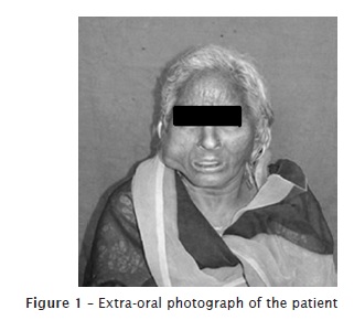
A 55-year-old woman referred to the Department of Oral Medicine and Radiology with an asymptomatic swelling on right side of her face of 9 years duration. There was no history of bleeding, pain or sensory changes or trauma (figure 1). Her past dental/medical history was not significant.
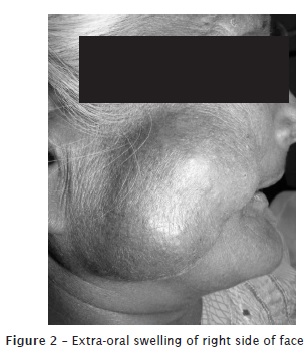
Physical examination revealed facial asymmetry due to swelling on the right side of face. The swelling was irregular in shape and had a smooth surface (figure 2). The skin over the swelling appeared normal. The well defined swelling extended from the lateral border of the right eye to the right lower border of the mandible; anteroposteriorly, 2 cm lateral to ala of the nose to 2 cm in front of the lobule of the right ear, approximately 4.5 × 3 cm in size. The right ear lobule was not raised. There was no discharge or any other secondary findings.
On palpation, the swelling was non-tender. The swelling was firm in consistency, non-fluctuant, non-reducible, compressible and non-pulsatile. The temperature over the swelling was normal. The skin over the swelling was pinchable.
Panoramic radiographic examination revealed edentulous maxillary and mandibular ridges. No abnormality was detected.
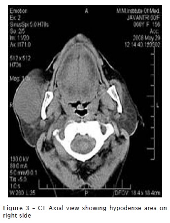
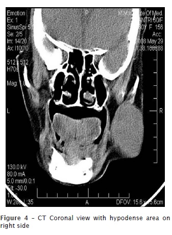
Computed tomography of the lesion showed a large well defined encapsulated homogeneously hypodense soft tissue swelling of size 3.3 cm (ML) X 5.6 cm (AP) X 4.4 cm (SI) in the minor salivary gland of buccal mucosa near the superficial lobe of parotid gland in the antero-inferior part of the right parotid gland extending more anteriorly. It was extending into the soft tissue of cheek (figures 3, 4).
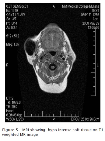
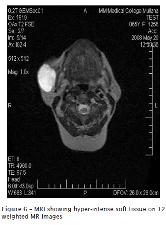
Magnetic Resonance Imaging showed hypo-intense soft tissue with few hyper-intense areas on T1 weighted MR image. Hyper-intense soft tissue was seen with hypo-intense areas on T2 weighted MR images (figures 5, 6).
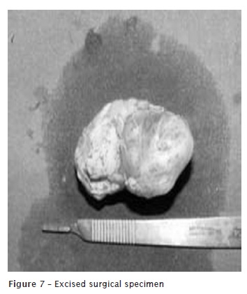
Based on the clinical and radiological appearance, a provisional diagnosis of benign pleomorphic adenoma of minor salivary gland was made. Surgical excision for tumor was performed under general anesthesia (figure 7). Histologically, the features were consistent with pleomorphic adenoma.
Discussion
The categorization of salivary gland tumors as suggested by WHO pathological classification has proved to be most useful in daily practice and has been adopted worldwide 11. According to Vicente et al. in 2008, most studies of salivary neoplasms include both the major and the minor salivary glands, and few articles focus only on minor salivary gland tumors 9.
Tumors originating in the minor salivary glands are uncommon neoplasms of upper aero digestive tract 10. In 2002, Jansisyanont et al. suggested that a total of 80 minor salivary gland tumors were identified in 49 female patients and 31 male patients and the ratio range from 1.2:1 to 1.9:1 5. In 2009, Oliveira et al. reported that in his review of 599 salivary gland tumors, 78.3% cases were benign 6. Pleomorphic adenomas are usually well demarcated or encapsulated but extension of tumor into the capsule is a common feature and sometimes lobules of tumor may appear to be completely separated from the main tumor mass 7.
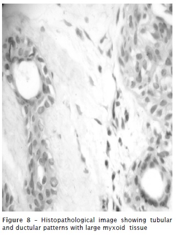
Histopathological features of pleomorphic adenoma are a variable pattern of epithelium in a loosely fibrous stroma which may be myxoid, chondroid or mucoid. The epithelium is usually arranged in sheets or strands, and ductal structures, often bilayered, are typical. In the minor glands,lesions are often more solid or cellular than those seen in the major glands, and the myoepithelial cells are often polygonal with a pale eosinophilic cytoplasm giving an epithelioid or plasmacytoid phenotype 7.
Our diagnosis was confirmed by tubular and ductular patterns lined by single to double layered cuboidal cells with large darkly stained nucleus in centre and eosinophilic cytoplasm interspersed between large myxoid areas. Connective tissue capsule is absent (figure 8).
Conclusion
The salivary glands may show a diverse range of lesions presenting a challenge to even the most experienced clinician and pathologist. Pleomorphic adenoma of minor salivary gland is a tumor of rare occurrence and a diagnosis should be made carefully lest a major salivary gland be resected. A point to be noted here is that the ear lobule was not raised, thus clinically indicating that the swelling may not be of parotid gland origin. High index of suspicion and an adequate clearance of the tumor with a cuff of surrounding dispensable normal tissues is the key to successful treatment of such tumors. Long-term multicenter studies for comparison of treatment strategies are needed in the coming decades to guarantee further optimization of tumor management on a profound clinical and scientific basis.
References
1. Dhanuthai K, Sappayatosok K, Kongin K. Pleomorphic adenoma of the palate in a child: a case report. Med Oral Patol Oral Cir Bucal. 2009 Feb;14(2):73-5. [ Links ]
2. Dalati T, Hussein MR. Juvenile pleomorphic adenoma of the cheek: a case report and review of literature. Diagnostic Pathology. 2009 Mar:4(32):1-5.
3. Fernández JR, Micas MM, Tello FJ, Berjón J, Montalvo JJ, González GF et al. Metastatic benign pleomorphic adenoma. Report of a case and review of the literature. Med Oral Patol Oral Cir Bucal. 2008 Nov;13(3):193-6.
4. Hakeem AH, Hazarika B, Pradhan SA, Kannan R. Primary pleomorphic adenoma of minor salivary gland in the parapharyngeal space. World Journal of Surgical Oncology. 2009 Mar;7(85):1-4.
5. Jansisyanont P, Blanchaert RH, Ord RA. Intraoral minor salivary gland neoplasm: a single institution experience of 80 cases. Int J Oral Maxillofac Surg. 2002 Sep;31:257-61.
6. Oliveira FA, Duarte ECB, Taveira CT, Maximo AA, Aquino EC, Alencar RC et al. Salivary gland tumor: a review of 599 cases in a Brazilian population. Head and Neck Pathol. 2009 Sep;3:271-5.
7. Speight PM. Update on diagnostic difficulties in lesions of the minor salivary glands. Head and Neck Pathol. 2007 Jan;1:55-60.
8. Thoeny HC. Imaging of salivary gland tumors. Cancer Imaging. 2007 Apr;7:52-62.
9. Vicente OP, Marqués NA, Aytés LB, Escoda CG. Minor salivary gland tumors: a clinicopathological study of 18 cases. Med Oral P Patol Oral Cir Bucal. 2008 Nov;13(9):582-8.
10. Vuhahula EAM. Salivary gland tumors in Uganda: clinical pathological study. African Health Sciences. 2004 Sep;4(1):15-23.
11. Valizadeh Y, Mohagheghi MA. Review of 103 cases of minor salivary gland tumors. Acta Medica Iranica. 1995 Nov;33(1):22-6.
 Correspondence:
Isha Singla
Post graduate student – Department of oral medicine and radiology
M.M. College of Dental Sciences and Research
Pin code 133203 – Mullana – Ambala – Haryana – India
E-mail:drishasingla@gmail.com
Received for publication: April 14, 2011.
Accepted for publication: July 11, 2011.
